FF: Pilus

Anatomy Def: Pilus is the scientific term for a single hair; pili means multiple hairs

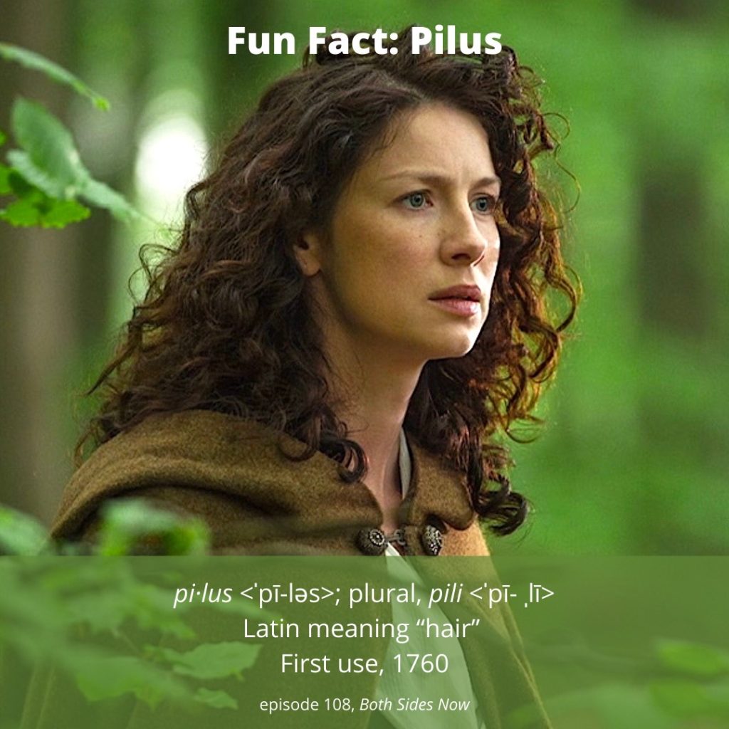
Outlander Def: Claire-hair forms a halo of curly pili! (psst… Jamie loves it!)

Learn about hair in great detail in Anatomy Lesson #6, “Claire’s Hair, Jamie’s Mane or Jesus H. Roosevelt Christ! 

Hey, anatomy students! How about a brief pilus quiz? No grading, I promise.

What type of pili inhabit your head? Turns out, there are different systems of classifications, but the following one is practical and easy. 

Shape of your pili:

    • Straight?
    • Wavy?
    • Curly?
    • Coily?
    • None? 😉

Type of pili strand: 

    • Fine?
    • Medium?
    • Coarse?

Amount of hair? (based on circumference of “full hair ponytail”). If you don’t have a ponytail, then guess.

    • Thin (ponytail is 2” or less)
    • Normal (ponytail is 2”-4”)
    • Thick (polytail is > 4”)

How did you do? Wouldn’t you know it, all such characteristics have been described and worked out. 🤗 There are even subcategories of hair if you want to read more! 

Pili are fascinating for many reason. First and foremost, pili are products of skin, our body’s largest organ! The skin of “average adults” weighs about eight pounds, with a surface area of 22 ft²!  😲 The larger one is, the more weight and more surface area is taken up by skin.

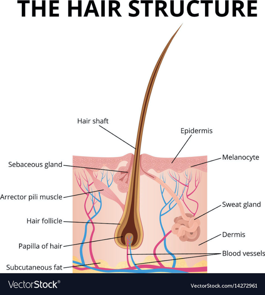
The skin is equipped with various appendages including pili, erector pili muscles , nails, and various glands. 

Turns out, hair is far more complex than one might imagine. Read on for more fascinating deets!

Hair growth differs depending on body region (duh 😉):

    • Glabrous: Regions sans hair – palms, soles, external genitalia, lips, back of ear, and scars.
    • Terminal: Thick, coarse hair – beard, pubic area, eyelashes, brows, scalp.
    • Vellus: Thin, fine, light-colored hair typical of childhood and adult women. In female adults found on eyelids, face, chest, etc.  Vellus hair can convert to terminal hair under the influence of androgens.

Next, anatomy divides the pilus into two parts:

    • Follicle: Part embedded in the dermis – the only living part of a hair.
    • Shaft: Thin filamentous part that extends beyond skin surface – non-living part

Follicle: A follicle is the part of a pilus that lies below the skin surface. Pull out a strand of head hair, and observe a pale enlargement on the end that was embedded in skin – this is the bulb or root of the follicle. The shaft is produced by the root.

Follicles are lined with skin stem cells that can re-grow a hair after it is lost. It may also regrow skin after various wounds, such burns. Here, stem cells produce new skin cells that grow out of the follicle and spread across the damaged surface to help cover the injury.  This is effective if the wound is relatively small; larger wounds may require skin grafts. Lastly, the new skin is a type of scar tissue which does not regrow appendages.

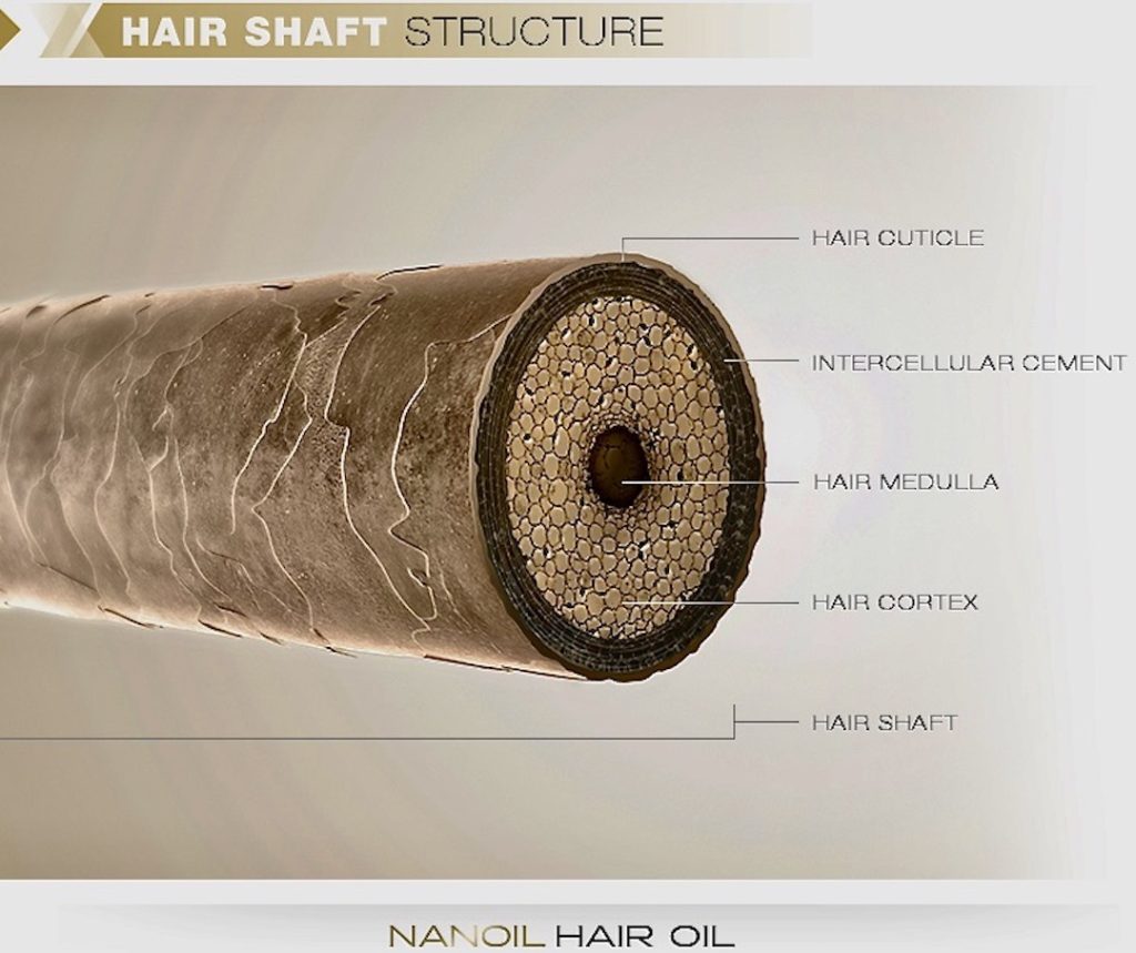
Shaft: The shaft is 2-3 layers of non-living material:

    • Cuticle: consists of thin, flat cells overlapping like shingles of a roof.
    • Cortex: Rod-like bundles of alpha keratin, a protein that strengthens the shaft. This layer also gives hair its color.
    • Medulla: Unstructured area in the center – only present in large pili.

People with straight hair have round shafts. People with wavy, curly or coiled hair have oval or flattened shafts. The follicle itself determines the shaft shape and genetics orchestrates the follicle to do its unique thing🤓!  

Growth: Each human hair follows its own cycle, at its own pace, including periods of growth and times of quiescence. Think about it! If all our pili were on the same cycle, we would molt! 😳

Angle: You should also know that the shaft does not grow upright; it emerges at a slant. 

Try this: Check the angle of growth of your hair: place your forearm on a flat surface with the palm down. Examine your forearm hairs and see that they are angled toward the little finger side of the forearm. That’s the slant!

Arrector Pili Muscle: Microscopic bundles of smooth muscle (meaning these cannot be voluntarily contracted) are attached to the follicle. If these muscles contract, they pull on the follicles causing shafts to stand upright, creating “goose bumps.” Watch this video about the arrector pili muscle for perspective.  Contraction happens when we are cold or creeped out! 🥶😱

Contraction of arrector pili muscles also causes oil glands to release their product (sebum) into their respective follicles following the pilus shaft. 

Pili are highly valued in many societies which explains the vast sums of money spent on hair products each year; almost 80 million dollars in US in 2019 –  down from 90 million spent two years earlier.

Read about Claire’s-Hair in Outlander book. Diana has provided us with ample descriptions of her follicles and shafts.  Here are three iconic descriptions of her amazing pili!!! 😲

The wind was rising and the very air of the bedroom was prickly with electricity. I drew the brush through my hair, making the curls snap with static and spring into knots and furious tangles!

… “Mo duinne?”…“It means ’my brown one.’ ”He raised a lock of hair to his lips and smiled, with a look in his eyes that started all the drops of my own blood chasing each other through my veins. Rather a dull color, brown, I’ve always thought,”….”No, I’d not say that, Sassenach. Not dull at all.”  He lifted the mass of my hair with both hands and fanned it out. “It’s like the water in a bern, where it ruffles over the stones. Dark in the wavy spots, with bits of silver on the surface where the sun catches it.”

…”Fretful porpentine, was it?” he asked. He tilted his head, examining me inquisitively. “Mmm,” he said, running a hand over his head to smooth down his own hair. “Fretful, at least. You’re a fuzzy wee thing when ye wake, to be sure.” He rolled over toward me, reaching out a hand. “Come here, my wee milkweed.”  🥰

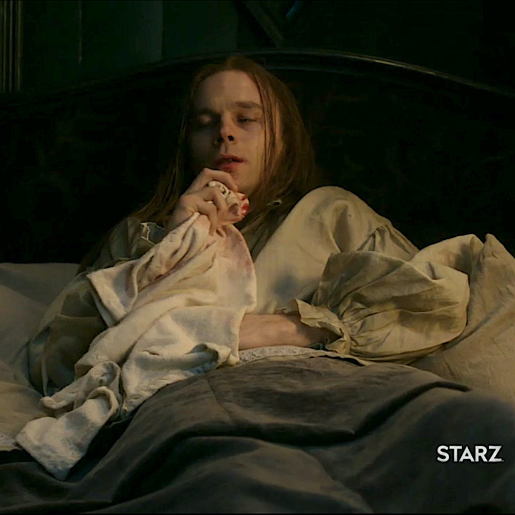
See Claire glorious crown of pili in Outlander, episode 109. Both sides Now!

Grateful for each and every one of my pili! How about you?

The deeply grateful,

Outlander Anatomist

Follow me on:

Photo Credits: Sony/Starz; www.vectorstock.com; nanoil.com

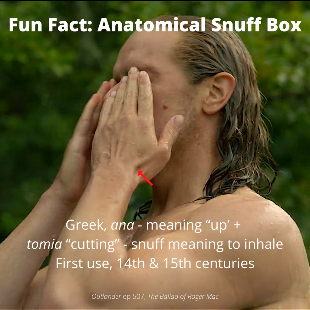
Fun Fact: Anatomical Snuff Box


Anatomy Def: A triangular hollow on the back of hand, at the base of thumb 

Outlander Def: Up-to-snuff divot of Jamie’s hand – holds Claire’s kisses! 💋

Students, before we delve into our mini-lesson, it is worthwhile to consider the word snuff, which enjoys many meanings – some old, some new:

    • charred part of a candle wick
    • umbrage or offense
    • huff (chiefly Scottish)
    • extinguish 
    • execute or kill
    • a type of film
    • forcible inhalation
    • sniff
    • powdered tobacco

The last definition refers to a smokeless tobacco made of pulverized tobacco leaves. In the late 15th century, members of Christopher Columbus’s crew observed indigenous peoples of the Lesser Antilles inhaling ground tobacco. Labeled snuff, the practice of sniffing the snuff-stuff took hold in Europe in the 1500s. 👃🏻

Warning: Similar to other tobacco products, snuff contains nicotine and numerous carcinogens. Snuffing is also addictive and associated with increased risk for certain cancers. 🚫

Now, onward and upward with our Fun Fact!

Learn about the awesome hand and its components in Anatomy Lesson #22: Jamie’s Hand – Symbol of Sacrifice and Anatomy Lesson #23: Harming Hands – Helping Hands – Healing Hands, both lengthy and detailed lessons. Why? Because, the hand is one of the most elegant and detailed regions of human anatomy, and the anatomical snuff box (ASB) is a fascinating bit of its story. 

Found on the back of the hand, at the base of the thumb, the ASB was used to snort snuff, hence its name! A pinch of the stuff was placed into the divot and then brought to one nostril and forcibly inhaled. Ditto on the other side. Reportedly, it gave the inhaler an instant and significant nicotine ‘hit’.

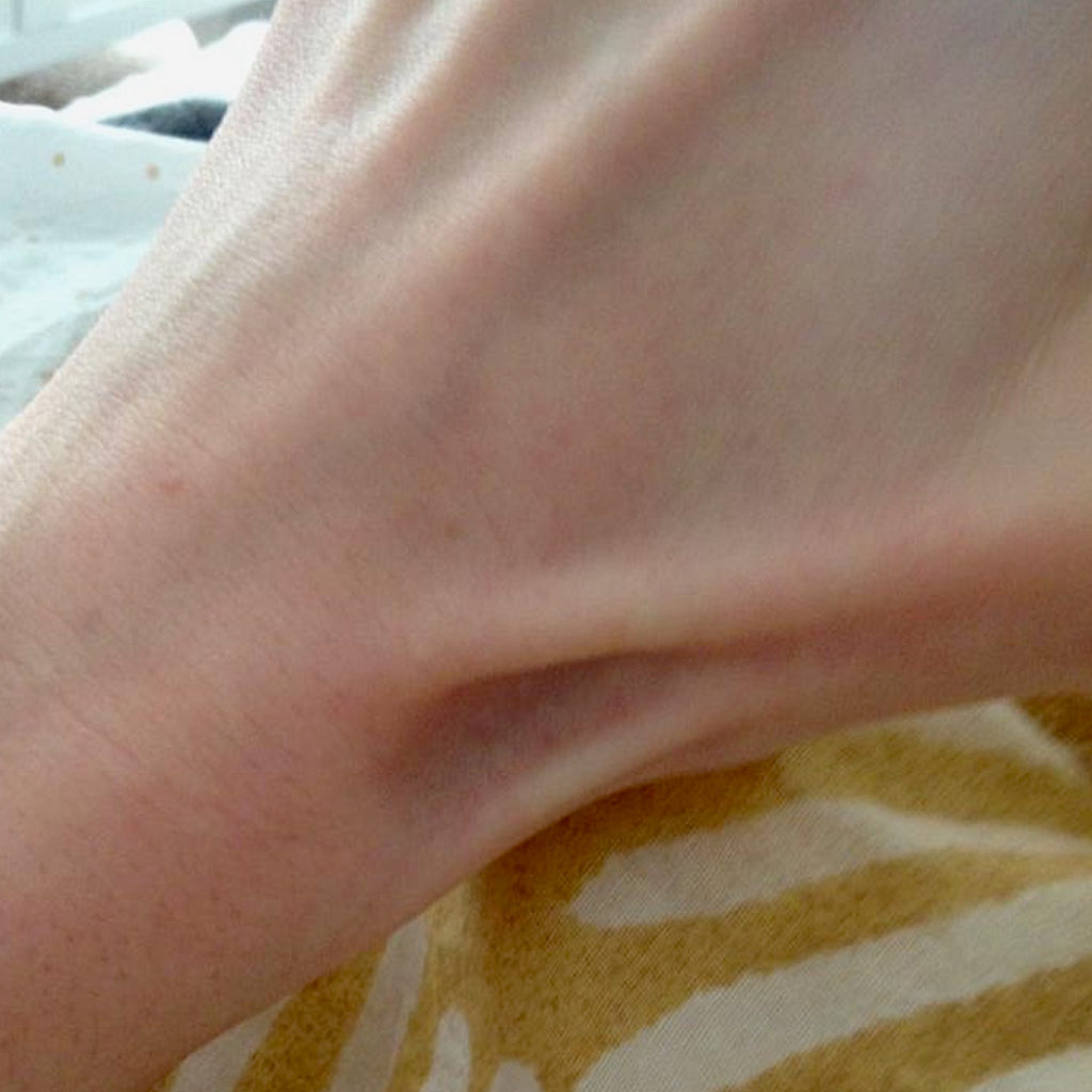
The ASB has a range of appearances. In some people, the divot is deep and obvious (below image); in others, it is less noticeable. Either way, the divot is most evident with the thumb extended (as in hitchhiking). 

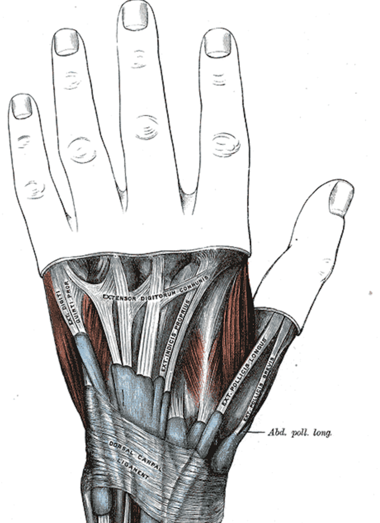
ASB is formed by three tendons of the back of the hand, extensor pollicis longus, extensor pollicis brevis, and abductor pollicis longus, as shown on this image from Grey’s Anatomy:

Why is the ASB important, other than for snuffing? Because, two crucial structures are associated with it: 

    • radial artery
    • scaphoid bone

The all-important radial artery passes through the floor of the ASB, where its pulse can be detected; it is a major contributor to blood circulation of the hand. Its loss compromises roughly half of the hand’s the blood supply.

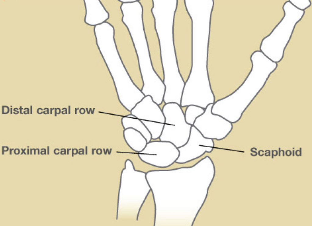
The scaphoid bone (Latin, meaning boat-shaped), a bone of the wrist helps form the base of the ASB. This oddly-shaped, small bone confers mobility but not stability, to the wrist. 

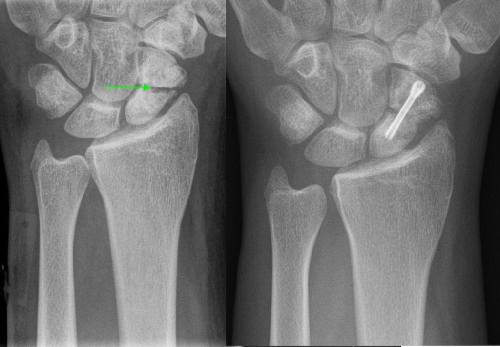
A person who “falls onto an outstretched hand” experiences a FOOSH, wherein the heel of the hand is forcefully driven into a surface by the body weight. Thus, the scaphoid bone is at high risk for fracture. 

Below, the L x-ray shows a broken scaphoid (green arrow); the R x-ray shows a screw securing the fragments. Scaphoid fracture is common and is a leading cause of medico-legal challenges.

x-ray by Hellerhoff 

Try This #1: Want to see your own ASB? Lay your non-dominant hand on a flat surface, palm down. Fully extend your thumb. You should see two tendinous ridges on the back of your hand. The tendon nearest your little finger is extensor pollicis longus, the other will be both extensor pollicis brevis and abductor pollicis longus. 

Try This #2: You may be able to feel the pulse of your radial artery by placing the tip of the middle finger into the ASB and pressing toward the thumb tip. Can you feel it? Do not use your index finger to test the radial pulse as the index finger has its own pulse which may fool you.

Try This #3: You may also palpate the mound of scaphoid bone by pressing the same finger into the ASB but toward the wrist. This is the wee bone often injured by a FOOSH.

Hopefully, you now understand the history and importance of the Anatomic Snuff Box!

Read about Jamie’s ablutions in preparation for war in Diana’s fifth big book, The Fiery Cross:

I had done it often enough to recognize this particular ritual when I saw it. Jamie was not merely washing; he was cleansing himself, using the cold water not only as solvent but as mortification. He was preparing himself for something, and the notion made a small, cold trickle run down my own spine, chilly as the spring water.

Sure enough, after the third bucketful, he set it down and shook himself, droplets flying from the wet ends of his hair into the dry grass like a spatter of rain.

He took his dirk from its discarded sheath, and with no hesitation, drew the edge across the fingers of his right hand. I could see the thin dark line across his fingertips, and bit my lips. He waited a moment for the blood to well up, then shook his hand with a sudden hard flick of the wrist, so that droplets of blood flew from his fingers and struck the standing stone at the head of the pool.

See Jamie’s anatomical snuff box as he conjures up his uncle, War Chief, Dougal MacKenzie! Visibility of his ASB indicates the tension in his hand as he prepares for battle in episode 507, The Ballad of Roger Mac!

Anatomy, Anatomy, everywhere,

So exciting when we share! 🤗


The deeply grateful,

Outlander Anatomist

Follow me on:

Photo Credits: Sony/Starz; Grey’s Anatomy; www.assh.org; www.oceanortho.com; www.teachmeanatomy.info; www.wikipedia.org;

Fun Fact: Necrosis


Anatomy Def: Necrosis means tissue death, as caused by disease, injury, or loss of blood supply.

Outlander Def: Jamie’s dreadful, blackening wound courtesy of a fang-bang! 🐍

Doubtless, Jamie’s horrific thigh wound in Outlander, episode 509, Monsters and Heroes, caused each of us to cringe! 😣

Learn about necrosis in Anatomy Lesson #37: Outlander Owies – Mars and Scars!

Depending on the species of snake, venoms cause predictable signs and symptoms. Within minutes to hours after the bite, Jamie exhibits most of the following:

    • Puncture marks at the wound ✔︎
    • Redness, swelling, bruising, bleeding, or blistering around the bite ✔︎
    • Severe pain and tenderness at the site of the bite ✔︎
    • Increased salivation and sweating ✔︎
    • Labored breathing (in extreme cases, breathing may stop altogether) ✔︎
    • Rapid heart rate, weak pulse, low blood pressure ✔︎
    • Numbness or tingling around face and/or limbs ✔︎
    • Disturbed vision (?)
    • Nausea, vomiting, or diarrhea (?)
    • Metallic, mint or rubber taste in mouth (?)
    • Muscle twitches (?)

Back at the surgery, Claire quickly ascertains that Jamie’s wound is infected!

What might be the source(s) of the infection? Here are some ideas:

    • Bacteria from the snake’s fangs and saliva
    • Bacteria from Roger’s knife
    • Bacteria in Roger’s saliva, as he drains Jamie like a vampire (Sorry, Bree)
    • Dirty bandage
    • Forest floor
    • All the above? 😲

As recorded in The Fiery Cross (TFC),  Claire determines that Jamie’s infected wound shows signs of septicemia, broadly known as blood poisoning.

The foot and ankle on the injured side were still warm and pink—or rather, red. That was a good sign, insofar as it meant the deeper circulation was intact. The problem was to improve circulation near the wound, enough to prevent a massive die-off and sloughing of tissue. The red streaks bothered me very much indeed, though; they could be only part of the hemorrhagic process, but it was more likely that they were the early signs of septicemia—blood poisoning.

Claire must halt the infection quickly because sepsis is life-threatening! Using the serpent’s fangs she injects Jamie with home-brewed penicillin to kill the blood-borne organisms.

Add insult to injury: the wound  also exhibits necrosis! Claire evaluates the red-black tissue at the bite-site (see above image) –  this tissue is dead. 

What causes necrosis? Most likely, loss of blood supply. Necrotic tissue must be removed to avoid loss of muscle mass, gangrene, and possible amputation! 🦵Removing  the dead tissue is a process known as debridement.  

Alrightie, then. How might Claire debride the wound to remove dead tissue but spare the living? Surgical removal is always an an option but difficult to perform with the precision required. If she accidentally leaves necrotic tissue, it can still turn gangrenous. If she accidentally removes living tissue along with the dead, it further compromises Jamie’s thigh tissues. Hum…. 🤔

Well, here’s an idea….. 💡 How about someone rustles up some fly maggots, blow-fly maggots to be precise – Bree (in TFC) or Josiah (in ep 510)??? 

So, into the wound go the maggots and its suppertime! 😋 As gross as it seems, not only will maggots clean Jamie’s wound of necrotic tissue while sparing the living,  his wound will heal faster. Truth! 

“Nothing but a mouth and a gut,” declares Claire (TFC)!

So, where did Claire get the idea of using maggots in Jamie’s wound? From her medical training, of course.

Maggot therapy is the introduction of live maggots into human or animal wounds to remove necrotic tissue but spare the living. (Psst….Just so you know, in today’s medicine, maggots are disinfected before use.) 🤫

Understand, maggot therapy is not new. Aboriginals of Australia and Mayan tribes in Central America frequently used fly larva to clean wounds. Clearly, these folks were observant of the world around them! 👀

When did Western medicine start to employ maggots? In the 1500s, some field surgeons (e.g. Ambroise Paré) observed that wounds infested with maggots healed faster but failed to credit the wee beasties as healers. The first documented therapeutic use of maggots in the US is credited to Confederate medical officer, Dr. J.F. Zacharias, who reported during the American Civil War that:

“Maggots … in a single day would clean a wound much better than any agents we had at our command … I am sure I saved many lives by their use.

Today, some modern hospitals employ medical maggots (Maggots, LLC? 😜) to solve otherwise vexing cases that do not respond to traditional therapies.

One last note – do we believe that a poisonous snake bit Jamie? Nope.

I love Jamie’s (Diana’s) succinct and accurate comparison of venom vs. poison; from Diana’s fourth book, Drums of Autumn:

“Venemous,” Jamie corrected him. “If it bites you and makes ye sick, it’s venomous; if you bite it and it makes ye sick, it’s poisonous.”

Read about Jamie’s horrific snake bite injury and Claire’s therapy in Diana’s fifth book, The Fiery Cross.

“It looks nastier than it is,” I said reassuringly, hearing Marsali’s unguarded gasp at the sight. That was true, but the reality was nasty enough. The slash marks were crusted black at the edges, but still gaped. Instead of the sealing and granulation of normal healing, they were beginning to erode, the exposed tissues oozing pus. The flesh around the wounds was hugely swollen, black and mottled with sinister reddish streaks. I bit my lip, frowning as I considered the situation. I didn’t know what kind of snake had bitten him—not that it made much difference, with no antivenin for treatment—but it had plainly had a powerful hemolytic toxin. Tiny blood vessels had ruptured and bled all over his body—internally, as well as externally—

See Jamie receive maggot therapy for his necrotic wound in Outlander, episode 509, Monsters and Heroes.

Thank you, Claire!

Be well, Jamie! 🙏🏻

The deeply grateful,

Outlander Anatomist

Follow me on:

Photo and Video Credits: Sony/Starz