Anatomy Lesson 15: “Crouching Grants –  Hidden Dagger”

Greetings everyone! Welcome to Anatomy Lesson #15: The Thoracic Cage. Now, don’t leave this lesson just because the title doesn’t include Jamie! He’s up next time. Promise.

This lesson follows the fascinating self-defense session from Starz episode 8, Both Sides Now, wherein Angus teaches Claire how to kill using the sgian dhu  or hidden dagger. Is Angus’ lesson accurate and are his descriptions anatomically correct? Read on and find out. It will be interesting, I assure you.

A wee note of explanation: I analyze the images and self-defense lesson as an academic. If my comments seem cavalier, please know that the topic matter is difficult for me, too.

Now, knives flash everywhere in this episode. Mayhap RDM wrote it?


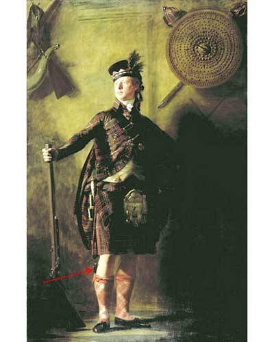
Knives are not my specialty (although I’m, um, good with a scalpel) but let’s start the topic with a brief intro to the sgian dhu. Apparently, 17th and 18th century Scots carried this dagger hidden in the sleeve or lining of a jacket. As a guest, however, proper courtesy and etiquette required that Highlanders remove the weapon from hiding and display it in a stocking top (Photo A – red arrow) as depicted in this elegant and manly portrait of Alasdair Ranaldson MacDonell of Glengarry (Henry Raeburn, artist – 1812).
image

Photo A

If ye recall, an important event leads to Claire’s sgian dhu lesson. Just blame it on the crouching Grants! Aye, those sneaky dudes were hidden in the rocks under veil of darkness with pillage on their minds. Springing from their hiding place, they attack the MacKenzie party and ruin Rupert’s mellifluous waterhorse tale; what a voice that man has!

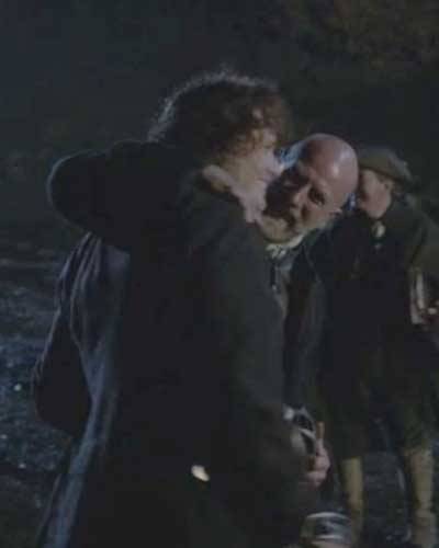
Happily, Dougal and his fighting warriors quickly prevail and Uncle is so delighted that, wonder of all wonders, he hugs his nephew! I’ve burned this image on my retinas thinking it may be the only time we see Dougal show affection for the lad. Wait ‘til Jamie hears that his uncle has the hots for his new wife…probably no hugging after that.
image

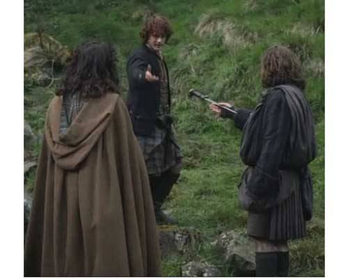
Next morning the search is on for Jamie’s dirk, lost by Claire during the nighttime scuffle. Ah, gie it to my wife. But alas, the dirk willna work for the lass.
image

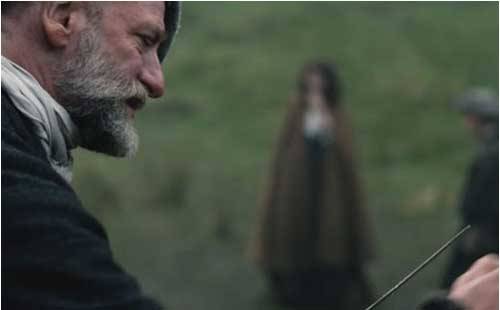
Ned Gowan thinks that someone should teach this leddy how to defend herself. Jamie agrees and war chief Dougal sits honing his dirk with a stone. Ever dapper in his wool bonnet and tidy beard, he intones that “the lass needs a sgian dhu.”Claire is – huh? – until her ruadh gallant translates: “sgian dhu, hidden dagger!”
image

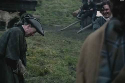
So, does anyone have a sgian dhu for Claire? To be sure, that crusty solicitor Ned, does. A lot of folks keep theirs hidden in a stocking, but he keeps his “in another more private place”… ahem, who knew? Willie (what a cutie) thinks Ned’s hiding spot is verra clever. Careful Ned, ye need all those body parts and passions lest you lose face time with the strumpets!

Herself writes in the Outlanderbook:

“The tiny sgian dhu, the sock dagger, was deemed acceptable, and I was provided with one of those, a wicked-looking, needle-sharp piece of black iron about three inches long, with a short hilt.”
image

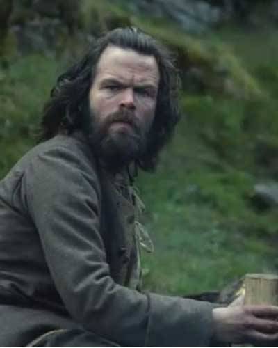
OK, she’s got a knife but who will give her a lesson? Turns out Angus is “a good man with a blade!” and he’s been waiting to teach Claire a thing or two. I recall him thrusting his dirk while watching Claire in the surgery at Castle Leoch (Starz episode 3, The Way Out) and trying to get a wee “keek at her breests” on her wedding night (Starz episode 7, The Wedding). Naughty Angus!

Herself records in the Outlander book (Here, Rupert is the teacher):

“So I was marched out into the center of a clearing and the lessons began…  After a good deal of amiable discussion, they agreed that Rupert was likely the best among them at dirks, and he took over the lesson.”
image

Again from Herself’s own hand (Outlander book):

“He found a reasonably flat spot … in which to demonstrate the art of dagger-wielding … “Generally, ye want to use the underhand; overhand is only good when ye’re comin’
down on someone wi’ a considerable force from above.”

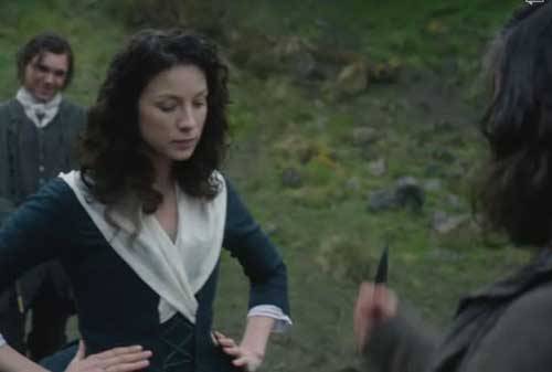
Angus demonstrates to dubious Claire several fierce upward jabs with the wee wicked weapon (dare ye to say that rapidly three times in a row).
image

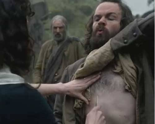
“Aim straight up and as hard as you can into the heart” is Angus’ genteel advice. First try, Claire stabs upward near Angus’ breast bone and gets a warning! Noooo……. Dinna ye love the look on Angus’ face? He’s such an avid teach!
image

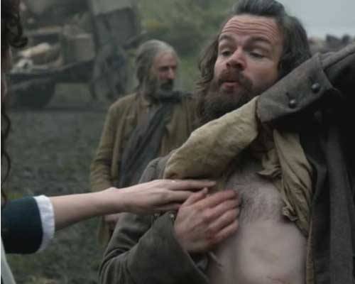
“Uh, oh, avoid the breast bone – you get your knife stuck in that soft part at the top and you’ll be without a knife.”

Whoa, there’s a tip-top issue here. Angus, did ye really say “at the top?” Ah, well, now, the adult human breastbone doesn’t have a soft spot at the top. After all, we aren’t chickens (well, maybe sometimes) …. I checked the CC script and sure enough the word is top. But, I think Angus says tip not top in which case, he is correct because the breast bone tip can be soft (see below). Mayhap he was misunderstood? Who cares? Anatomists to be sure. Otherwise, Angus’ anatomy lesson is flawless.  Bravo professor Angus!
image

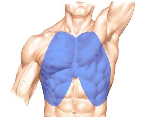
Lest Angus and Claire get too far ahead of us, let’s halt the lesson and start our own. Gruesome as it may seem, this lesson presents us with a fine opportunity to explore the thoracic cage and vital organs it protects. The thorax is the upper part of the trunk easily distinguished from abdomen because the former contains ribs. From the front, the thorax occupies the region shown in blue (Photo B) and is partially overlaid by chest and abdominal muscles.
image

Photo B

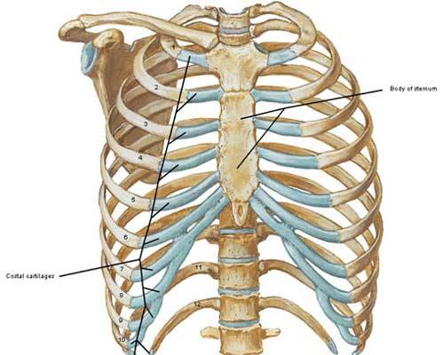
The bony foundation of the thorax is the thoracic cage composed of thoracic spine (Anatomy Lesson #10), sternum, ribs and rib cartilages (Photo C). Let’s examine these structures.
image

Photo C

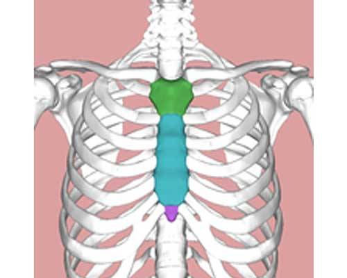
Being verra imaginative men, early anatomists visualized the sternum as a sword and named its components accordingly. Can ye see the sword in Photo D? The sternum is divided into three parts:

  • manubrium (grip of the sword – green),
  • body (blade of the sword – turquoise)
  • xyphoid process (tip of the sword – purple).

At birth, the sternum is typically six separate pieces of cartilage and bone. By 18-22 years, the segments ossify (become bony) into a single bone although the tip may remain soft (cartilaginous) until about age 40. So, yes, Claire’s knife could get stuck in the soft tip – not top!
image

Photo D

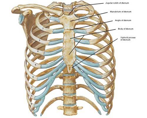
The manubrium (Latin meaning handle) is fused with the body and articulates with costal cartilages of 1st and 2nd ribs (Photo E).  It bears the midline jugular or suprasternal notch (Anatomy Lesson #12) and articulates with the clavicles (Anatomy Lesson #2) at paired sternoclavicular joints.

Try  this: place a finger in your jugular notch and feel bony knobs at each side, the sternoclavicular joints. These are usually visible in a mirror; the thinner the person, the more prominent the joints. With a finger  on one joint, lift, lower, pull forward, pull backward and rotate the same  shoulder; feel the movement? This is truly amazing because the entire skeleton of each upper limb articulates with the thorax only at this single bony contact point!

The body or gladiolus (Latin meaning sword – as in gladiator) is the largest section of sternum; it is fused with manubrium, above and xyphoid process, below. It also articulates with costal cartilages
of ribs 2 – 7. Please palpate yours.

The xyphoid process (Greek meaning sword-like) is fused above with the body but terminates as a free end. Highly variable in shape, it may be curved, depressed, forked, deviated or pierced by a hole. Rarely it remains cartilaginous beyond 40 years.

Try this: suck in your breath as much as possible, hold your breath and lift the chest. Palpate the free tip of your xyphoid process – press gently as it may be tender.
image

Photo  E

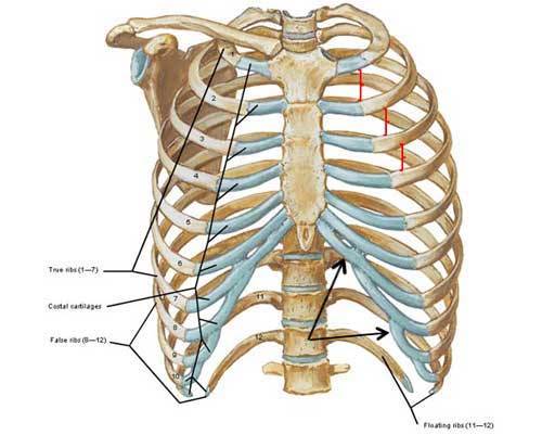
Now, for the ribs: humans typically have 12 pairs of ribs (Photo F). Ribs 1-7 are true ribs because their costal cartilages directly join the sternum; ribs 8 -10 are false ribs because their costal cartilages join with costal cartilages of ribs above (Photo F – black arrows); ribs 11 and 12 are false and floating ribs because they do not join either costal cartilages or sternum. The joined costal cartilages of ribs 7 -10 form right and left costal margins.

Try this: holding your breath, run fingers along the free edge of the left ribs – this is your left costal margin.

Variations in rib count do occur: some individuals may have cervical or lumbar ribs or they may have only 11 pairs. Because of location, cervical ribs can compress arteries and nerves in the neck producing neurological and vascular symptoms. Otherwise, such variations are rarely a source of concern.

Between adjacent ribs are intercostal spaces, 11 pairs total (Photo F – red brackets). Although they appear large and empty in the image below, in life they are narrow and filled with intercostal muscles and fibrous tissue.
image

Photo F

Ribs vary in size and shape but each has a head (Photo G) which articulates with the bodies of two adjacent vertebrae and the intervening IV disc (Anatomy Lesson #10). Interestingly, if an individual rib is placed on a flat surface, it is seen as a twisted arch that will not lie flat. This twist affects the shape of thoracic cage and intercostal spaces.

image

Photo G

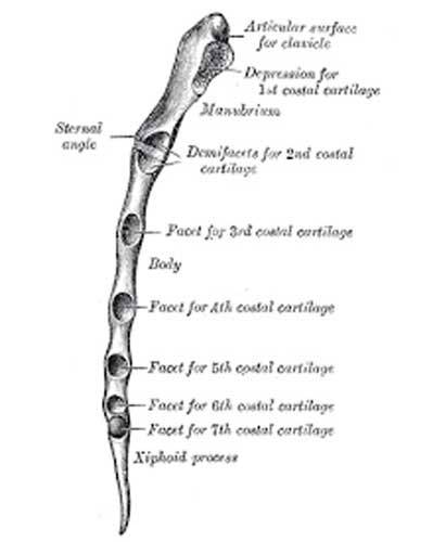
Another important feature of the rib cage is the manubriosternal joint also known as the sternal angle of Louis. Here, the union between manubrium and body creates a palpable bump (Photo H – side view) and is the level where the costal cartilages of the second ribs attach to the sternum. It is of clinical importance as various structures and anatomical boundaries occur at this joint.

Try this: place fingers in your jugular notch and work downward (about 2” or 5 cm) until you feel a bump; this is the sternal angle of Louis, a very useful landmark for health care workers to count ribs and determine  locations of various thoracic structures. Men have more pronounced sternal angles than do women.

Occasionally, my acquaintances wish to wear heavy necklaces but cannot because of neck pain. I explain that they can likely wear heavy necklaces but only if the jewelry doesn’t fall below the sternal angle. If a heavy necklace is short, then clavicles and manubrium help support weight and relieve neck strain. Try it…
image

Photo H

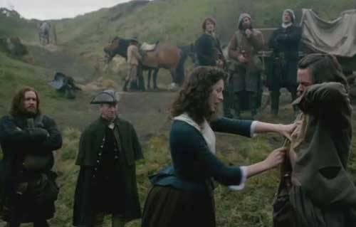
Now, back to Angus’ lesson… Next, he redirects Claire’s knife from his sternum to left rib cage. He tells her to stab straight up and into the heart. Having some acumen with a scalpel too, Claire “follows a man’s orders for once” (or wait, isn’t that twice?) with a thrust so convincing that Rupert laughs: “Oh, dinna kill him yet mistress. Wait ‘til the lesson is over!”
image

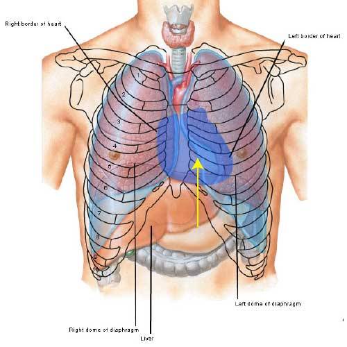
Now, is Angus correct: can a 3” blade reach the heart? Oh, aye – Angus scores again! The thoracic cage protects vital organs including heart, lungs and great vessels. In the following image (Photo I – anterior view) we see the heart in blue  overlay with left and right borders shown in silhouette. Although the respiratory diaphragm divides thoracic and abdominal cavities, it rises surprisingly high under cover of the ribs. The heart sits atop the diaphragm and the abdominal liver is just below. Much of the heart lies protected behind the sternum but its point or apex reaches to the left (model’s left – not your left). A backward thrust with a sgian dhu pierces liver, too far left and it pierces lung. To reach the heart the blade must be thrust upward as hard as possible under the left ribs near the sternum so it pierces thoracic diaphragm and heart (yellow arrow).
image

Photo I

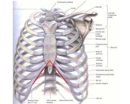
Next, does the shape of the rib cage matter to a sgian dhu wielder? Weel, it should. Angus, for example, has a narrow rib cage and a narrow subcostal angle (Photo J – red lines) formed by union of costal cartilages and xyphoid process. The subcostal angle varies considerably from person to person. A typical angle is roughly 70° but some individuals have a wide rib cage and subcostal angle making a thrust to the heart easier to, erm, execute. In such people, the angle may exceed 100° allowing for a more successful stab.
image

Photo J

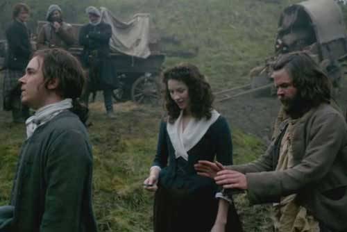
Taking the lesson very seriously, Angus next teaches Claire how to “kill from behind.” Sweet Willie is the model (Murtagh in Outlander book). Poor guy, he gets his share of abuse on this, his first rent trip.

And another fabulous Outlander quote from Herself (again, Rupert is teacher):

“I was timid and extremely clumsy at first, but Rupert was a good teacher, very patient and good about demonstrating moves, over and over.”

Aye, Angus is an excellent teacher as he patiently explains: “Now, this is the spot in the back. Either side will do. Now, you see all the ribs and such? Very difficult to hit anything vital when you stab in the back.”
image

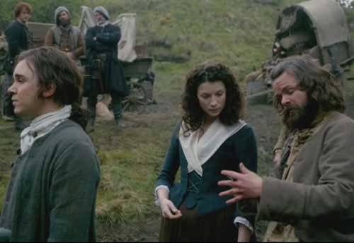
He continues: “Slip the knife between the ribs, huh? That’s one thing but a lot harder than ye might think.” Now, is Angus correct about stabbing between the ribs, and if so, why? Stay with me now…
image

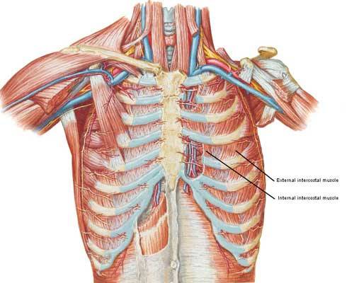
Yes, Angus is right! There are three reasons why stabbing between the ribs is difficult. As I explain, please recall that intercostal spaces are filled with intercostal muscles and fibrous tissue (Photo K).

  • The intercostal spaces are not horizontal: most take a downward slope in the back and an upward slope in the front making it difficult to judge where to thrust a blade. Although a few medical papers report stab wounds between the ribs, these are often in the 1st or the  2nd intercostal spaces which are more horizontal than the rest.
  • The intercostal spaces are narrow with an average width of about 2/3” (1.7   cm) so the chance of placing a knife in such curved, thin spaces is slim.
  • Width of the intercostal spaces changes during each breath cycle so it presents a moving target. 

Try this: place a finger in an intercostal space and breathe in as deeply as possible. Feel the change in width at deepest inspiration and again with complete expiration. During deep inspiration, the greatest width is discernible; with deep expiration, you’ll be lucky to feel an intercostal space at all. This reality greatly diminishes the chance of a successful stab into an intercostal space.
image

Photo K

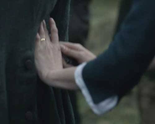
Returning to the knife lesson, Angus instructs Claire to place her dagger “Here just under the last rib, you stab upward and into the kidney. Straight up – They’ll drop like a stone.” Whew, talk about a backstab! So, Nurse Claire delivers a well-placed thrust “straight up and in. See? Got it.”
image

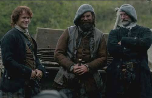
So how did Claire do with her lesson? Was she a good student? Jamie’s proud of her knife skills and he can’t wait to get her alone so he can prove it; Murtagh’s thinking that mayhap Claire’s best weapon isn’t poison; and Dougal, well, that’s an admiring eye to be sure! Not sure what’s on yer mind Uncle, but ye best not share it wi’ Jamie!
image

So, is Angus right – can a 3” blade reach the kidneys from behind? Oh, aye, he scores again! Angus, you are on a roll, man!

Let’s see why. From behind, the thorax includes the area shown in blue (Photo L) and here, the underlying thoracic cage is formed of posterior ribs and thoracic vertebrae (Anatomy Lesson #10).

image

Photo L

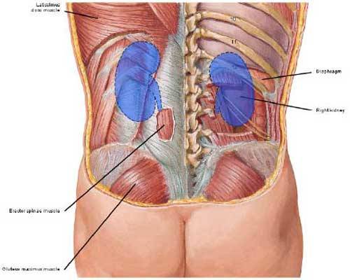
In the back, latissimus dorsi and erector spinae muscles (Anatomy Lesson #10) cover the 11th and 12th ribs of the thoracic cage and deeper yet are the paired abdominal kidneys flanking the spine (Anatomy Lesson #8). A posterior view with muscles removed shows upper poles of right and left kidneys (Photo M- blue overlay) protected by 11th and/or 12th ribs. Note that the right kidney sits lower than the left making it more vulnerable to a knife thrust. This occurs because the right lobe of liver is considerably larger than the left lobe, displacing the right kidney downwards.
But, Angus is correct, either side will do: a 3” blade thrust under either 12th rib near the spine will surely strike a kidney and mayhap its renal artery. Ye may recall that the kidneys receive roughly 22% of the cardiac output (Anatomy Lesson #8)? This extremely high blood flow causes quick and profuse bleeding in the event of a significant laceration. Shock alone from such a wound could cause a person to “drop like a stone”. Although not always in the back, kidney stabs are fairly common injuries seen in urban ERs.
image

Photo M

Harry a redcoat deserter soon provides Claire with a chance to apply her sgian dhu lesson. Talk about coitus interruptus! Those deserters sure know how to rain on a parade.Warning: If you dinna wish to see or read details about the stab wounds, please skip the next paragraph and three subsequent images.

Herself records the horrific, teeth-jarring attack in the Outlander book (Rupert and Murtaugh – not Angus and Willie):

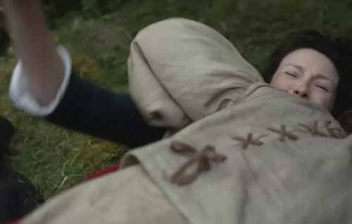
“I lay back, breathing heavily. I concentrated on my objective, trying to erase everything else from my mind. It would have to be in the back; the quarters were too close to try for the throat….I could see Rupert’s blunt finger stabbing at Murtagh’s ribs, and hear his voice, “Here, lass, up under the lowest ribs, close to the backbone. Stab hard, upward into the kidney, and he’ll drop like a stone……..I slipped my left arm around his neck to hold him close; holding the knife hand high, I plunged it in as hard as I could……… Unable to see, I had aimed too high, and the knife had skittered off a rib. I couldn’t let go now…”
image

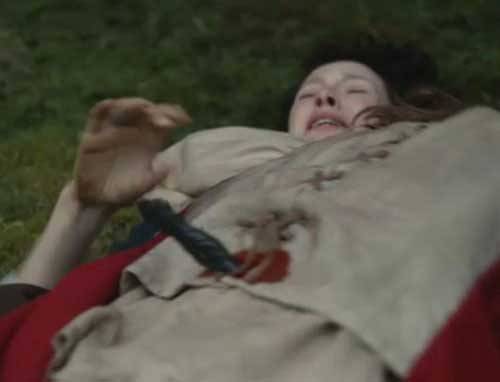
“I stabbed again, with a desperate strength, and this time found the spot. Rupert had been right. Harry bucked in a hideous parody of the act of love, then collapsed without a sound in a limp heap on top of me, blood jetting in diminishing spurts from the wound in his back.”

BTW, happy is the anatomist to see the bright red color of arterial (not dark venous) blood from the stab wound in Harry’s back.
image

Now, its Jamie’s turn…… his dirk flashes and Arnold instantly joins his pernicious pal in death! Oh, aye, knives are everywhere in this episode!

From the Outlander book, Herself shares:

“Arnold’s attention had been distracted for an instant by the spectacle on the ground, and an instant was more than long enough for the maddened Scotsman he held at bay. By the time I had gathered my wits sufficiently….. Arnold had joined his companion in death, throat neatly cut from ear to ear by the sgian dhu that Jamie carried in his stocking.”

Whew! Jamie’s kill is so swift that I canna capture even one still image of the carnage!
image

And, not to be outdone by the other card-carrying knifers in episode 8, that crazy bastard, BJR, uses his own blade to harass and torment a terrified Claire. Not only does he destroy her pristine white fichu (how can that be after weeks of camping in mud and dirt?) but he cuts her lacings and tears her shift (aye, he’s a bodice ripper!). But, BJR isna really interested in women or their soft bodies; he wants intel and likes torture.
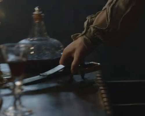
image

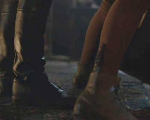
And, where is Claire’s hidden dagger when she needs it? Och! Safely hidden in the top of her lace-up leather boot whice she can’t reach (not bent over a desktop face-first with her skirts up)! Too bad Claire, otherwise ye could teach BJR your own wee dagger lesson! He’s earned it!

Pssst: just between you and me, I am fairly sure that Claire’s boots at Fort William are not the same ones she wears while climbing Craigh na Dun; the latter look a bit like UGGs! Mayhap there’s a Zappos near the fairy stones??
image

After all the blood and gore, let’s end this anatomy lesson on a happy note! Here’s the funny sequence between Jamie, Claire and Rupert over Jamie’s lost dirk after the crouching Grant’s attack. It happens so fast that ye may have missed the full effect. So here it is in slow-mo…

Ye recall Claire sighing that Jamie’s dirk is too long and heavy for her? With a huge, cheeky grin plastered on his face, Rupert chuckles “all the lasses say that to me.” Claire looks faintly disgusted. But lass, what’s wrong? Have ye never heard a man make a joke before? Aye, this time he’s the witty one!

image

Oops! Not sure Jamie likes the double entendre… Suddenly, a wary Rupert isn’t grinning. I wish the camera had caught Jamie’s face as he swings toward the jokester.
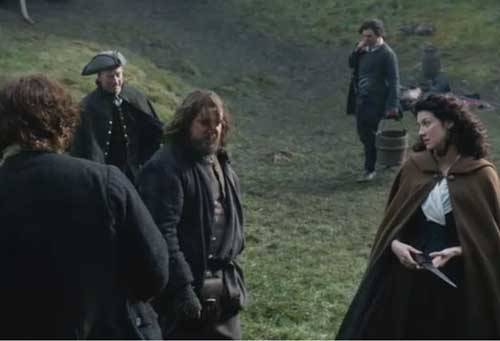
image

Um, Rupert, ye best get while the getting’s good! Jamie doena like other men making ribald jokes to his wife. It’s one thing to pummel Jamie when he willna fight back, but ye likely won’t win if and when he decides ye are fish bait.
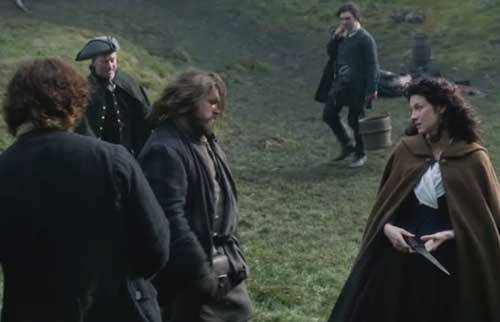
image

Aye, Rupert gets the message. Off he goes!
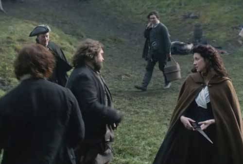
image

Claire finds the joke a wee bit amusing but Jamie isna smiling. Bye, bye, Rupert. For a big man, ye move pretty fast!
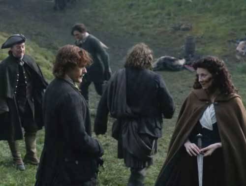
image

So, Jamie gets back his knife and rewards Claire by showing off. It’s not the first time we’ve seen him, erm, play with his blade. As Claire kens well, he’s darn good with a blade!
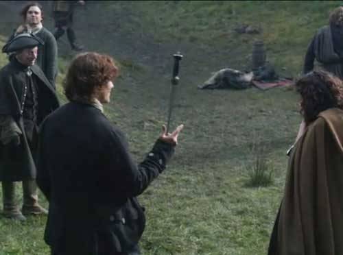
image

In closing, remember that the thoracic cage is a bony envelope formed of sternum, ribs, costal cartilages and thoracic vertebrae; it is engineered to protect vital organs including heart, lungs, great vessels and kidneys. Although vulnerable to trauma, a stab attack to the thoracic cage and its enclosed organs must be executed effectively to impose lethal harm. Mayhap Angus should teach the next Anatomy Lesson, you say? If I need guidance, I’ll ask. Snort!


A deeply grateful,

Outlander Anatomist

You can now follow me on Facebook and Twitter!

Photo creds: Starz, Gray’s Anatomy,
39th ed., Netter’s Atlas
of Human Anatomy, 4th ed., Clinically
Oriented Anatomy, 5th ed., Hollingshead’s
Textbook of Anatomy, 5th ed., www.Wikipedia.org

Anatomy Lesson 14: “Jamie and Claire” or “Anatomy of a Kiss“

Happy Valentine’s Day!

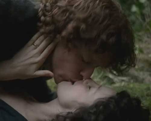
Confession time: I have wanted to do a lesson on the anatomy of a kiss for a very long time. Today is that day!  Welcome to Anatomy Lesson #14: The Lips (and a little tongue too).

Now, you may think there is little anatomy of the lips but there is so much info and complexity that we’ll be lucky to cover it in a single lesson!

I need models for our lesson and who better than Claire getting her first kiss (Starz episode 101, Sassenach). NO, not this kiss!! Claire usually has to urge Frank but she seems to like this kiss just fine and I’m betting there is a group of folks out there that give two thumbs for any face time with Frank.

image

Alrightie then, how about this kiss (Starz episode 104, The Gathering)? NO, again? Geez, some folks are so hard to please!

Clearly Claire is not pleased with this kiss. Dougal’s efforts garner him a kick in the stones, a sharp slap and a wallop over the head with a stool! Dougal, best get ye gone man before you end up “paying a greater price!”

image

Ok, how about this one? Better? Ha! Getting down to business, Jamie and Claire seal the wedding vows with their first kiss (Starz episode 107, The Wedding). In Outlander book, Herself records the moment:

“More mumbling from the priest and Jamie bent to kiss me. It was clear that he intended only a brief and ceremonial touching of lips, but his mouth was soft and warm and I moved instinctively toward him. I was vaguely conscious of noises, Scottish whoops of enthusiasm and encouragement from the spectators,but really noticed nothing beyond the enfolding warm solidness. Sanctuary.”

image

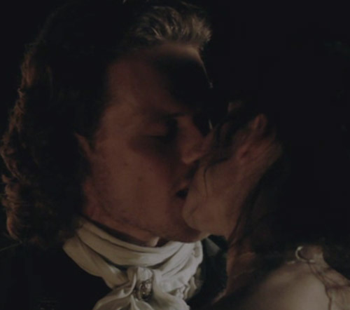
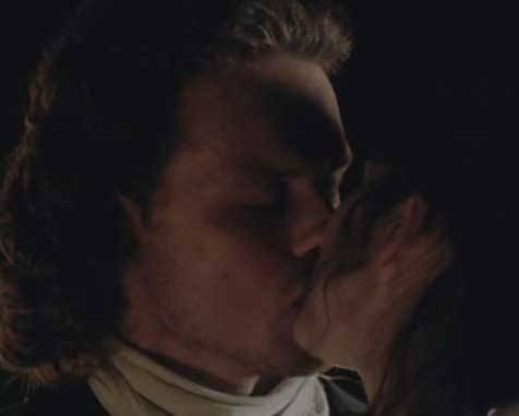
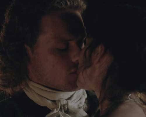
Whoa, Prof! This wedding kiss lasts waaay longer than one brief frame! How about some time lapse photography? OK, students, not to dwell on the point, but here’s Claire as she accepts Jamie’s kiss.

image

A wee release of the lips and more kiss! Aye, she gives as good as she gets.

image

More lip action; she really leans into it. Good lass!

image

Finally, at the end of the lengthy lip-lock, Claire is on her tippy-toes panting for more. No ceremonial peck here. Well, when she kisses him like that maybe she isna so sorry to be marrying him after all.

Gasp! Mayhap we all best come up for a wee gulp of air!

image

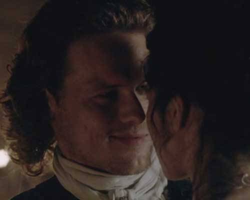
Can I ask you a question? “Did you like it?” He he – Jamie did!

image

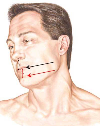
Topography of Lips: Now, on with the anatomy lesson!  First, let’s begin with topographical (surface) anatomy of the lips.  The lips are the part of the face surrounding the oral fissure, the external opening to the oral cavity (Image A, black arrow).

In anatomy, the upper lip (black bracket) extends from nasal septum all the way inside  to the web of tissue near the upper gums. Similarly, the lower lip (red bracket) extends from the groove of chin (red arrow) inside to the web of tissue near the lower gums.

Lips are soft, pliable and mobile and almost prehensile in their movements. Consider that lips are active in biting, chewing, drinking, licking, swallowing, speaking, singing, screaming, sucking, crying, humming, etc. Wow, not too many areas of the body can lay claim to such a vast range of services! And, lastly, the lips are a tactile sensory organ and thus classified as an erogenous zone.

image

Image A

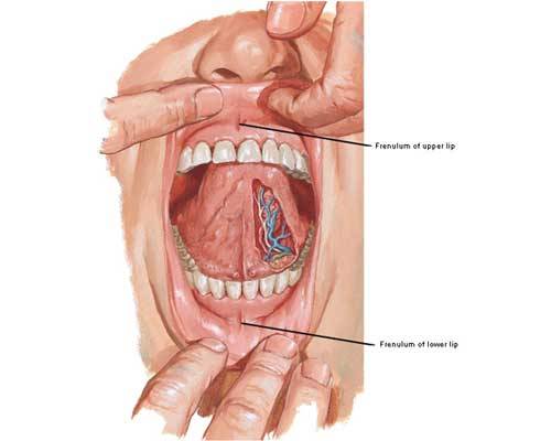
Image B shows upper frenulum and lower frenulum, webs of tissue that mark the
internal terminus of upper and lower lips. Frenula (pl.) help anchor lips
to gums.

Try this: Grasp your upper lip and lift locate the upper frenulum. Repeat with the lower lip.

image

Image B

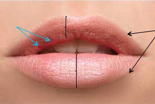
Vermillion Border:  The vermillion border is the margin where facial skin meets upper and lower lips (Image C, black arrows) – the interface where lip liner is applied.

Vermillion Zones: In anatomy, the areas commonly called upper and lower lips are vermillion zones (Image C, black brackets) – the areas where lipstick is applied.

From vermillion zone to oral cavity, lips transition from thin, dry hairless skin to wet mucosa  (blue arrows).

Try This:  Look in a mirror and identify vermillion border and vermillion zones.  Next, pull down your lower lip and find where the skin of the vermillion zone turns into wet mucosa of the inner lip. Got it?  Good!

image

Image C

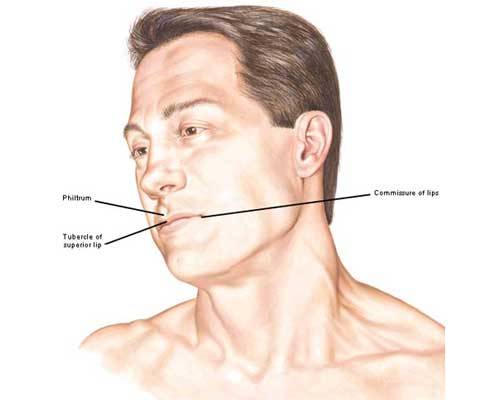
More Lip Structures (Image D):

  • tubercle:  lip-blip in the midline of upper lip – it varies in size
  • philtrum: vertical groove from nasal septum to lip tubercle
  • philtral columns: paired ridges of skin flanking philtrum
  • commissures: corners where upper and lower lips meet (Anatomy Lesson #11 and Anatomy Lesson #13)

Try this: Return to the mirror to locate these features of your own lips or,
better yet, use a partner!

image

Image D

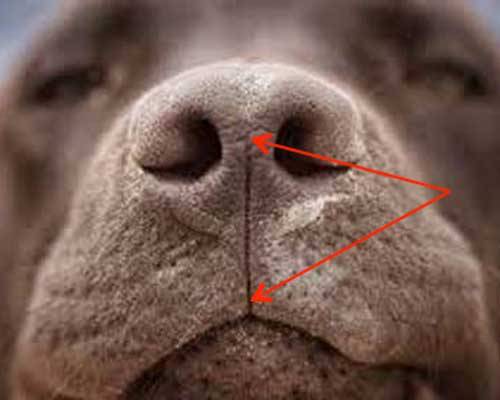
Always fun to throw in a wee bit of comparative anatomy…in mammals such
as the dog, the philtrum is more extensive, a midline cleft extending from nose through upper lip (Image E – red arrows) that may act as a channel to direct dissolved odorants towards the nasal septum.

image

Image E

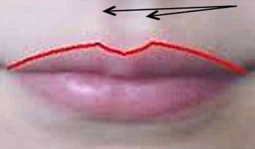
Cupid’s Bow: The vermillion border of our lower lip is an even line, but not so with the border of the upper lip which bears twin peaks, the so-called Cupid’s bow (Image F – red line) in honor of the weapon born by Cupid, Roman god of erotic love. And, the peaks of the bow coincide with philtral pillars (black arrows). The more prominent the peaks of Cupid’s bow, the more obvious are the philtral pillars.

image

Photo F

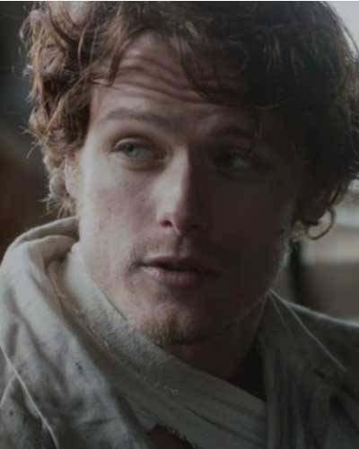
No surprise here, but Jamie’s upper lip demonstrates a prime and manly example of Cupid’s bow and its associated philtrum (Starz episode 102, Castle Leoch) but I wager you already ken this. Wink, wink. His bow is well-defined (mmphm) and his upper and lower lips are picture perfect. Studies show that lip size in men is linked to sexual appeal. Subjects in these studies preferred men with masculine lips, neither too large nor too small. Sound like anyone you favor?

image

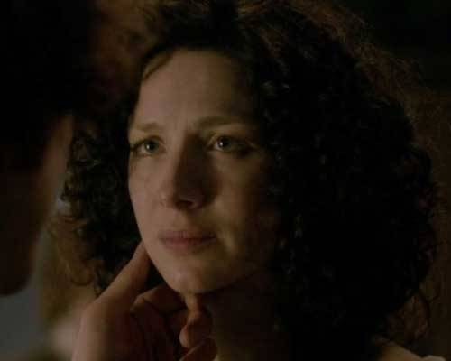
Starz Claire has absolutely beautiful lips that are full, expressive and verra feminine!
They define her face to a large extent and along with her eyes were features I
noticed first (Starz episode 109, The Reckoning). Again, psychology studies have
shown that the larger a woman’s eyes and the fuller her lips, the more desirable and feminine she was perceived by participants of these studies. Lipstick is thought to enhance this underlying perception.

image

Microscopic Anatomy of Lip: Let’s turn from surface anatomy to the lip as seen in a microscope. Microscopic anatomy is AKA histology (Greek meaning tissue knowledge). Considerable technology is required to prepare tissues for microscopy: samples are imbued with paraffin (sometimes plastic), sliced very thin, picked up on glass slides and stained before examination. How thin is the slice? Well, it would be roughly 6 µm (.006 mm or .00023 in) – thin enough for light to pass through the slice.

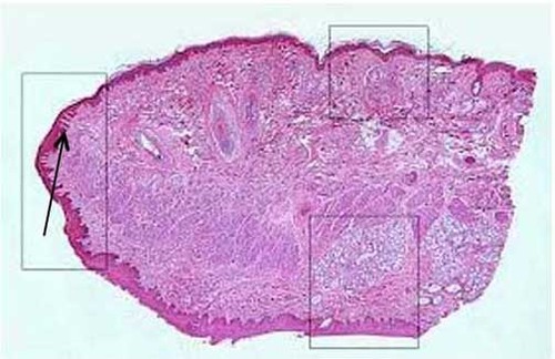
Image G is a tissue section of lip as view with a microscope (Image G). It takes
some skill to recognize subtle differences, but the top box surrounds a patch
of facial skin with hair follicles, sweat and sebaceous glands (Anatomy Lesson
#5
and Anatomy Lesson #6); the left box encloses the vermillion zone of thin
skin sans hair and glands; the bottom box surrounds a patch of wet mucosa
(living cells throughout the surface layer). Look closely: the black arrow
points to dovetails (interdigitations) between the dark red thin skin and the deeper
pink-blue dermis. Let’s take a closer look:

image

Photo G

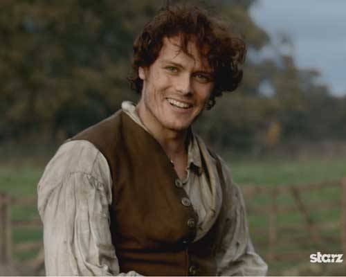
Hey, students, wake up! Stay with me now – I can see you nodding off! Here, this might help (Starz episode 102, Castle Leoch). Oh, weel, I see that got your attention. Talk about lips doing their job and giving us a full wattage smile!

image

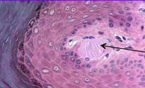
Photo H is a much higher magnification of the dovetail region indicated by the arrow in Photo G. See the odd pink oval structure that resembles shredded wheat or
venetian blinds (Photo H-black arrow)? This is a special sensory body first identified by Georg Meissner, a German anatomist and physiologist (1829-1905). These structures, named Meissner’s corpuscles are excited by light touch. The dermis of
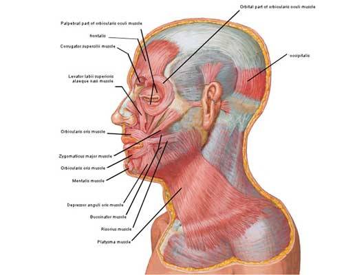
erogenous zones, such as the lips, contains higher concentrations of Meissner’s
corpuscles than non-erogenous regions. When excited by light touch, the nerve
endings fire and information is transmitted along nerves to the brain.
image

Photo H

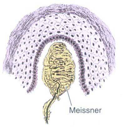
Photo I is an artist’s rendition of a Meissner’s corpuscle. The lavender and black region is overlying thin skin; the gold and black structure is the corpuscle. Ergo, increased numbers of these touch-sensitive structures explains the basis of the so-called erogenous zones of the body.

image

Photo I

Break time for practical
application of our knowledge about the lips: From Outlander, Herself quotes the Latin poet, Catullus:

“Then let amorous kisses
dwell
On our lips, begin and
tell
A Thousand and a Hundred
score
A Hundred, and a Thousand
more.”

Well, Jamie and Claire dinna disappoint. There are many amorous kisses shared by these two in Starz episodes 107 and 108. I was actually surprised by the frequency as I worked on this post. Gah! Messed with my sleep patterns!

image

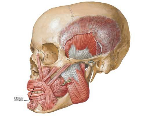
Now let’s move to gross anatomy of the lips. Do you recall from Anatomy Lesson #11 the name of the muscle surrounding the oral fissure? If you said orbicularis oris, you get 100 points! For years, anatomists taught that orbicularis oris was a circular, sphincter-like muscle that contracted like a camera diaphragm to close the oral fissure (Photo J). Well, recent studies have proven this is not the case.

image

Photo J

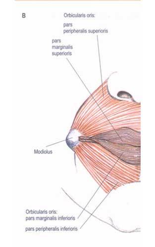
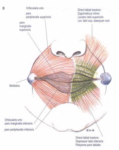
Orbicularis oris is composed of four interlacing quadrants of muscle (Photo K – right side only): upper left, upper right, lower left, lower right. Further, each quadrant is divided into two parts: fibers of the vermillion zone form pars marginalis (Latin meaning marginal part) and fibers outside the vermillion border form pars
peripheralis
(peripheral part). Doing simple math, this means our oral fissure is surrounded by eight interactive layers of orbicularis oris. This arrangement lends major control over shape so orbicularis oris is proficient at closing and puckering the lips.

Also, notice the mass labeled modiolus (Photo K)? This is a paired fibromuscular node just inside the commissures of each lip.

Try this: slip a clean thumb inside a corner of your mouth about 0.5 in (1.25 cm); close your index finger against the thumb. You should feel a thickening; this is the modiolus. At least seven facial muscles connect to or pass through each modiolus (one on each side) making it extremely important to facial symmetry. While performing surgery of the lip area, reconstructive surgeons strive to symmetrically align the modioli (pl.); once these are secured, associated facial muscles of each side are more likely to balance. Notice that orbicularis oris fibers attach to the modiolus.

image

Photo K

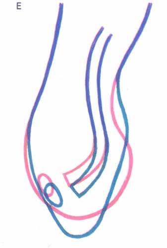
The following line drawing (Photo L) shows how the upper lip changes shape during contraction of orbicularis oris. Imagine a midline vertical slice (midsagittal plane) through the upper lip, then view it from the side; the stick drawing is a simple rendering of that slice. The pink line shows the lip shape at rest; the blue line shows the lip flattened during a pucker. The shape-shifting is due to contraction of orbicularis oris.

image

Photo L

Here’s a verra good example of lip puckering by Jamie’s orbicularis oris as he plants a lovely one on Claire’s lips. Good job, lad! He’s a terrific anatomical model as I keep saying (over and over and over). Ha! Well, it’s true!

image

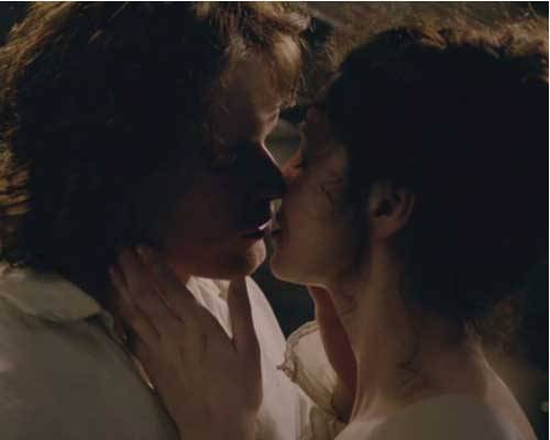
Time for another interlude: how about this image from Starz episode 107, The Wedding at the end of the first amorous kiss between Jamie and Claire? Oh, Jamie, your lip looks a wee bit swollen. Did you get thumped by a horse? Those fillies can be
dangerous! Whinny!

image

And yet another drop-dead wonderful kiss between our hero and heroine! The next quote doesn’t exactly fit wi’ the image (Start episode 107, The Wedding) but I’m betting ye can suffer through it. Herself writes in Outlander book:

“He leaned down and gently fitted his mouth over mine. I had kissed my share of men…Jamie, though, was something different. His extreme gentleness was in no way tentative; rather it was a promise of power known and held in leash; a challenge and a provocation the more remarkable for its lack of demand. I am yours, it said. And if you will have me, then…I would, and my mouth opened beneath his…”

image

Hey, yoo hoo, back to anatomy. I think I lost ye again, ha ha! It’s OK, I lost it myself…So, you may recall that several muscles of facial expression insert into orbicularis oris (Anatomy Lesson #11 and Anatomy Lesson #13) adding their tug on the lips. LLSAN, levator labii superioris and zygomaticus minor act as retractors to lift and curl the upper lip; platysma and depressor labii inferioris pull down and curl the lower lip
(Photo M). The left side of the drawing (on your right) shows the pull of the three elevator and two depressor muscles acting as retractors on orbicularis oris.

All of this detail reveals that our lips have a complex muscular framework, an arrangement lending impressive control over the lips and how they might engage in kissing: pucker, no pucker, curl, no curl, suction to produce a smack. Aye, if the lips are slightly curled and mild suction applied, the kiss produces an audible smacking sound. Re-watch (for the umpteenth time) Starz episode 107, The Wedding, and listen to the end of the wedding kiss; you hear a soft smack. Verra, erm, satisfying! Now ye know how that happens.

image

Photo M

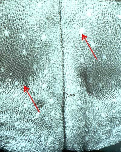
Many would argue that a kiss will be neither complete nor completely satisfying without a wee bit of tongue. I dinna have space to cover all tongue anatomy which is complex and lengthy. But a brief keek will suffice. The tongue core is very nearly all muscle; it participates in swallowing, taste, and speech. Its free surfaces are covered
with wet mucosa similar to that of the inner lips. Its top and sides are embellished with wee bumps, the papillae, some of which house taste buds. Photo N is an image of the tongue taken with a scanning electron microscope (SEM – Anatomy Lesson #5 and Anatomy Lesson #6), an instrument that greatly magnifies an object because it illuminates using an electron beam rather than light rays. Papillae on the top of the tongue are very apparent (red arrows). Check out your own papillae in a mirror; they come in 3 or 4 different shapes. Your taste buds are microscopic so ye cannot see them.

image

Photo N

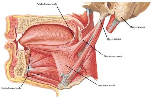
For the tongue to join in a kiss, its muscles must activate. Hard to believe but the tongue contains nine different muscles: four paired and one unpaired (Photo O)! Paired muscles are extrinsic meaning they arise from surfaces outside the tongue but end within it. The unpaired muscle, consisting of four layers, is intrinsic meaning the layers arise from and end within the tongue itself.

  • Genioglossus arises from inside the mandible, fans out and ends
    near the midline of the tongue. Acting with its mate, it protrudes the tip of
    the tongue. Look in the mirror and stick out your tongue – genioglossus muscles working!
  • Hyoglossus arises from hyoid bone (Anatomy Lesson #12) and inserts
    at the sides of tongue. Together they depress (pull down) the tongue.
  • Styloglossus arises from the styloid processes (Anatomy Lesson #12
    –  aye, those gothic spikes!) and blends with the above two muscles. Together, they retract the tongue – pull your tongue back in your mouth – styloglossus muscles working!
  • Palatoglossus arises from the palate and inserts into sides of tongue.
    Together with its mate, it lifts the sides of tongue.

But, perhaps the four layers of intrinsic muscle are most appreciated in kissing. They arise from various parts of the tongue and blend with the extrinsic muscles. Acting alone or together the four layers curl, shorten, dome, narrow, elongate, widen or fatten the tongue. Whew, that’s quite a list! Intrinsic muscles give the tongue precise and highly variable mobility for speech, swallowing and (although not listed anatomy texts) for kissing! So, consider this is why a kiss with tongue can be so remarkably creative. Try it out! Ye have my permission.

image

Photo O

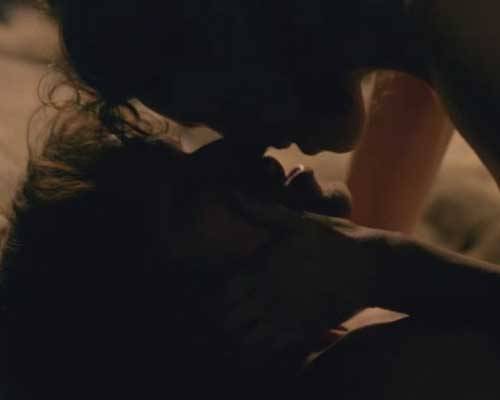
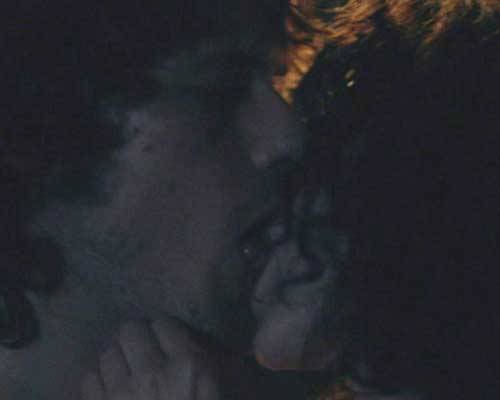
This last image (Starz, episode 107, The Wedding) is dark, but here Jamie serves once again as our anatomical model; the camera clearly loves him! This is just after Claire wraps him in a lover’s shroud of his own plaid. Oh, aye, his tongue is involved in this kiss. If ye canna see it here, go watch the episode again. OK, ye can all get up off the floor now!

image

So kisses involve lips and mayhap tongue and given the number of muscles involved
humans are granted considerable creative license in their use. Hope ye get to use yours on this beloved day and that ye all enjoyed learning about those busy lips and tongues and the anatomy of a kiss.

And if ye dinna have a partner today, then remember that your body contain trillions of cells each whispering “Tha gaol agam ort!” Gaelic for “I love you!” If our cells didna care, we probably wouldn’t exist.

image

Let’s end the lesson with a wonderful quote from Les Miserables by Victor Hugo (1802–1885):

“How did it happen that their lips came together?

How does it happen that birds sing, that snow melts,

that the rose unfolds, that the dawn whitens behind the stark shapes of trees on the quivering summit of the hill?

A kiss, and all was said.”

And for those who are new to my blog, a wee note of explanation: I follow the convention of using Outlander character’s names rather than actor’s names. This gives the cast at least one degree of separation and a wee bit of respect as I
dissect their bodies with words! I hope ye all understand!

The deeply grateful,

Outlander Anatomist

Follow me on Facebook and Twitter!

Photo Credits: Starz, Gray’s Anatomy 39th ed., Netter’s Atlas of Human Anatomy, 4th ed., Clinically Oriented Anatomy, 5th ed., Hollingshead’s
Textbook of Anatomywww.emedicine.medscape.comwww.elementsofmorphology.nih.govwww.britannica.comwww.instruction.cvhs.okstate.eduwww.siumed.eduwww.Wikipedia.orgwww.iup.ui.eduwww.bio.rutgers.eduwww.wallpapers.brotherssoft.com

Anatomy Lesson 13: “Frank and BJR” or “Face Off”

Welcome colleagues! Today, Frank’s handsome visage gets the spotlight and everyone kens that BJR deserves to be on the dissection table for a change. Welcome to Anatomy Lesson #13: Anatomy of the Face. Today, we will expand on muscles of facial expression (Anatomy Lesson #11) as well as other facial anatomy; Frank and BJR will aid us in our quest.

Warning! If ye are a wee bit squeamish, this lesson includes several illustrations of the face minus skin.

Let’s start wi’ Claire as she stumbles upon Johathan Wolverton Randall, Esquire, Captain of his Majesty’s Eighth Dragoons (Starz episode 101, Sassenach). A doppelganger for Frank if I ever saw one! Herself writes:

“…the man himself might have been Frank’s brother. There was the same lithe, spare build and fine-drawn bones; the same chiseled lines of the face; the level brows and wide hazel eyes; and the same dark hair, curved smooth across the brow.”

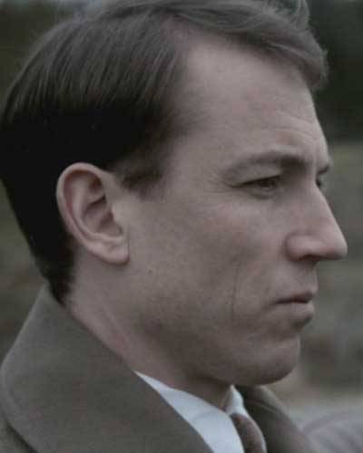
His deeply-grooved skin is weathered to a “deep-baked tan.” His eyes…..…cold, calculating, assessing, dispassionate and perhaps weary? Well, it’s exhausting being a cold, bloody bastard! Burrrr!

image

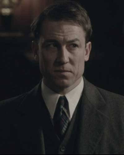
Compare and contrast BJR’s features wi’ Frank’s. Here, Frank leaves Inverness for Oxford on December 21, 1945 (same day General George S. Patton dies). He stops his roadster near the entrance to Craigh na Dun, his face a blank canvas (Starz episode 108, Both Sides Now). His scholar’s mind entertains Mrs. Graham’s explanation of Claire’s disappearance so he takes a wee road trip up the craigh. Verra interesting how our Starz Frank can make subtle changes in his facial expression to differentiate between his evil ancestor and his current, pensive self.

image

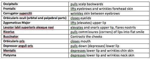
Ye now ken that muscles (and nerves) control facial expression. So, let’s do a quick and brief review of the twelve muscles of facial expression studied in Anatomy Lesson #11 – aye, there were many (Photo A). All are bilateral (paired) except the unpaired orbicularis oris. Here’s a wee chart of those muscles and their actions:

image

And, here’s a visual review of the same twelve muscles covered thus far (Photo A):

image

Photo A

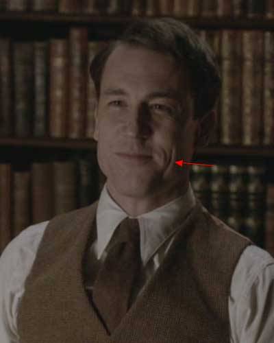
And just to get us in the mood, here Frank is seated in Rev. Wakefield’s study, looking at Claire (Starz episode 105, Rent). His flat smile is largely due to contraction of powerful rizorius muscles such that his laugh lines or skin creases assume a vertical orientation (red arrow) rather than the more common nasolabial (nose-to-lip) fold that characterizes most smiles. BTW, he asks Claire: what she was doing out there on the desert wi’ Uncle Lamb (Quentin Lambert Beauchamp)?

image

Weel, Herself explains in Outlander book what Claire was doin’ in the desert wi’ Uncle Lamb; what any young girl does after the death of her parents at age 5:

“…to the Middle East, to South America, to dozens of study sites throughout the world. Had learned to read and write from the drafts of journal articles, to dig latrines and boil water, and to do a number of other things not suitable for a young lady of gentle birth.”

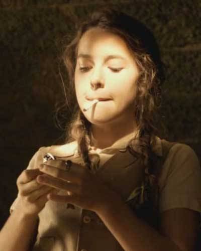
Apparently, she was also rollin’ and lightin’ cigarettes (Starz episode 5, Rent) and gettin’ her first kiss from a nine-year-old dragonman’s son (Outlander book). Ooooh, what Mrs. Fitzwould say aboot that; the French corset was bad enough!

image

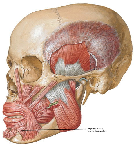
Now, let’s add a few new muscles of facial expression. Here’s a new lip depressor, the paired depressor labii inferioris muscles (Photo B). Each attaches from mandible to orbicularis oris of the lower lip. Try this: conjure the deepest frown possible. Feel tension or firmness at the sides of your neck, in the chin, and below the lower lip because four pairs of muscles are contracted:  platysmas, mentalis, depressor anguli oris and depressor labii inferioris. Aye, it takes four pairs of muscles to frown!

image

Photo B

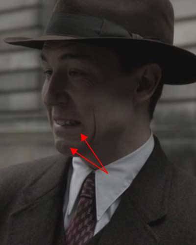
We see Frank contracting  depressor labii inferiorisdepressor anguli oris and mentalis muscles as he tells Claire “So?” It doesn’t matter that his parents are stuck waiting for them at the restaurant (Starz episode 107, The Wedding); he wants to marry her right then and there! So, off to the justice of the peace (or whoever did the marryin’ back in 1940s England) with Claire slipping and sliding on her heels!

image

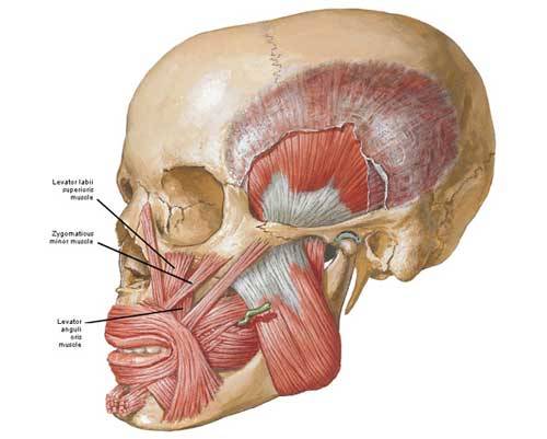
But, wait here’s three new lip-lifters for you and all are paired (Photo C). Levator labii superioris muscles span from maxillary and zygomatic bones to orbicularis oris. Zygomaticus minor muscles arise  from the zygomatic bone to end on orbicularis oris. Finally, levator anguli oris muscles arise from maxillary bones and end on orbicularis oris near the angle of the mouth.

Try this:  Grin your biggest grin ever and feel tension under cheeks and at sides of nose. Levator labii superioris, levator labii superioris alaequae nasi (LLSAN), zygomaticus major, zygomaticus minor, rizorius and levator anguli oris are all contracted. It takes six pairs of muscles to smile, ye ken.

Yes, I know about the fable that it takes more muscles to frown than to smile. Not so. Just to be thorough, a genuine smile also incorporates contraction of orbicularis oculi to make eyes crinkle. A fake smile leaves out the eyes!

image

Photo C

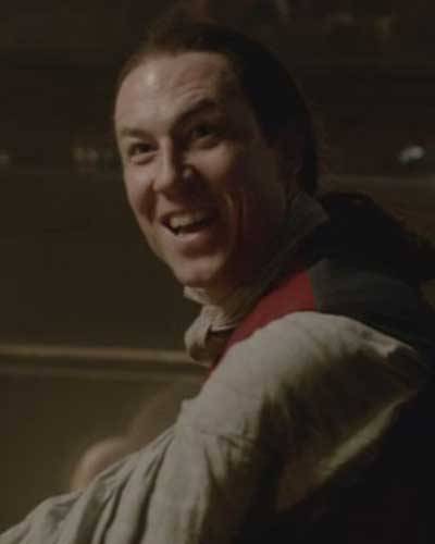
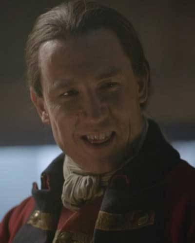
Now, here’s BJR in full grin – mayhap better described as wicked glee (Starz episode 108, Both Sides
Now
)! Herself writes:

“The smile had not left Randall’s features since Jamie’s appearance. Now it broadened enough to show the pointed dog teeth.”

Yup, his canines are showing (hummm… he would make a good vampire!). Claire and Jamie just made his day! Oh, aye, they delivered themselves into his power and he’s more than thrilled. All six pair of lip lifters are  activated along with his frontalis muscles. Fort William isna a hotbed of humor, ye ken? At least not wi’ this sadistic bastard stalking the tiles and wielding a nine-tailed cat!

image

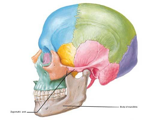
Next, let’s review a couple o’ skull bones (Photo D). The paired zygomatic arches or cheek bones receive contributions from temporal (pink) and zygomatic (orange) bones (Anatomy Lesson #8). The large unpaired mandible (Anatomy Lesson #11) is the movable part of the jaws that houses the lower teeth.

image

Photo D

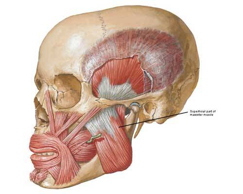
Now for another new pair of facial muscles: although classified  as muscles of mastication, the  masseters add to facial expression (Photo E). Each masseter (Greek meaning to chew) spans from zygomatic arch to mandible. Contraction of masseters closes the mandible exerting 200 lbs (90.7 kgs) of pressure on the molars! Based on weight, masseters are the body’s strongest muscles. Very powerful!

image

Photo E

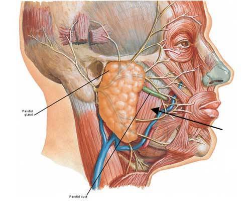
The parotid glands, major salivary glands, overlap the upper part of each masseter (black arrow) and empty saliva into the mouth via a pair of parotid ducts (Photo F).

Try this: place your fingers on the chin and move them back to the angles of the mandible. Now, bite down firmly; feel the tissue harden under yer fingers?  These are your masseters! Now, relax the masseters suddenly – did the  mandible pop open? Aye, this is due to tissue recoil and pull of gravity. Now with fingers in the same place, move them upward a wee bit and press. Ye may feel warmth and the flow of saliva in your mouth because you just pressed on one or both parotid glands! You may not know this, but the mumps virus targets the parotid glands.

Last maneuver: open your mouth wide and say ahhhhh….. (as in the dentist’s office) – a very deep pair of muscles just contracted to perform that movement but we’ll puzzle those out in a later lesson.

image

Photo F

Now, hereeeeee’s Jamie! I know, I know, I just can’t write a lesson without him as an anatomical model – he’s just way toooo perfect! Steady. Steady. Staying completely scientific, he provides the best example of masseters in the series. Here’s Jamie and Claire at the stables (Starz episode 102, Castle Leoch) sharing a delightful lunch made of UST (a.k.a. unresolved sexual tension).

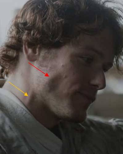
Jamie has quite an appetite, ye  ken? Snort! He’s telling Claire the grim story of how he became outlawed. There’s a swelling (mmphm) over his right mandible; jaw is clenched because both masseters are contracted! Besides chewing, contraction of these muscles conveys intense emotion such as frustration, concern or anger. Och! I almost forgot…do ye ken the prominent ridge on Jamie’s neck (gold arrow)? Can ye name it?

image

Oh, aye, if ye said right external jugular vein (Anatomy Lesson #12), a gold star for you! Good work!

image

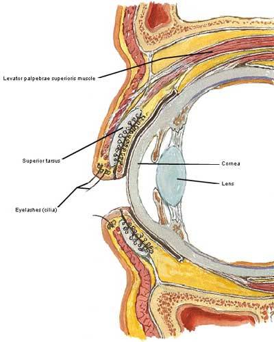
Here’s another muscle adding ta facial expression: classified as muscles of the orbit, levator palpebrae superioris muscles lift the upper eyelids. They run from bony orbits to superior tarsal plate (Photo G).  As the levators contract, the upper eyelids lift.

Try this: gently pinch an upper lid between thumb and index finger. Feel a rubbery mass? This is the
superior (upper) tarsal plate. The lads in my elementary school loved to evert their upper lids to gross out the lassies – doable because of the tarsal plates. Ye can also feel a wee version in each lower lid but no muscles to move them; gravity and tissue recoil open the lower lids.

image

Photo G

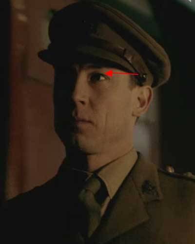
Movements of levator palpebrae superioris muscles are easy to demo on both Jack and BJR because both like to tweak their upper lids. Starz episodes capture the movements best, but here Frank tweaks both eyelids as Claire’s train leaves for the front lines while he stays behind to do WW II intelligence work (Starz episode 103, The Way Out). She will not have him use his influence to keep her home! Bye, bye Frank.

image

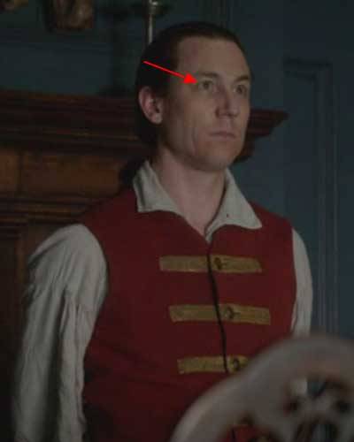
Not to be outdone, BJR contracts his levator palpebrae superioris muscles after tellin’ Dougal that Claire must be returned for further questioning by next day or he will suffer the consequences even unto death (Starz episode 610, The Garrison Commander)! Speaking of death, did ye notice the dead look of his eyes? Understand that his irises are hazel and in this light we cannot distinguish the boundary between pupil and iris. In effect, this enlarges the pupil lending a more detached and threatening appearance to the eyes.

image

Now, let’s use Frank and BJR expressions for more review. Frank (Starz episode 108, Both Sides Now) has just beaten two guys within an inch of their lives and now his hand is closed around the
hyoid bone (Anatomy Lesson #12) of not-her-name Sally! His grin is flat and eyelids are slightly closed. And, oooh, his cruel smile doesna reach the eyes. In urban speak, this is known as smizing. Gives me the shivers! Remind ye of someone? Which facial muscles are contracted? If ye said rizorius and palpebral parts of orbicularis oculi then ye are correct! If ye said nothing, that’s OK too.

image

Call it weapon anatomy, but I must comment on the objet d’art that Frank pulls from his pocket in the alley off Drummond Lane (Starz episode 108, Both Sides Now). In the US, such a  weapon is called a blackjack, perhaps a not-so-subtle reference to his sadistic ancestor? Frank worked in the Intelligence Unit at MI6 during WWII. He thoroughly beats and stomps his attackers suggesting he learned a wee bit about interrogation of a darker nature. Or, mayhap violence runs in his family line? After all, “evil knows but one cup.”

image

Wonder of all wonders: Claire makes BJR grin, a dangerous gambit to be sure! She tells him that we MacKenzies are all loyal subjects to the King – the funniest thing he has heard all week (Starz episode 108, Both Sides Now). Apparently, not much comedy goes on at Fort William.

Which facial muscles are contracted? This time, it is palpebral part of orbicularis oculi, rizorius and zygomaticus major because the flat grin is slightly lifted and the nasolabial (nose-mouth) fold as well as the vertical skin crease are now in play although the vertical creases are much more evident.

image

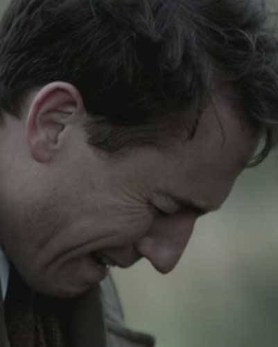
Next, Frank expresses grief at Craigh na Dun (Starz episode 108, Both Sides Now). He misses his wife even if he’s more interested in dusty old papers about his ancestors and she has to coax him into physical intimacy.  Which muscles are contracted? The palpebral and orbital parts of orbicularis oculi, rizorius and zygomaticus major and likely mentalis as his chin skin is wrinkled.

image

Now, this image of Frank is absolutely priceless (Starz episode 108, Both Sides Now). Funny man, Reverend Wakefield, explains his favorite theory of Claire’s disappearance: she gets disoriented,
falls into the Findhorn River and is swept 20 miles downstream all the way to Dunnaway Forest where she lives in a cave for seven weeks eating fish and frogs. Erm…Frank looks as if his best pal has gone bonkers! Which  facial muscles are contracted?

image

Well, obviously the frontalis muscles because his forehead is wrinkled, and corrugator supercilii (Anatomy Lesson #11) fer the deep wrinkle between the brows. Rizorius is contracted pulling the mouth into a grim line along with a slight downward tug by depressor anguli oris. Better the Findhorn River than the arms of another man! Another gold star to you for playing along!

image

Did you find the Reverend’s theory laughable? Well, turns out something like this happened to an Australian woman last fall. She disappeared from a swimming hole in a Queensland rainforest. Gone for 17 days, she lost 17 kg (37.4 lbs) during her ordeal; surviving on insects, fish and river water. And, ye will not believe it but her last name is Fraser! Aye, ’tis the truth! You can not make this stuff up! Below is a photo of the area where she was lost.

image

Now for an Outlander Anatomy PSA about accepting differences

Robert  J. Oppenheimer once claimed that “understanding is a lot like sex. It’s got a practical purpose but that’s not why people do it normally…” Here is a wee bit of knowledge to open perspectives and loosen sphincters while studying the body.

Ye ken that each of us is different on the outside? Even identical siblings with the same genetic
material enjoy subtle nuances in body form. Well, years in the dissection lab has taught me that we are each different on the inside too! Every person has their own unique expression of muscles, bones, blood vessels, organ shapes and so on making every human different inside and out. This is cause for
celebration, not for shame.

A heartfelt thanks to Diana’s books and the Outlander cast for providing a platform whereby we may explore this simple truth.

The deeply grateful,

Outlander Anatomist

Follow me on Facebook and twitter!

photo creds: Starz, Netter’s Atlas of Human Anatomy, 4th ed., Clinically Oriented Anatomy, 5th ed., Hollingshead’s Textbook of Anatomy, 5th ed., The Guardian, Australia, Daily Mail Australia, www.wikipedia.org, www.commons.wikimedia.org, www.medical-dictionary.thefreedictionary.com