Anatomy Lesson #59: Complete Feet

Hello, anatomy students! Hello feet! Today’s anatomical offering derives from the Old English fot, meaning “foot.” 

You might think Outlander has little to say about feet, but not so. Outlander has fleet feet, sweet feet, trick-or-treat feet, body heat feet, upbeat feet, mincemeat feet, eek feet, beat feet and indiscrete feet. As always, Outlander images and quotes are sprinkled throughout the lesson. Yay!

You might also think society has little to say about feet, but not so. Dozens of feet adages accent life’s little highs and lows:

  • Foot in both camps (fence sitter)
  • Ankle deep (in trouble)
  • Back on your feet (on the mend)
  • Bound hand and foot (hampered)
  • Cold feet (lost interest)
  • Get your feet wet (get involved)
  • Drag your feet (unenthusiastic)
  • Feet of clay (flawed)
  • Foot in mouth (oops!)
  • Keep feet on the ground (be sensible)
  • Footloose (6 degrees of Kevin Bacon)

As a grad student, my gross anatomy prof declared feet the ugliest body part! Beauty being in the eye of the beholder aside, our foot is far more specialized than our hand as no other animal has a foot quite like ours! I hold with the master, Leonardo DaVinci who opined: 

The human foot is a masterpiece of engineering and a work of art.

Yes!    

Understand, the human foot is a truly complex mechanical structure consisting of 26 bones, 33 joints, and more than a hundred muscles, tendons, and ligaments! Despite this complexity, our feet usually work pretty well, especially if we care for them.  Also, know this lesson is geared for general readers, so the most complex features and smallish details are not covered.

Before we begin the lesson, let’s take a gander at Claire’s fleet feet (Outlander ep 101 Sassenach) as she retreats from redcoat muskets firing live rounds! Her poor heels are rubbed raw from running – leather shoon sans sox. Not a good plan, but did the lass have another choice? Och!
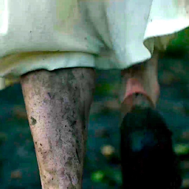
Foot: Always prudent to begin a lesson with definitions. In anatomy, the foot is the lower limb below (distal to) the ankle joint (Image A –  blue line).

Orienting ourselves further, the top of foot is the dorsal surface; the bottom (sole) is the plantar surface. Inner side is the medial surface and the outer side is the  lateral surface.
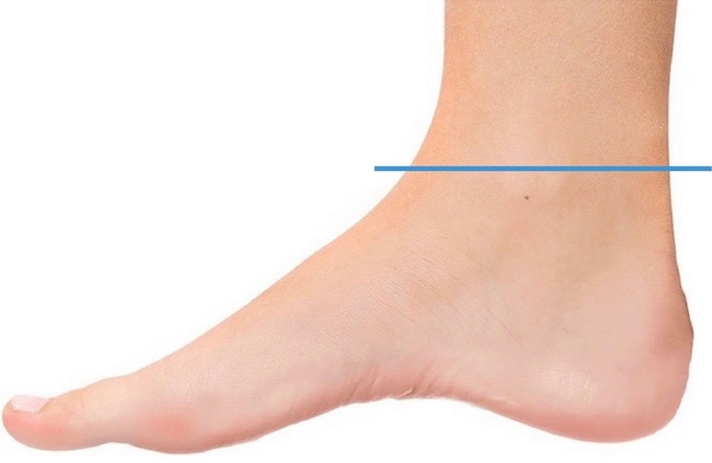
 

Image A 

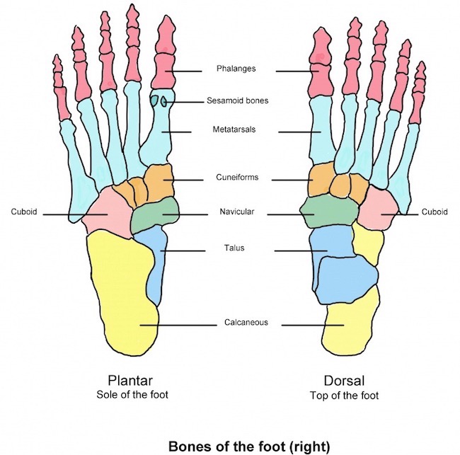
Skeleton: We begin with the foot skeleton, a foundation of 26 bones. Image B shows bones of a right foot, 26 in all. The left panel shows bones from the bottom or plantar perspective. The right panel shows foot bones from above, the dorsal view.

Toe bones are homologous to digits of the hand (Anatomy Lesson #22, Jamie’s Hand – Symbol of Sacrifice). They are numbered 1-5 beginning with the big toe and working to the little toe.

Phalanges: Toes contain 14 phalanges (Image B, pink) – three bones per each toe, except the big toe which, like the thumb, only contains two phalangeal bones. And, the big or great toe goes by the scientific name, hallux.

Metatarsals: Five metatarsal bones (aqua) are homologous with metacarpals of the hand. 

Tarsals: Seven tarsal bones are homologous to eight carpals of the wrist, although more massive and oddly-shaped than the small carpals.  The largest, the calcaneus, is the heel bone. Atop it is the talus which helps form the ankle joint. 

There won’t be a quiz of tarsal bones <g>, but, just so you know, their names are (Image B):

  • three cuneiforms – 1, 2, & 3 (start counting on big toe side – orange)
  • cuboid (pink)
  • navicular (green)
  • calcaneus (yellow)
  • talus (blue)


Image B 

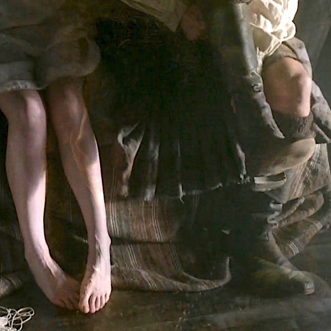
Need another Outlander hit? My pleasure. No, really! The morning after (he, he), Claire sports a pair of verra sweet feet (Starz ep 107, The Wedding)! Aw, look at those wee tootsies, shyly nesting. It was a big, big night! ’Nuf said! 😉

Oops, not quite, ‘nuf said. A lovely foot passage from Dragonfly in Amber book. Herself even mentions metatarsals!

“I’m honest enough to say that I dinna care what the right and wrong of it may be, so long as you are here wi’ me, Claire,” he said softly. “If it was a sin for you to choose me … then I would go to the Devil himself and bless him for tempting ye to it.” He lifted my foot and gently kissed the tip of my big toe. I laid my hand on his head; the short hair felt bristly but soft, like a very young hedgehog. “I don’t think it was wrong,” I said softly. “But if it was … then I’ll go to the Devil with you, Jamie Fraser.” He closed his eyes and bowed his head over my foot. He held it so tightly that I could feel the long, slender metatarsals pressed together; still, I didn’t pull back. I dug my fingers into his scalp and tugged his hair gently.
Divisions: Foot bones are divided into three regions. Such divisions aren’t whimsy, they are important in issues such as evaluating trauma, surgical amputation of part of all of a foot or evaluation of foot mechanics. 

  • Forefoot: Includes phalanges and metatarsals (Image C – white, left side). 
  • Mid-foot: Includes three cuneiforms, cuboid and navicular (Image C, turquoise). 
  • Hind-foot: calcaneus and talus (Image C – white, right side). 
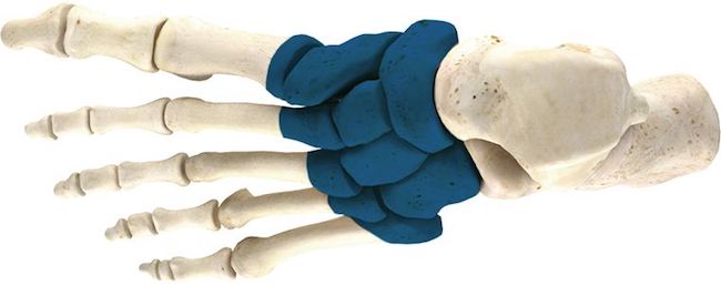
Image C 

Ligaments: Now, the 26 foot bones don’t just hang out under the skin. They are firmly bound to each other and to our leg bones via dozens of ligaments (Image D)!

Ligaments are fibrous tissues binding bone to bone and they are critical for foot integrity because feet bear our weight against gravity! Loose or torn ligaments give folks many problems because these compromise the integrity of the skeletal system! 

Image D shows only some of the numerous ligaments anchoring and stabilizing foot bones; here, we see lateral and dorsal ligaments. The plantar and medial ligaments are not visible.

The lesson won’t discuss these ligaments in detail because they are complex and tedious, but the image dramatically emphasizes some of the many ligaments needed to stabilize the foot skeleton!
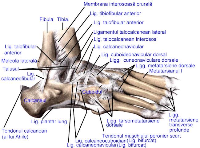
Image D 

Arches: We all know the foot has an arch, but did you know it actually has three arches (some count a 4th partial arch)? Two are longitudinal and one is transverse. The arches are maintained by interlocking tarsal and metatarsal bones, supported by ligaments and very strong tendons (image E).

  •  medial longitudinal arch (Image E – blue dashed line) extends from heel bone to first three metatarsals. Typically, it curves above the ground. When barefoot at the beach, it does not leave an imprint in sand (unless one is seriously flatfooted!).
  •  lateral longitudinal arch (Image E – green line) is a low arch arch between calcaneus and 5th metatarsal. When barefoot, it typically leaves an imprint in sand.
  • transverse arch (Image E – red line) runs across the foot at the tarsometatarsal joints (defined below).  

Although these arches are supported by strong ligaments and tendons, they exhibit some mobility when weight is applied to or removed from the foot. This springiness makes walking and running more economical in terms of energy.
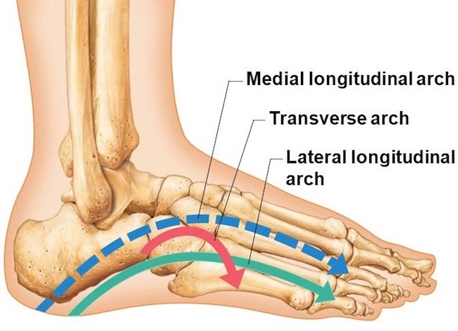
Image E 

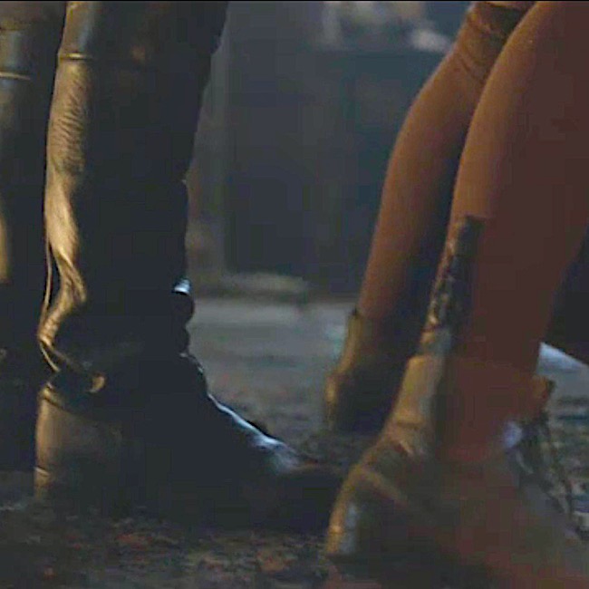
Speaking of arches, how ‘bout some booted ones! Yep, another dose to wake you students! BJR’s booted “trick or treat feet” (Starz, ep 108, Both Sides Now) will do the trick, nicely! The blackguard throws Claire over his desk preparing to further assault her. Darn! She canna reach the sgian dubh in her boot! No treat here – all vicious tricks!

Diana describes Claire’s toes just after Jamie squats in the prison window (Outlander book). 

Randall bent and scooped up the gun in a quicksilver motion. As soon as the knife left my throat, I tried to sit up, but he placed a hand on my chest and shoved me flat again. He held me down with one hand, using the other to aim the pistol at Jamie. The discarded knife lay somewhere on the floor near my feet, I thought. Now, if only I had prehensile toes.… The dirk in my pocket was as unreachable as if it were on Mars.
Plantar Aponeurosis: Remove plantar skin (very difficult on a cadaver) and a triangular sheet of connective tissue is revealed, the plantar aponeurosis. It is anchored to the calcaneus, flares in the mid-foot and ends as five (or more) bands radiating toward bases of the toes (Image F). 

The tough, fibrous aponeurosis is made mostly of collagen fibers. As such, it is a shock absorber when the foot strikes the ground. It also stabilizes arches of the foot and allows flexion at the first metatarsophalangeal joint, which carries the majority of body weight during ambulation.

If the plantar aponeurosis becomes injured or inflamed, it may cause plantar fasciatis. A painful condition common to athletes, it causes stinging foot pain that can lead to further leg injuries if untreated.
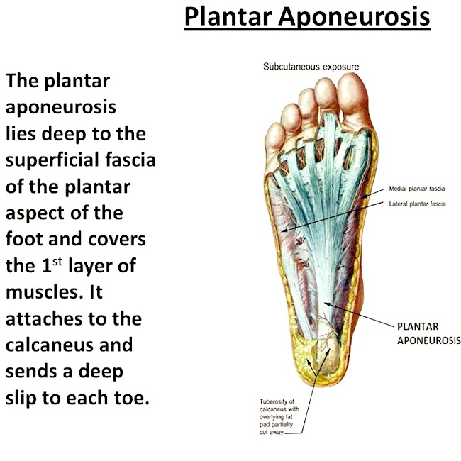
 

Image F 

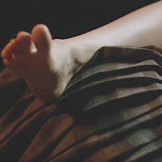
Another break for Outlander! This is a splendid example of body heat feet (Starz, ep 109, The Reckoning). Things are on broil up at Castle Leoch! Claire’s right heel hooks over Jamie’s Fraser plaid….hum…. Talk about a foothold! Snort!
Joints: We have covered joints (the anatomical type) in prior lessons (e.g. Anatomy Lesson #2, When Claire Meets Jamie or How to Fall in Love While Reducing a Dislocated Shoulder Joint!).  

To reiterate, joints are sites where two or more bones meet; some are moveable and some are not. Moveable joints allow for motion and there are several types. Our 33 foot joints fall into the following categories:

  • TC Joint (1): between distal tibia, fibula and talus, a.k.a. talocrural or ankle joint
  • IT Joints (13): between tarsal bones 
  • TM Joints (5): between metatarsals and tarsals
  • MP Joints (5) : between proximal phalanges and metatarsals
  • IP Joints (9): between phalangeal bones

Reducing the technicality of this topic, we will only cover the superbly designed TC or ankle joint! The ankle joint is a mortise joint, a term used in carpentry. Here, the talus projects upwards and fits inside a three-sided bone box formed by tibia and fibula of the leg  (Anatomy Lesson #27, Colum’s Legs and Other Things, Too!). Thus, our “ankle bones” are not separate bones, they are parts of tibia and fibula.  The inner ankle bone is actually the medial malleolus of the tibia; the outer ankle bone is the lateral malleolus of the fibula. This is a highly stable hinge joint that allows movement (see below).
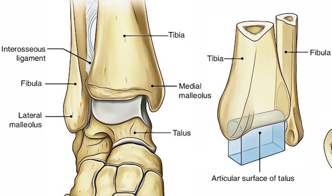
Image G 

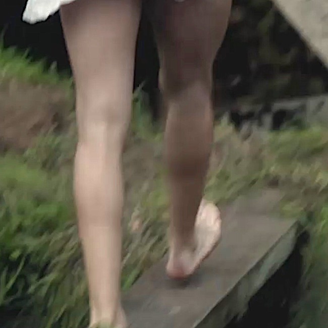
Before the lesson turns to movements, let’s take a quick keek at Jamie’s upbeat feet! Cheerfully dressed in nothing but a sark, he strides to the freezing mill stream to sleuth out a prob with the water wheel (Starz ep 113, Lallybroch). Seems it is producing gritty bannocks! Upbeat feet, that is, until a mess ‘o Redcoats arrive! Notice: his right foot is lifted at the ankle, a movement known as dorsiflexion. Yep! Read on to learn more about this term.
Movements: Various movements occur at the foot joints. Some are slight; others are more generous and important for ambulation. The talocrural joint (ankle joint) allows for six movements at the ankle; the first four are demonstrated in Image H: 

  • dorsiflexion: lifting foot at the ankle
  • plantar flexion: pointing the foot at the ankle
  • inversion: turning sole medially (toward midline)
  • eversion: turning sole laterally (toward the side)
  • abduction: turning foot to side (slight)
  • adduction: turning foot toward midline (slight)

Psst…..Practitioners often prefer the terms, supination for inversion and pronation for eversion.
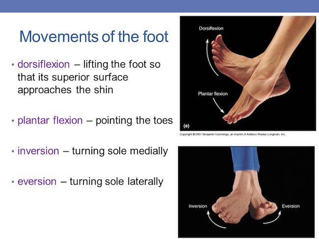
Image H 

Next, there are toe movements which can occur independent of the ankle joint. These involve IP and MP joints: 

  • flexion: curling the toes 
  • extension: lifting the toes
  • abduction: spreading the toes
  • adduction: returning the toes to a resting position
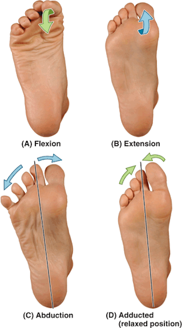
Image I 

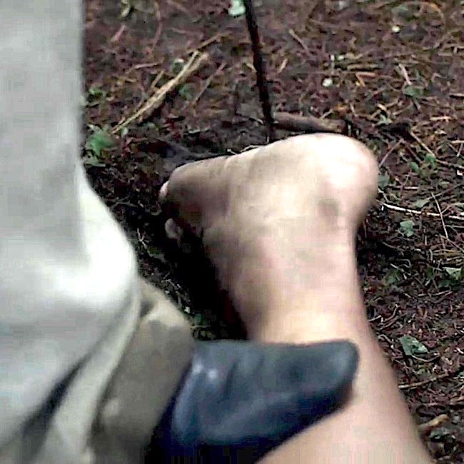
Back for an Outlander scene and a collective gasp!  Jenny takes a hot poker to the sole of a redcoat captive. Ouch! The poor man now has mincemeat feet. What ya doing Jenny?

Spill, messenger! Where is my bro??? In no uncertain terms, Big Sis declares to Claire: 

Love Forces a Person to choose!
Extrinsic Muscles: First, a wee definition…long time students will remember that in anatomy the leg is the lower limb between knee and ankle joints; thigh is between hip and knee joints. Most people use the term lower leg for the anatomical leg.

Muscles acting on the foot are classified as extrinsic muscles, those originating in the leg, and intrinsic muscles, those originating in the foot.

Amazing Fact: all leg muscles, excepting one, actually act on the foot! These are so complex, they must be simplified. Yes, Image J is simplified!!!

Extrinsic muscles in Fig. J (anterior and side muscles – left panel):

  • tibialis anterior – dorsiflexes & supinates foot
  • extensor hallucis longus – extends big toe & dorsiflexes foot
  • extensor digitorum longus – extends toes 2-5 & dorsiflexes foot
  • fibularis longus & fibularis brevis – pronate & plantar flex foot

Extrinsic muscles in Fig. J (superficial posterior muscles – middle panel):

  • gastrocnemius – plantar flexes foot (cut away in image)
  • soleus – plantar flexes foot

(Note: gastroc and soleus jointly share the massive achilles tendon which inserts into calcaneus. Normally, these are extremely strong plantar flexors)

Extrinsic muscles in Fig. J (deep posterior muscles – right panel):

  • flexor hallucis longus – flexes big toe & plantar flexes foot
  • flexor digitorum longus – flexes toes 2-5 & plantar flexes foot
  • tibialis posterior – plantar flexes & supinates foot
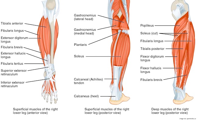
Image J 

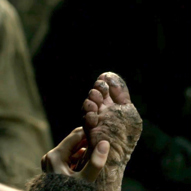
Whew. That was scary! Time for another Outlander treat to lower the blood pressure. Oops, this is pretty scary, too (Starz ep 209, Je Suis Prest) – yuk! It’s eek feet for puir Angus – the lad has been careless with his feet. Nurse Claire warned him to keep them dry! 
Back to anatomy. No, we are not finished with muscles. Gasp!

Intrinsic Muscles of Dorsum (top) of Foot: As stated above, intrinsic muscles arise from foot bones. There are two smallish muscles on the dorsum of the foot, not shown in Image K. 

  • extensor hallucis brevis – extends the big toe
  • extensor digitorum brevis – extends toes 2-5

Intrinsic Muscles of Plantar Foot: Believe it or not, 18 muscles are located deep to the plantar aponeurosis. Who would have thought??? These are arranged in four layers (Image K –  from left to right). If you think these look challenging, you are right. Outside the head, the foot is one of the most difficult body parts to dissect! 

First structure in Image J (left panel) 

  • Plantar aponeurosis – not a muscle

1st Layer of Intrinsic Muscles in Image J ( 2nd panel):

  • abductor hallucis – draws big toe towards midline of body
  • abductor digiti minimi (I love this name!) – draws 5th toe away from foot
  • flexor digitorum brevis – flexes toes 2-5

2nd Layer of Intrinsic Muscles in Image J (3rd panel)

  • Lumbricals – both flex & extend different phalanges of toes 2-4
  • Quadratus plantae – flexes toes 2-4

3rd Layer of Intrinsic Muscles in Image J (4th panel)

  • flexor hallucis brevis – flexes big toe
  • adductor hallucis – draws big toe towards foot
  • flexor digiti minimi brevis – flexes 5th toe

4th Layer of Intrinsic Muscles in Image J (5th panel –  horizontal)

  • dorsal interossei – abduct toes 2-4 (spreads toes)
  • plantar interossei – adduct toes 3-5 (returns toes to resting position)
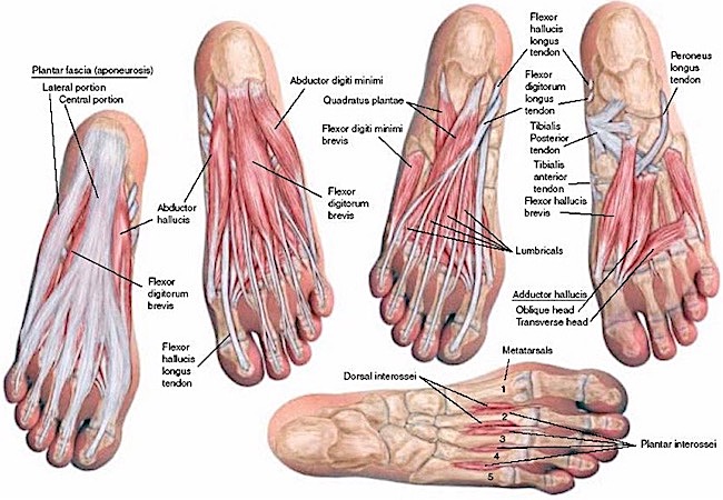
Image K

Now, given that mess of muscles, you probably appreciate how complex foot movements can be achieved. With some 20 intrinsic and 10 extrinsic muscles controlling our feet, they are quite capable, indeed!

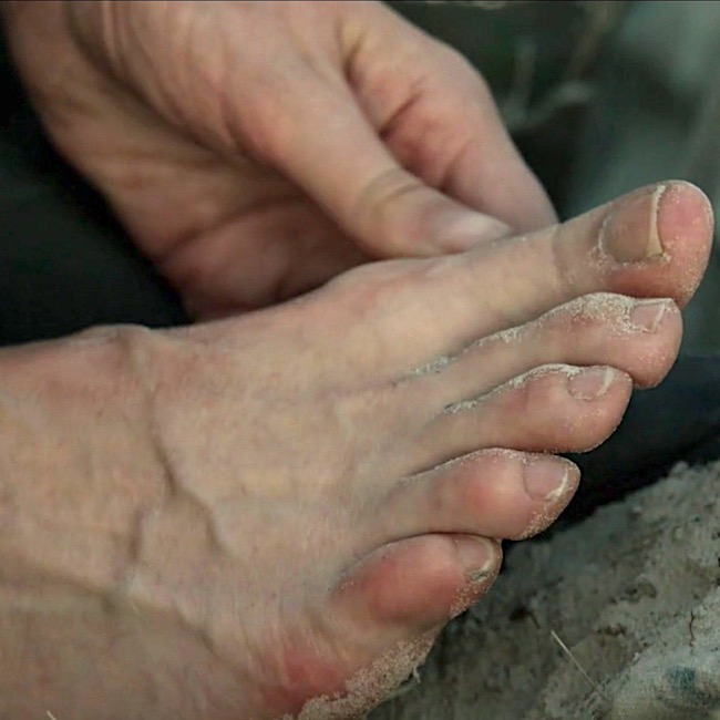
Back to Outlander! We see Claire’s poor, weary beat feet, exposed to sand, surf, sun and formicidae (Anatomy Lesson #55, Formidable Formicidae) in Outlander ep 311, Uncharted! Trudging in wet shoes, dealing with festering ant bites, surviving snake slithers…. Diana explains Claire’s feet in Voyager book:

Squads of tiny purple crabs ran off in profound agitation at my approach. My feet sank into the mud to the ankles, and I thought better of putting on my shoes, wet as they were… My feet were bruised and sore, and punctured by fallen palmetto fronds, but the path before us looked relatively smooth.
We really must take good care of our feet if we want them to last. Exercise, weight control, healthy diet, wearing supportive shoes all help ensure the feet bear our weight for a lifetime. Even to the most careful, our feet suffer many assaults: bone spurs, athlete’s foot, plantar fasciitis, corns, bunions, Morton’s neuroma, flat feet, hammer toe, warts, stress fractures, etc. Or, we lose toes because of poor circulation, trauma, or developmental issues –  such problems have plagued us since ancient times. To the point, the oldest known functional prosthesis is an Egyptian wooden mummy toe  (Image L). It actually articulates at the laced surfaces. So clever! 

Hey, wait! How do we know it is not a daddy toe? He, he!
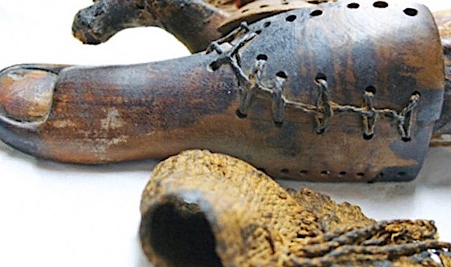
Image L 

Speaking of toes, Voyager book describes a splendid battle with pirates aboard ship. Sadly, the scene did not make it into the TV version. But, here is a shocking tidbit from the bloody fight:

Cursing incoherently under my breath, I ran to the bottom of the ladder, and reaching up, swung the long-handled amputation knife at his foot, as hard as I could. There was a high-pitched screech from the pirate. Something flew past my head, and a spray of blood spattered across my cheek, wet-hot on my skin. Startled, I dropped back, looking down by reflex to see what had fallen. It was a small brown toe, calloused and black-nailed, smudged with dirt.

Alrightie then! <G>

Let’s consider how you can hurt your neat, sweet, elite, complete feet… In your wildest dreams, do you think shoes such as these are good for feet (Image M)? Wear high heels for long, and one guarantees that later in life, the wearer will have foot problems. The foot is not designed to walk around on the metatarsophalangeal joints (ball) of the foot, which is why wearing spikes hurt!!!

Gentle admonition: if you wear this type of footwear, you should stop.

Image M 

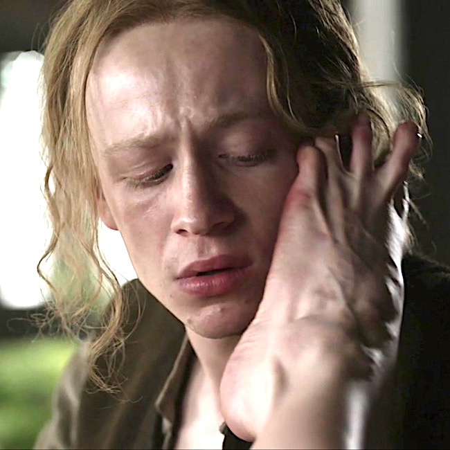
Closing this lesson with indiscrete feet: no Outlander lass is quite as indiscrete and raunchy as Geillis (Gillian) Edgars Duncan Abernathy. Puir young Ian; that dear lad’s cheek is no place for a witch’s foot (Outlander ep 313, The Bakra)! 

BTW, the prominent ridges passing to 2nd to 5th toes are tendons of extensor digitorum longus! Yay!
Bottom line: The complete and complex human foot is truly an anatomical work of art. Let’s vow to take good care of ours!

The deeply grateful,

Outlander Anatomist

Follow me on:

Photo Creds: Sony/Starz; www.cba.ca (Image – L); www.DHgate.com (Image M ); www.digikalla.info (Image J); www.earthslab.com (Image G); www.footankle.strker.com (Image C); www.goodfeet.com (Image A); www.heatheappointments.com (Image I); www.nurseslab.com  (Image K – ); www.slideplayer.com (Image E, Image F, Image H); www.teachmeanatomy.com (Image B); www.wickimedia.commons.org (Image D)

Anatomy Lesson #59: Claire the Knife!

“Claire the Knife” is not a usual anatomy lesson. Rather, let’s explore the many images of Claire with a honed edge either in her hands or directed at her person! Such images are taken from S1-S3 of Outlander. There are even more examples in Herself’s books that weren’t translated onto the screen. Please notify me if I miss any fine examples of “Claire the Knife!”  Let’s get started!

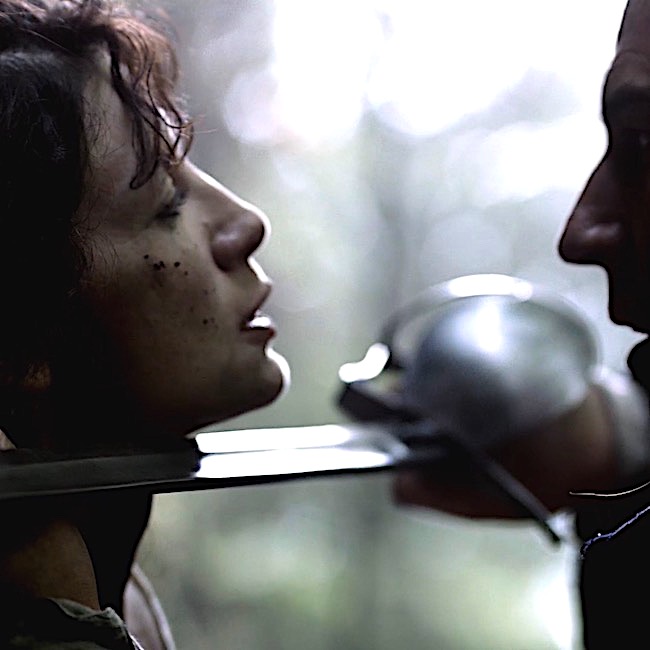
Early in Outlander episode 101, Sassenach, Claire accidentally encounters Frank’s ancestor, Captain Jonathan Wolverton Randall, Esq. From Outlander book:

“Who the bloody hell are you?” I demanded again. 

… He bowed sardonically, hand over his heart. “I am, madam, Jonathan Randall, Esquire, Captain of His Majesty’s Eighth Dragoons. At your service, madam.”

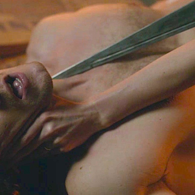
Not an officer-and-a-gentleman, she is soon backed against a stone ridge with his sword pressed to her lovely throat (Anatomy Lesson #12, Claire’s Neck or The Ivory Tower). 

Your honor, I submit, Exhibit #1! <g>

Then, in Outlander episode 104, The Gathering, Claire twice encounters a knife, once in her hand and once shoved into her beautiful face!

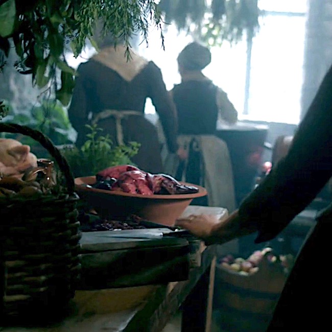
Swiftly gliding into Mrs. Fitz’s kitchen, Claire grabs for a butcher knife amid bins of potatoes (Hey, I thought Jenny introduces those to Scotland?), chicken and other delectables. She is gathering provisions for a furtive flight to the stones. Outlander book explains: 

During my hours helping in the kitchens and orchards, I had managed to stow away sufficient food to see me provided for several days, I thought… I had stout boots and a warm cloak, courtesy of Colum. I would have a decent horse; on my afternoon visit to the stables, I had marked out the one I meant to take.

Her knife grab is thwarted by Ms. Fitz: 

“Claire! You aren’t wearing that to the ceremony, are ye lass?”

No, Mrs. Fitz, she’s not wearing that butcher knife! Hee, hee.

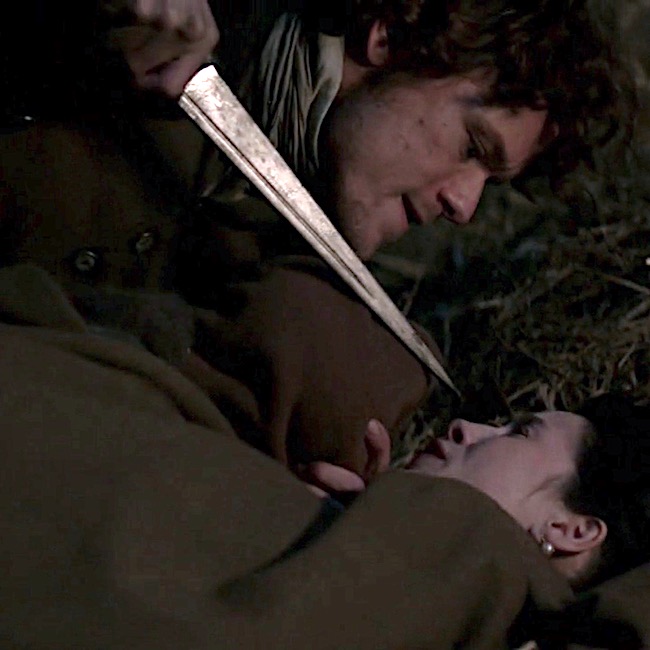
Later in evening as the entire ensemble is either oath-taking or stinking drunk, Claire sidles into the stables to steal away on Brimstone.

Oops! She steps on Jamie cocooned in hay (yay!).  Mistaking her as a threat, he threatens her with his dirk! (wink, wink) 

“Jesus H. Rosevelt Christ!” 

“No, sassenach, just me.” 

(We all ken he is divine, just not that kind of divine!)

From Outlander book: 

The obstruction rolled over with a startled oath and grasped me hard by the arms. I found myself held against the length of a sizable male body, with someone’s breath tickling my ear.

Then, Dougal and his band of merry men head off to collect taxes and conduct other clan “business” in  Outlander episode 105, Rent.  Dougal explains in Outlander book:

“Through the MacKenzie lands. Colum doesna travel, so visiting the tenants and tacksmen that canna come to the Gathering—that’s left to me. And to take care of the bits of business here and there.”

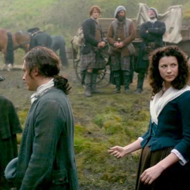
On the road, Claire witnesses “fowl” play by Dougal and his men. Declaring she will not eat yummy-looking birds she thinks were roasted by thieves, Claire quickly faces the point of Angus’ dirk.  Angus is having none of it that kind of name calling, especially from an English sassenach spy! Fortunately, Jamie talks him down and Claire flounces away with left eye intact. 

By episode’s end, Claire and Dougal are accosted by Lt. Jeremy Foster.

In Outlander episode 106, The Garrison Commander, both are delivered to the garrison at Brockton.  There, she enjoys a sumptuous meal (excepting the claret) with Brigadier General Lord Oliver Thomas, et al, until, horror of horrors, Captain Black Jack arrives. Thereafter, things take a distinctly downward turn! 

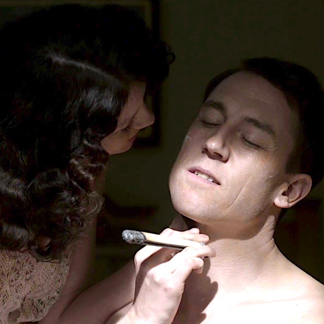
After aiding the amputation of a soldier’s arm, she returns to the dining room to find puir Corporal Hawkins performing ablutions on BJR! Claire recognizes the stropped blade as a family heirloom – the very one she used to shave first hubby, Frank.

If only, she held that blade to BJR’s neck – could have saved she and Jamie a world of hurt! Another blade – another time. From Outlander book: 

“Yes, I thought so. It had to be you, from MacKenzie’s description.” The door closed behind me, and I was alone with Captain Jonathan Randall of His Majesty’s Eighth Dragoons. He was dressed this time in a clean red-and-fawn uniform, with a lace-trimmed stock and a neatly curled and powdered wig.

But the face was the same—Frank’s face. My breath caught in my throat.

This time, though, I noticed the small lines of ruthlessness around his mouth, and the touch of arrogance in the set of his shoulders. Still, he smiled affably enough, and invited me to sit down.

Escaping BJR’s clutches, Dougal stops off at St. Ninian’s spring for a wee nip of sulfurous water. Known in the highlands as a liar’s spring, he waits to see if the magical waters will burn Claire’s gullet (Anatomy Lesson #45, Tremendous Tube – GI System, Part 2)!  He draws a dirk to finish her off should massive heartburn set in. From Outlander book: 

“No!” Something occurred to me. “So at least you believe me when I say I’m not an English spy?”

“I do now.” He spoke with some emphasis. …

“St. Ninian’s spring.

…They call it the liar’s spring, as well. The water smells o’ the fumes of hell. Anyone who drinks the water and then tells untruth will ha’ the gizzard burnt out of him.”

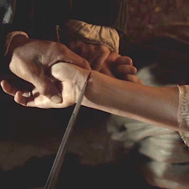
Fast forward one episode, Dougal finally gets his wish as his dirk bites into Claire’s wrist!

Blood vow at a wedding she never sought, but well worth the scar!

Ye are Blood of my Blood, and Bone of my Bone. 

I give ye my Body, that we Two might be One.

I give ye my Spirit, ’til our Life shall be Done.

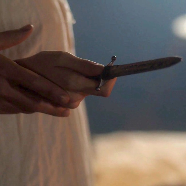
Outlander Episode 108, Both Sides Now: Still on rent-collection duty, the MacKenzie band is raided by Clan Grant! Rent, horses, food, what-have-you, everything is fair game. In the aftermath, Jamie is scolded for giving his wife a dirk for protection but not teaching her how to use one. Time for Claire to get a knife-life lesson. Ned Gowan extracts a small sgian dubh drawn from a more private part of his, ahem, breeks. So, she takes a knife-wielding lesson from TV Angus/book Rupert. Willie is the sacrificial lamb: 

He poked a blunt forefinger under the lower rib on the right, … “This is the spot in back—either side. See, wi’ all the ribs and such, ’tis verra difficult to hit anythin’ vital when ye stab in the back. If ye can slip the knife between the ribs, that’s one thing, but that’s harder to do than ye might think. But here, under the last rib, ye stab upward into the kidney. Get him straight up, and hell drop like a stone.” 

Fab advice as hours later, Jamie and Claire slip into the heather for a quick tryst, soon to be disturbed by two Redcoat deserters (coitus interruptus –  snort!). Thwarting her assaulter, Claire thrusts the sgian dubh – into the spot – as she was taught. The lass learned her lesson well!

Then, comes Outlander episode 109, The Reckoning: Captured by the Redcoats, assaulted (again) by BJR, rescued and then skelped by her second husband, they seek their way back to each other. As an act of penance, Jamie swears fealty to Claire, this time the dirk is pressed against his bosom (Outlander book):

I swear on the cross of my Lord Jesus, and by the holy iron which I hold, that I give ye my fealty and pledge ye my loyalty. If ever my hand is raised against you in rebellion or in anger, then I ask that this holy iron may pierce my heart.

And, they are off to the races! Oops – not so fast, my lad. In the midst of “reconciliation,” Claire threatens Jamie’s chin with his dirk (Anatomy Lesson #26, Jamie’s Chin – Manly Mentus).

“If,” I said through my teeth, “you ever raise a hand to me again, James Fraser, I’ll cut out your heart and fry it for breakfast!” 

Och! Parritch sounds much, much bettah!

Then, things quiet down for a bit and we see little of “Claire the Knife.” That is, until BJR gets his hands on Jamie. He has coveted the lad since the first flogging. After Murtagh scraps what is left of Jamie off the dungeon stones, they flee Wentworth, returning Jamie to his lawful wife.  There she mends his mangled hand, courtesy of BJR and giant Marley.

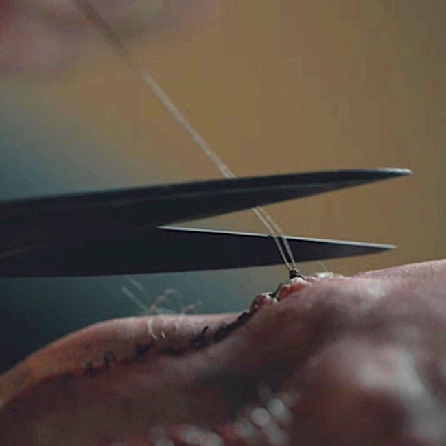
Blades again, this time in the form of scissors, clip sutures used to mend his broken skin (Outlander ep 116, To Ransom a Man’s Soul).

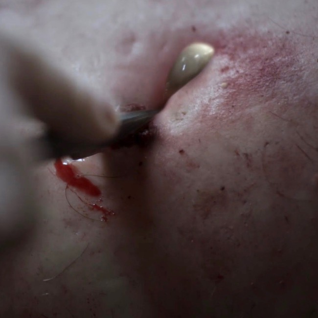
Up pops Season 2. Claire, being pregnant with Faith, isn’t into knife heroics.  An exception is her work at the Paris charity hospital, L’Hôpital des Anges. There, the scalpel blade slips nicely into her hands. Consider a workman’s festered wound due to an embedded splinter. She opens the wound and removes the offending object (Outlander episode 203, Useful Occupations and Deceptions). That knife has a life of its own!

Fast forward to Outlander episode 213, Dragonfly in Amber, wherein Jamie and Claire find themselves mired in a desperate struggle with Dougal – sly Uncle is intent on killing the sassenach witch!

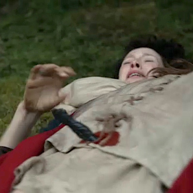
He should have known that Jamie would protect her with name, clan and body!  But, neither reason, threat, nor cajoling stops Dougal’s advance. A blade to the chest is the only remedy  – in which Claire takes an equal hand – she finishes what Jamie cannot do alone!

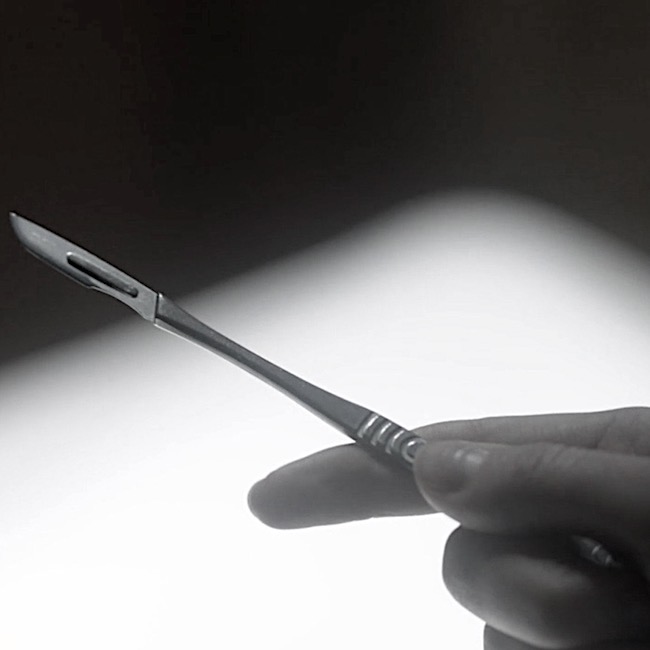
Then, comes season 3. The next time a knife creeps into Claire’s hand, it is at the teaching theater awaiting her first anatomy class as a medical student (yay!). Arriving early, she feels the heft of the professor’s scalpel and is pleased with the heft; it fits well in her surgeon-to-be hand (Outlander episode 302, Surrender). 

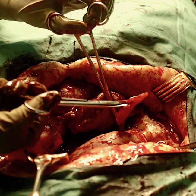
Following the swiftest medical education ever, Claire is soon a practicing surgeon, clipping “necrotic” tissue from a patient’s abdomen (Outlander ep 305, Whisky and Freedom). Time and training has given Claire the license to legally cut another human. Such is her special calling and she fulfills it with grace and skill. Snip, snip. (Pssst… necrotic tissue isn’t pinkish-red in color, but don’t tell anyone.)

Next thing we know, Claire has donned a bat suit and gone back through the stones to seek A. Malcolm of Edinburgh (Outlander ep 307, Creme de Menthe). Confronting an exciseman in nothing but her shift and a hidden knife, Claire stabs his leg. Have knife – will travel!! He falls, striking his head on the hearth.  

Jamie arrives and what to his wondering eyes should appear, but a dying man – oh such is his fear!

Sassenach, what the devil happened????

I step away to do a little smuggling and return to find my wife with her knife and a dying man!!!

Still counting? Trying to save the life of the SOB (she’s a doctor!), Claire sends for a trephine from the local barber surgeon. She opens her medical kit, retrieving her trusty-not-rusty scalpel. Yes!

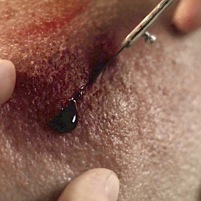
She makes a swift cut through the scalp – a space is opened through which the trepanning instrument is inserted. Then, she starts drilling for oil.  Not really, she drills to relieve pressure from an epidural bleed. 

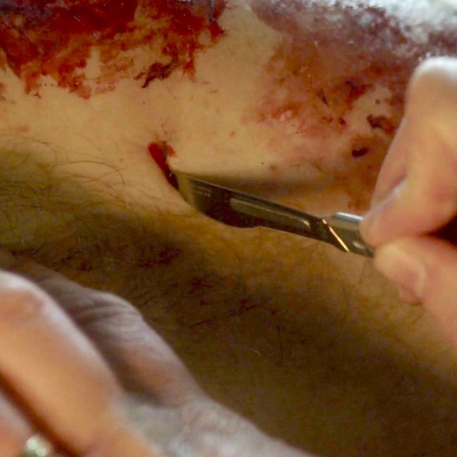
Hang on, Snoopy, hang on – we’re almost finished! Then, in episode 308, First Wife, Claire uses her magic scalpel to open a nick near Jamie’s left oxter (Anatomy Lesson #4, Jamie’s Chest” or “the 8th Wonder of the World!). Her goal is to retrieve bits of lead discharged from Laoghaire’s fowling piece. She grasps and retrieve the bits leaving Jamie’s axillary/brachial artery intact (Anatomy Lesson #56: Achy Brachial Artery)! She is super good with a knife!  

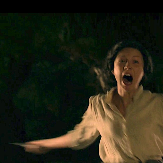
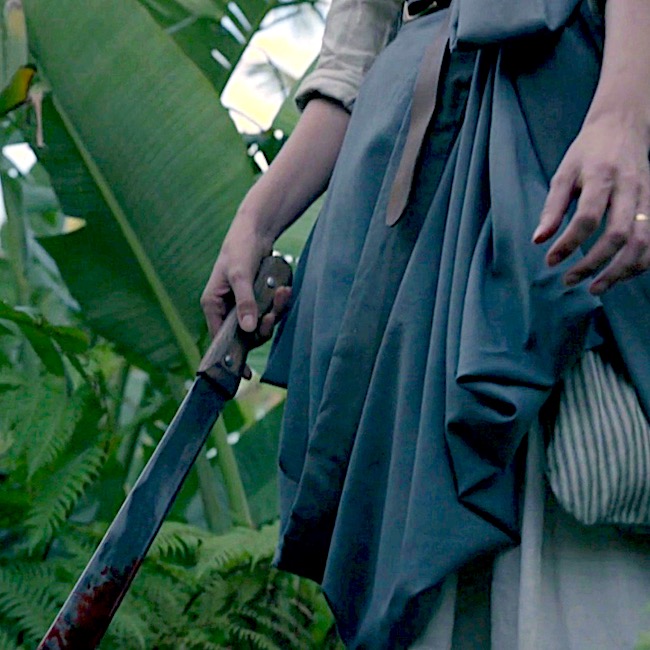
The last time we see Claire wield a blade in TV Outlander, she confronts the Bakra (Outlander episode 313, Eye of the Storm)! Picking up a machete (ax in Voyager book), she screams and advances on Geillis Abernathy! She must prevent the witch from her fulfilling her plan to kill young Ian and pass through the time portal to slay Bree. 

Whack! Claire’s blade finds its mark and the wicked witch lies deid! Graphically explained in Voyager book:

Reflections of the fire burned red in the pupils of her eyes. The red thing, Jamie had called it. I gave myself to it, he had said. 

I didn’t need to give myself; it had taken me.

After fleeing Abandawe cave with Ian and Jamie, Claire trembles in the jungle with gore dropping from the machete. 

It is then she realizes, the 200+ y.o. bones she examined in Boston with Dr. Joe Abernathy (Outlander ep 305, Whisky and Freedom) belonged to none other than Geillis Abernathy!  And, Claire herself, ushered in that well-earned fate! Like I said, Claire the Knife!

To summarize, over many episodes, Claire is at one end or the other of a blade; always has been – always will be. <G>

Bobby Darren sang the hit, “Mack the Knife.” I re-imagine it as, “Claire the Knife:”

Oh, the shark, babe, has such teeth, dear

And it shows them pearly white

Just a jackknife is Miss Claire, babe

And she keeps it, ah, out of sight.

Dinna mess with the Sassenach Surgeon!

The deeply grateful,

Outlander Anatomist

Follow me on:

Photo Creds: Starz, Outlander episodes 101, 104, 105, 106, 107, 108, 109, 116, 203, 213, 302, 305, 307, 308, 313


Anatomy Lesson #58: Needles, Nerves and Penicillin!

Welcome, all students to Anatomy Lesson #58, Needles, Nerves and Penicillin. What do these words share in common? Why, Jamie, of course!  In Anatomy Lesson #56, Achy Brachial Artery, we learned that birdshot from Laoghaire’s fowling piece almost ruptured one of Jamie’s major arteries. We also learned how Claire analyzed and repaired those wounds – based on human anatomy, of course!

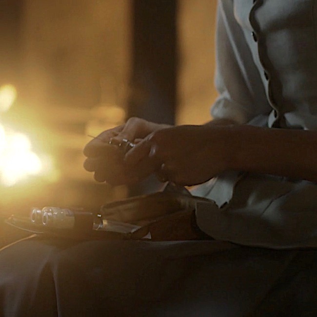
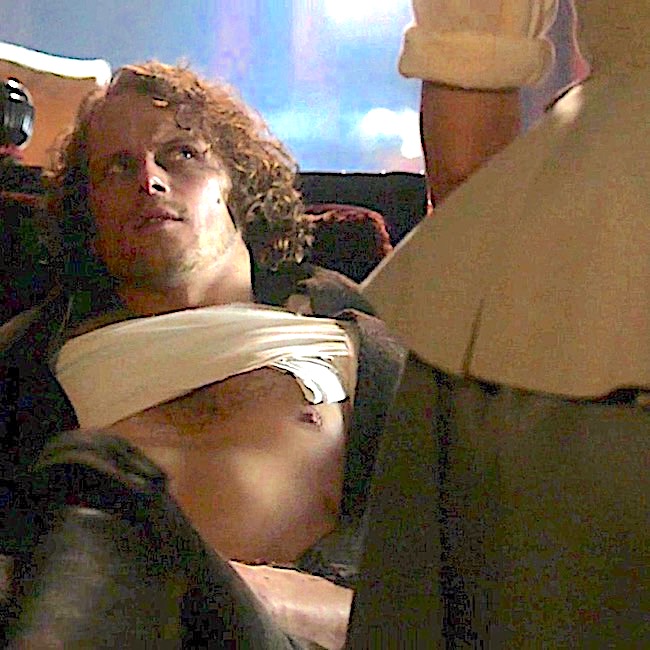
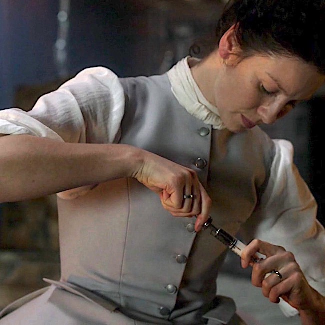
Today’s lesson will examine Claire’s final cure for what ails her man. In Outlander episode 308, First Wife, we see her open a small medical bag equipped with needle, plunger and bottles of white fluid. What is she doing? Diana explains in Voyager book:

I laid the small, flat case on the table and flipped the latch. “I’m not going to let you die this time either,” I informed him, “greatly as I may be tempted.” I carefully extracted the roll of gray flannel and laid it on the table with a soft clinking noise. I unrolled the flannel, displaying the gleaming row of syringes, and rummaged in the box for the small bottle of penicillin tablets.

Translation: Dr. Claire busily prepares a hefty dose of penicillin for our fever-ravaged, red-heided hero!  So, let’s begin the lesson with penicillin.

What is Penicillin? Penicillin is an antibiotic, from the English word, anti- meaning “against,” and the Greek word, bios meaning “life.”  In a nut shell, antibiotics are substances which retard the growth of and/or destroy bacteria (germs).

How does Penicillin Work? First, understand that human cells are surrounded by a fragile cell membrane, but bacterial cells are surrounded by a sturdy cell wall. Penicillins destroy bacteria by preventing synthesis of said cell walls or by promoting processes to break them down. They do not harm human cells because our cells lack cell walls. Make sense? Grand!

From Whence Cometh, Penicillin: Before penicillin, there were no effective treatments for common infections such as pneumonia or rheumatic fever; hospitals were filled with folks suffering blood poisoning contracted from nothing more than a mere cut or scratch!

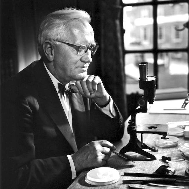
In 1928, Alexander Fleming (Image A), a British bacteriologist, returned from summer vacation in Scotland (yay!). Checking his lab specimens, he found that a mold (Penicillium notatum = P. notatum), probably from an open window, had contaminated his colonies of a common bacteria (Staphlococcus aureus or S. aureus).

Image A

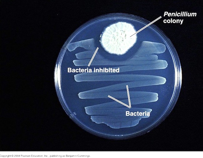
Examining the specimens via a truly primitive microscope (see Image A), he found the mold inhibited growth of S. aureus! Image B shows a modern version of what he observed: zigzagging ribbons of bacterial growth and a “halo” of no growth around the Penicillium colony. He correctly deduced his “mold juice,” which he later dubbed penicillin, inhibited bacterial growth!

Image B

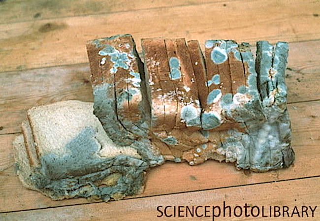
Stop! Now, we might imagine this was an entirely modern discovery, but others in the early 1900s had made similar observations although not much came of these. Even more amazing? Ancient Egyptians applied a poultice of moldy bread to promote healing of infected wounds. Very doubtful that ancient Egyptian bread looked anything like this loaf of sliced, white bread (Image C), but you get the idea. Icky!

Image C

Penicillin to the Rescue! Fleming published his experiments in 1929, but his paper focused mostly on uses for penicillin in the lab with only a brief remark about possible therapeutic benefits.

Ten years later, an Australian scientist and Oxford pathologists proved penicillin was an effective antibiotic. But, the mold was cranky and fastidious to grown and the yield so low, only small quantities could be produced.

Enter WWII with its urgent need for battlefield antibiotics! Various pharmaceutical companies teamed with US Department of Agriculture to mass produce penicillin (the chemical engineer who designed 10,000 gal. production vats was a woman!).

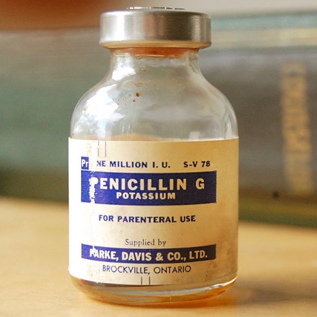
Simultaneously, a global search was launched to find a new mold that could be grown easier and yield larger quantities of penicillin. One hot summer day, a laboratory assistant arrived with a cantaloupe covered in  ”pretty, golden mold” which she (yes, another woman!) had found at a Peoria (IL) market. This mold was Penicillium chrysogeum, yielding 200 times the volume of penicillin as P. notatum!!! In 1954, using P. chrysogeum, US production of the drug jumped from 21 billion units to more than 6.8 trillion! And, penicillin was made available to allied forces (Image D).

A final note: Australia was the first country to make penicillin available for civilian use. Go, Aussies!

Penicillin does have drawbacks; it is ineffective against viruses and some types of bacteria (gram-negative), some people (0.03%) are allergic, and some bacteria have developed resistance due to over prescription or patients not following dosage instructions. Despite these drawbacks, penicillin can effectively treat the following short list of diseases (there is a long list, too):

  • Strep infections such as strep throat, scarlet fever
  • Staph infections such as post traumatic infections
  • Syphillus
  • Gonorrhea
  • Diphtheria
  • Lyme Disease
  • Typhoid Fever
  • Necrotizing Fasciitis (Flesh-eating disease)

If you remain curious about the fascinating history of penicillin, your thirst might be quenched by reading this article: https://www.acs.org/content/acs/en/education/whatischemistry/…/flemingpenicillin.html

Although I rarely move ahead of TV Outlander, this apropos quote from Diana’s book, The Fiery Cross, describes Claire’s work with penicillin. Herself wrote about molds from varied sources including the melon. Way to “break the mold,” Diana!

I twiddled the quill, rolling it between thumb and forefinger. I had kept a faithful account of my experiments with penicillin—the growing of cultures on media ranging from bread to chewed pawpaw and rotted melon rind, painstaking descriptions of the microscopic and gross identification of the Penicillium molds…

Image D

OK, now, back to Outlander episode 308, First Wife!

Nefarious Needles: With penicillin behind us (har, har) we broach the topic of needles. Ahhhh, yes, needles – a cruel device, with which most folks share a love-hate relationship! Claire prepares the penicillin and draws it into a syringe. Here, from Voyager book:

“What in God’s name are those?” Jamie asked, eyeing the syringes with interest. “They look wicked sharp.”

Psst … To me, he looks more wary than interested. Hah!

And more about needles:

I didn’t answer, occupied in dissolving the penicillin tablets in the vial of sterile water. I selected a glass barrel, fitted a needle, and pressed the tip through the rubber covering the mouth of the bottle.

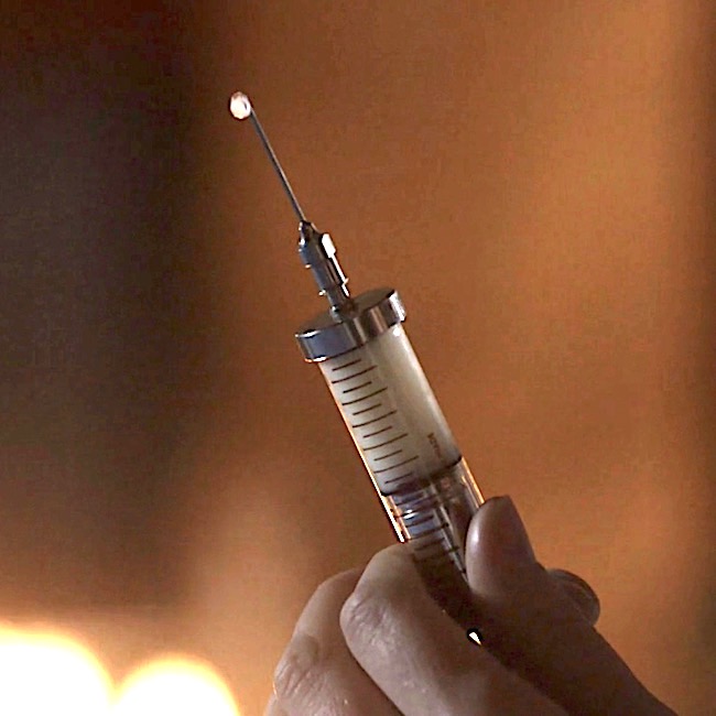
Holding it up to the light, I pulled back slowly on the plunger, watching the thick white liquid fill the barrel, checking for bubbles.

And more!

Then pulling the needle free, I depressed the plunger slightly until a drop of liquid pearled from the point and rolled slowly down the length of the spike.

Claire’s penicillin is a thick, white liquid, poorly received by Jamie’s remarkably regal rump. We might ask, why Claire didn’t carry oral penicillin for her return trip to 1700s Scotland? It was available in her time. I really don’t ken the answer, but the injectable type promises readers and viewers a much more rousing and carousing tale! <G>

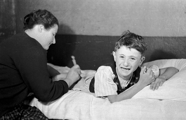
Understand, the first penicillins were injected, not ingested (Image E – puir wee laddie). Why? Because, when swallowed, stomach acid breaks down penicillin rendering it impotent. But, in the 1950s, types of oral penicillin which resisted stomach acid were developed. Today, many forms of penicillin include: capsule, chewable tablet, tablet for suspension, extended-release tablet, powder for suspension, powder for solution, capsule and syrup.

Image E 

Claire also brought her own 20th century syringes because they had not been invented in Jamie’s time. Almost a century later (1840s), Irish physician Francis Rynd invented the hollow needle to deliver the first modern-era subcutaneous (under-the-skin) injections! …”I’ve Got You Under My Skinnn!” ? Och!

Before we move on, let us honor resourceful Pre-columbian Native Americans who created early hypodermic needles and syringes using “hollow bird bones and small animal bladders!” Incredibly inventive and creative – using only nature’s gifts to presumably administer healing substances! No Spoilers: This relatively unknown fact should please book readers who will recognize something similar which Claire employs in The Fiery Cross.

I’ve said it before and I’ll say it again: if you haven’t read/listened to the books, you are missing one long joyous, glorious voyage!

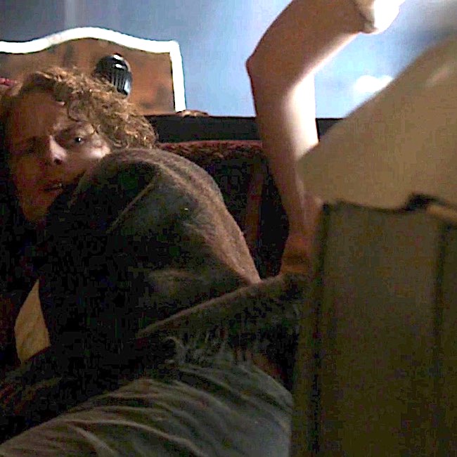
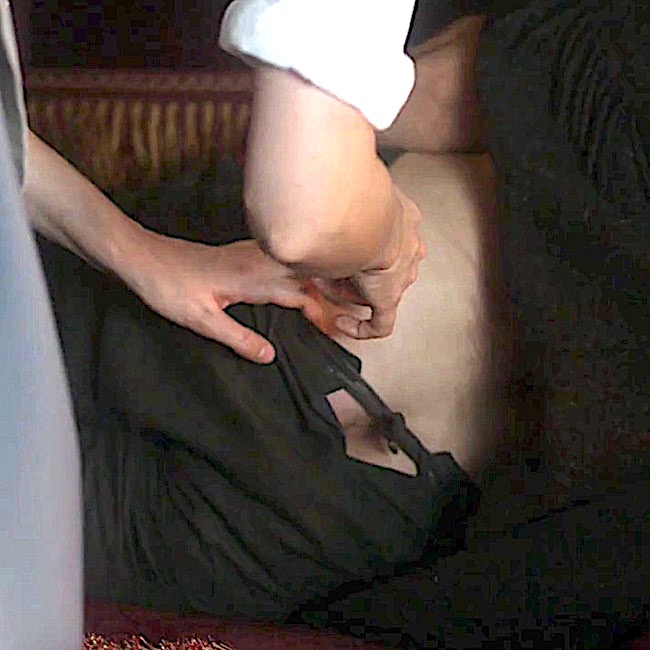
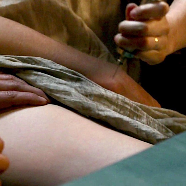
More penicillin adventures from Voyager book. Claire stabs Jamie in the backside with her wee needle spike. Hah!

“Roll onto your good side,” I said, turning to Jamie, “and pull up your shirt.” He eyed the needle in my hand with keen suspicion, but reluctantly obeyed. I surveyed the terrain with approval. “Your bottom hasn’t changed a bit in twenty years,” I remarked, admiring the muscular curves.

… I jabbed deftly and pressed the plunger slowly in.

“Ouch!” Jamie rubbed his posterior resentfully.

“It’ll stop stinging in a minute.” … Finally he said, “I thought ye stuck pins in ill-wish dolls when ye meant to witch someone; not in the people themselves.”

“It’s not a pin, it’s a hypodermic syringe.”

“I dinna care what ye call it; it felt like a bloody horseshoe nail. Would ye care to tell me why jabbing pins in my arse is going to help my arm?”

Splendid, splendid passage!

Here’s a few pearls from the above quote to consider: Why did Claire push the plunger slowly? Because, penicillin is a thick liquid which flows slowly and because it is viscous, it requires a large bore needle (18 gauge). The bigger the needle, the more it hurts while traversing the skin (yikes)! Because it is injected into muscle, the fibers are pushed apart by the fluid and that hurts, too!  I well remember penicillin shots from my youth. Not fun!

Crucial Question: Let’s get to the bottom of things (snicker)…..is Claire free to poke that needle anywhere in Jamie’s bottom? Absolutely not!!! Clarie carefully selects the area where she deftly jabs her needle spike.

Anatomy to the Rescue: Now, wait just a sec! Why does it matter where Claire sticks the needle? After all, it hurts regardless, right? Let’s find the answer by studying anatomy of the backside.

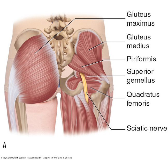
Remove skin and subcutaneous tissue and the buttock region looks like the left side of Image F: Each massive gluteus maximus muscle (Anatomy Lesson #1,  Jamie’s Tush) covers the back of one hip. Because the muscle is thick and meaty, injection into its fibers is a perfect destination!

Try this: Stand erect and place your palms on each buttock with fingers facing downward. Now, clench your bottom and feel it lift and tighten (hopefully)? Gluteus maximus muscles at work!

Sciatic Nerve: Butt, wait (he, he)! Remove the gluteus maximus and what lies underneath (Image F – right side)? One sees a host of smaller hip muscles and a very large yellow structure emerging from under cover of the right piriformis muscle . This is the sciatic nerve, largest and longest nerve of the human body – many adult sciatic nerves are as big around as an adult thumb! And, just to keep us crazy, although most sciatic nerves emerge from below the piriformis, there are five other variations that occur with less frequency.

Each sciatic is formed by contributions from five levels of the spinal cord (L4, L5, S1, S2, S3 – Anatomy Lesson #10, Jamie’s Back or Aye, Jamie’s Back!) as it traverses one buttock and descends through the back of thigh. In case you don’t know, L = lumbar and S = sacral.

Image F 

Protect the Sciatic Nerve! Heath practitioners who inject the buttock know this cardinal rule: Never inject penicillin (or other drug) into or near a nerve as this may result in permanent neurological damage! Such damage can include permanent paralysis, muscle inflammation, gangrene or necrosis (tissue death). Major arteries must also be avoided.

True Story: Years ago, I knew a woman with foot drop, meaning she could not lift her foot at the ankle joint (called dorsiflexion). Why? Because a careless health care provider injected penicillin into the wrong area of her gluteus maximus and it destroyed that part of the sciatic nerve responsible for innervating leg muscles which are required to lift the foot. And, yes, the injury was permanent!

Safe Zone: So, is there a safe zone for injections? Well, of course there is, otherwise, Claire would not have put her beloved Jamie at risk.  

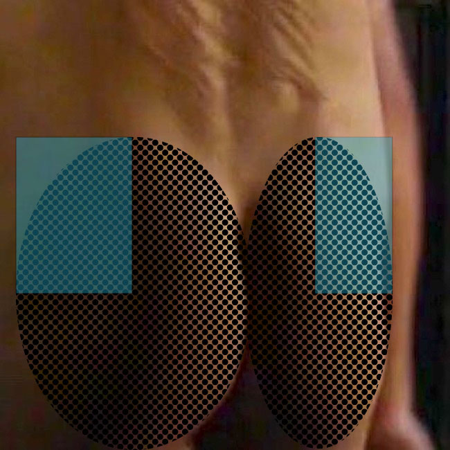
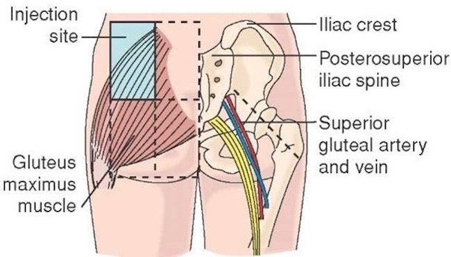
Let’s pretend this is Jamie’s bum. Hey, this is Jamie’s bum! <G> Black dots show regions corresponding to right and left buttocks. The blue rectangles are considered safe zones for intramuscular injections, including penicillin.


Using a graphic image might not be as much fun, but surely is informative. The buttock to be injected is divided into halves by a vertical line then divided into halves again by a horizontal line (Image G – left side). Injecting near the center of the upper outer quadrant, shown in blue, is considered the safe zone and avoids injuring sciatic nerve or nearby artery and vein (superior gluteal).

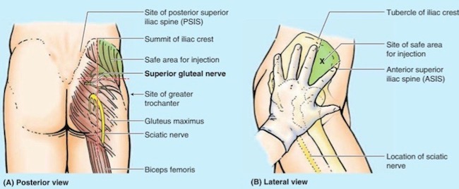
A newer technique uses a diagonal line as shown on the right side of Image G. Injecting above this line is also considered a safe zone. This line is harder to visualize because it requires a more thorough understanding of surface (topographical) anatomy.

Either technique is typically done in the mind’s eye of the practitioner, although, drawing actual lines on the buttock can also be done to verify safe zones.

Image G 

This excellent You Tube video, created by Dr. Nabil Ebraheim, explains both approaches.

The meaty quadraceps femoris muscles (Anatomy Lesson #7,  Jamie’s Thighs or Ode to Joy!) of the thigh present another acceptable site for penicillin injection. This explains why, much later on the Artemis, Claire injects herself in the thigh with penicillin after Jamie canna do the deed (Outlander episode 311, Uncharted).  Our braw hero is afeared of wicked needles!

But, arguably the safest approach involves injecting into the gluteus medius muscle which peeks above the gluteus maximus. The safe region is indicated as the green area in Image H. This technique avoids the huge and very important sciatic nerve!

Ubiquitous Germs! One final issue and then our lesson ends. How might Jamie get an infection from second-wife wounds? Both birdshot and his skin would be likely sources for the bacteria causing his raging fever! Claire explains it quite well in this passage from Voyager book:

I took a deep breath. “Well, do you remember my once telling you about germs?”

He looked quite blank. “Little beasts too small to see,” I elaborated. “They can get into your body through bad food or water, or through open wounds, and if they do, they can make you ill.” He stared at his arm with interest. “I’ve germs in my arm, have I?” “You very definitely have.” I tapped a finger on the small flat box. “The medicine I just shot into your backside kills germs, though. You get another shot every four hours ’til this time tomorrow, and then we’ll see how you’re doing.”

I paused. Jamie was staring at me, shaking his head. “Do you understand?” I asked. He nodded slowly. “Aye, I do. I should ha’ let them burn ye, twenty years ago.”

Nay, Jamie doesn’t mean this wee salvo aimed at first wife. Snort!

Thank you Diana, Jamie and Claire for helping us to understand needles, nerves and penicillin!

Everywhere: Bacteria, molds and yeast are ubiquitous, covering virtually every non-sterile surface on earth. Recent tests have shown, seven of the 10 germiest (is this even a word?) spots in our homes are found in kitchens – with the dirtiest being kitchen sponges or dish cloths! Imagine, these are more germy than bathrooms!

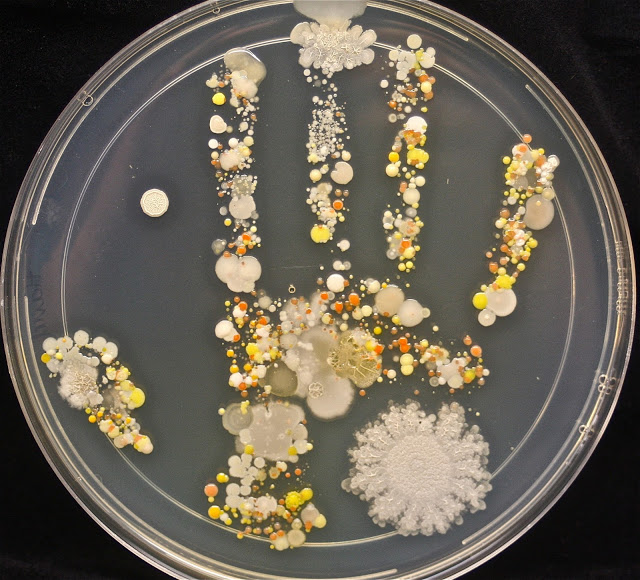
Our lesson closes today with this visual of bacteria and molds found on one person’s hands (Image H). The hand was pressed into growth medium filling a petri dish. After some time, colonies of mold, yeast and bacteria grew from the hand print. Beautiful in an weird way, but definitely GROSS! The bottom line? Our mums were right. Let’s wash our hands

Image H by Tasha L Sturm, microbiologist and photographer

The deeply grateful,

Outlander Anatomist

Follow me on:

Photo Creds: Starz, www.pbs.org/newshour Image E; www.Karsh.com  Image A;  www.learnmuscles.com Image F; www.free-stock-illustration.com Image C; www.medium.com Image B; www.prezi.com Image D; https://everythingmicro.blogspot.com Image H; www.teachmeanatmy.info Image G