Fun Fact: Tonsil

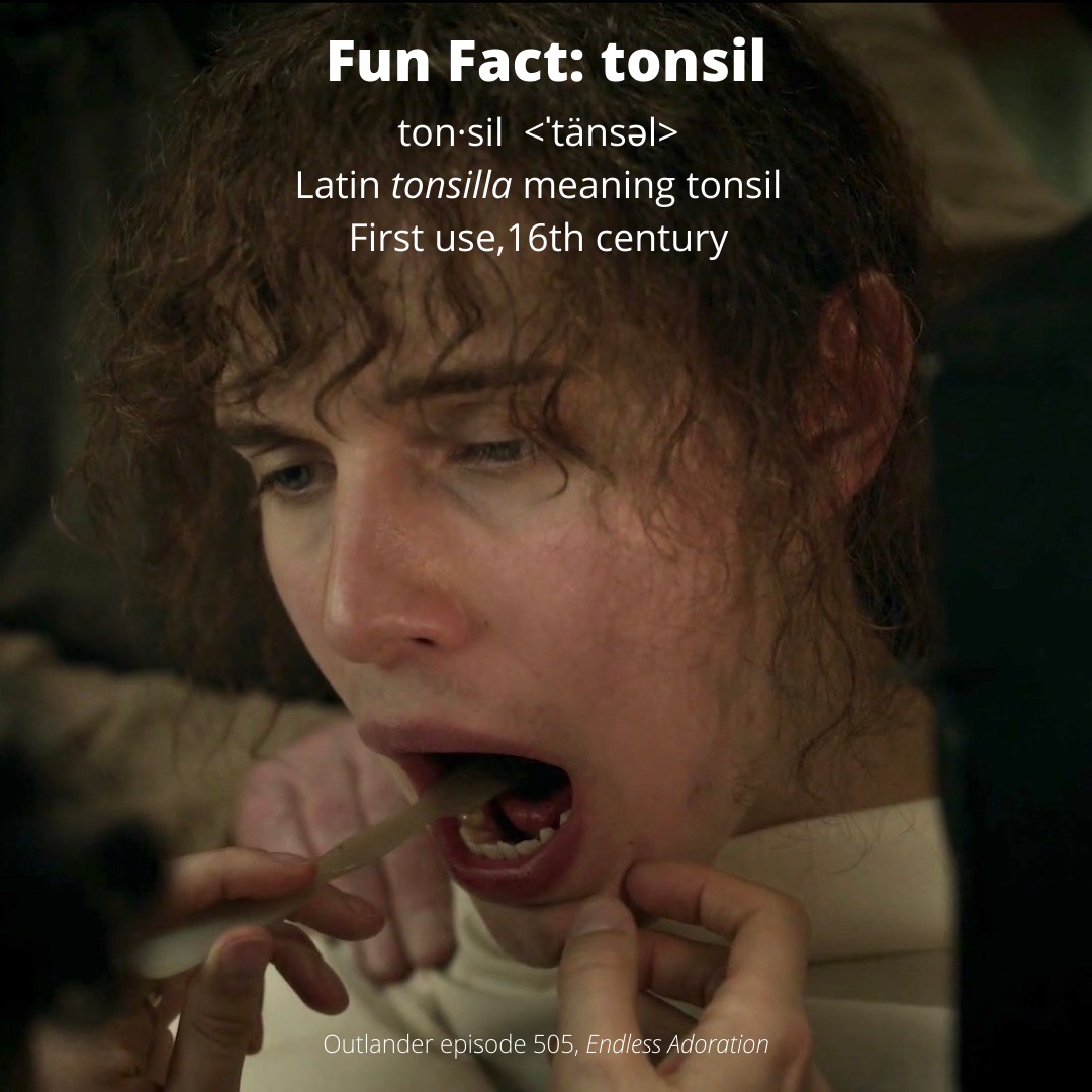
Anatomy Def: Tonsils are masses of lymphoid tissue located in oropharynx (behind mouth)  and nasopharynx (back of throat).

Outlander Def:  The “say “ahhhh” tissues!”

Learn about tonsils in Anatomy Lesson #34, G.I. Tract, Part 2 – Tremendous Tube!

Most folks are familiar with a pair of tonsils we see via an open mouth. But, our nasopharynx and oropharynx are equipped with several sets of tonsils.

Anatomists agree there are three sets of tonsils and many add a fourth (see L image👇🏻):

    • Palatine tonsils: Paired masses at back of mouth (blue)
    • Lingual tonsil: Unpaired mass embedded in back of tongue (green)
    • Pharyngeal tonsil: Unpaired mass embedded in back of pharynx (yellow)
    • Tubal tonsil: Paired masses surrounding opening of eustachian tube (violet)
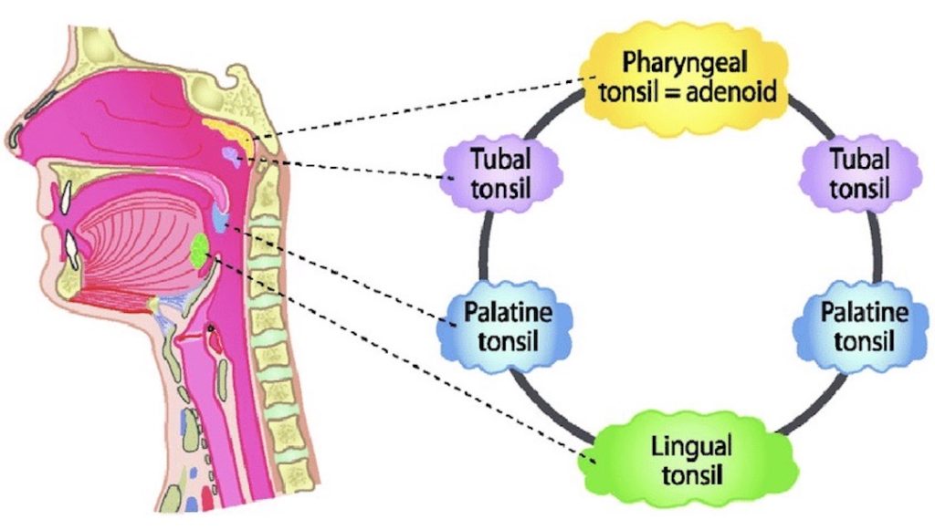
What are tonsils? Tonsils are masses of lymphoid tissue that produce defensive cells known as lymphocytes. If viewed from the front (see R image 👆🏻) , the four sets of tonsils form Waldeyer’s ring, a circle of lymphoid tissues.

What are tonsils for? Tonsils are strategically oriented in Waldeyer’s ring to encounter antigens we breathe in via the nose or swallow through the mouth. The tissues respond by mounting an immune response against the antigens, producing antibodies and pursuing other defensive tactics.

Fun Fact: Tonsillectomies were once the most common surgery done on US children. Today, they are performed mostly to treat breathing problems or chronic issues that are not resolved by other forms of treatment.

Read about Josiah’s tonsillectomy in Diana’s 5th big book, The Fiery Cross!

“All right, then?” I asked.

He couldn’t speak, with the tongue depressor in his mouth, but made a good-natured sort of grunt that I took for assent.

I needed to be quick, and I was. The preparations had taken hours; the operation, no more than a few seconds. I seized one spongy red tonsil with the forceps, stretched it toward me, and made several small, quick cuts, deftly separating the layers of tissue. A trickle of blood was running out of the boy’s mouth and down his chin, but nothing serious.

…The whole thing couldn’t have taken more than thirty seconds per side. I drew the instruments out of Josiah’s mouth, and he goggled at me, astonished. Then he coughed, gagged, leaned forward, and another small chunk of flesh bounced into the basin with a small splat, together with a quantity of bright red blood.

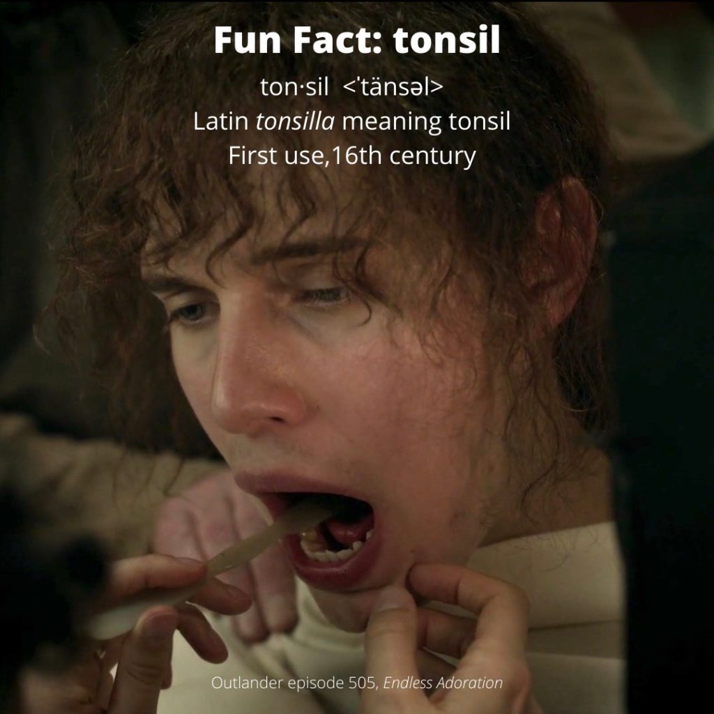
See Claire perform a tonsillectomy on puir Kezzie in Outlander episode 505, Perpetual Adoration! Let’s hear it for the brave laddies!

The deeply grateful,

Outlander Anatomist

Follow me on:

Photo credit: Sony/Starz; www.researchgate.net

Fun Fact: Depressor Labii Inferioris

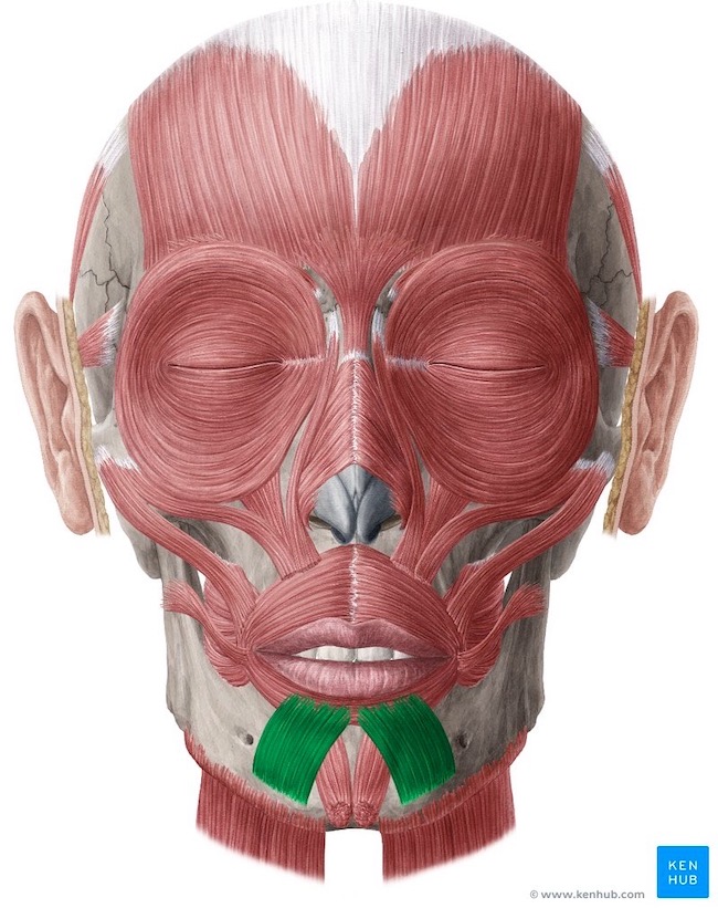
Anatomy Def: Depressor labii inferioris are paired muscles which together pull down (depress) the lower lip.

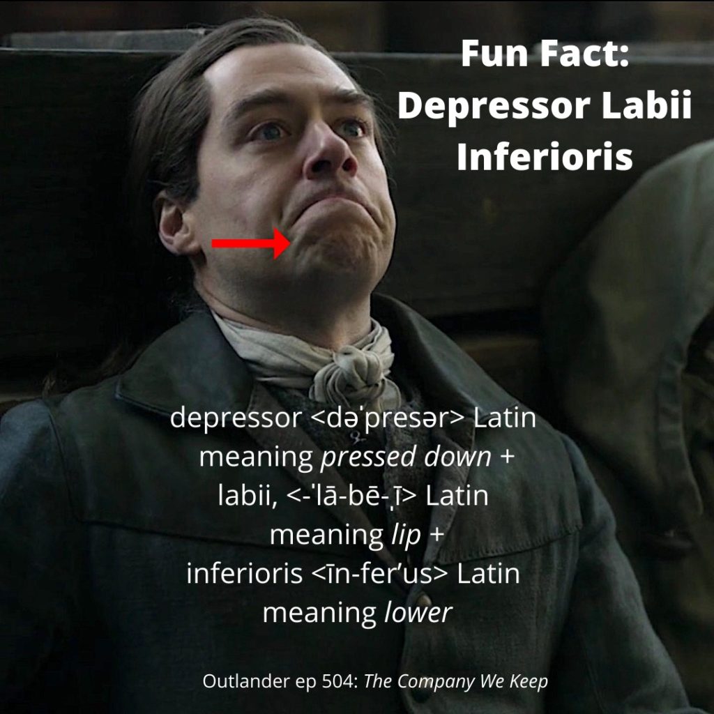
Outlander Def: Roger’s “oh shite – what do I do now?” muscles (red arrow)!

Learn about depressor labii inferioris (DLI) in Anatomy Lesson #11, “Jamie’s Face or Ye Do it Face-to-face?,” a lesson I wrote way back in 2015. Warning, it is a long one!

Roger has a pair of the most prominent DLI I have ever seen! His are contracted at this moment in the Brownsville standoff, drawing his lower lip down and producing two very prominent ridges of his chin-skin. The red arrow marks his right DLI but the left ridge is also visible. Can you see both?

Try this: Stand before a mirror and strongly depress the lower lip. See if you can produce similar ridges in your chin skin.

Anatomists classify DLI as muscles of facial expression. Most humans have about 20 pair of such muscles. If you ever ponder how it is we have such mobile faces, it is due to a total of 40 or so muscles moving the facial skin to achieve our amazing range of facial expression. Fear, anger, distrust, frustration, joy, etc., are all non-verbal forms of communication produced by this group of muscles.

DLI are relatively small, thin and flat. Like all muscles of facial expression, structure can vary from person to person. These are also innervated by the same pair of nerves which is why serious injury to one of these nerves causes paralysis of the same side of the face.

Read about lips in Diana’s fifth big book, The Fiery Cross; they are referenced many times in this tome. This excerpt is from the Brownsville standoff:

“What did he say?” Mrs. Brown whispered to her sister-in-law. The older lady shook her head, lips drawn in like a pursestring. “Mr. Morton is alive and well,”

Roger translated for them. He coughed. “Fortunately for you,” he said to the male Browns, with as much menace as he could contrive to put into his voice.

See Capitaine Roger MacKenzie’s DLI muscles in action in Outlander episode 504, The Company We Keep! Splendid episode! 👏🏻👏🏻👏🏻

The deeply grateful,

Outlander Anatomist

Follow me on:

Photo credit: Sony/Starz

Fun Fact: Mold or Mould?


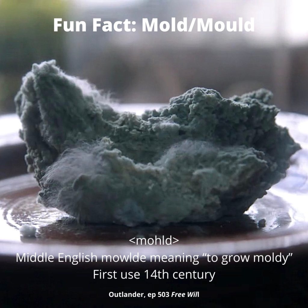
Anatomy Def: Mould/mold is a superficial, hairy growth produced by some fungal species; discoloration and woolly surfaces are common features.

Outlander Def: The icky layer of fuzz on Claire’s handmade bread which she hopes will yield penicillin! 🤞🏻

Learn about penicillin mold in Anatomy Lesson #57, Needles, Nerves and Penicillin.

Question #1: So, which is it, mold or mould? Ha, ha! Well, that depends on where you live. The entire English-speaking world, excepting the U.S., spells it, mould. Being somewhat contrarian, the US is the only English-speaking country that spells it, mold! 😉

Question #2: What is mold/mould? Molds are a large and diverse group of fungi (Pssst….. fungus is singular spelling, fungi is plural).

Question #3: What are fungi? In the past, fungi were classified as plants, but science marches on and fungi are now assigned to their own kingdom, placed on an even keel with plants, animals and two or three other kingdoms of life!

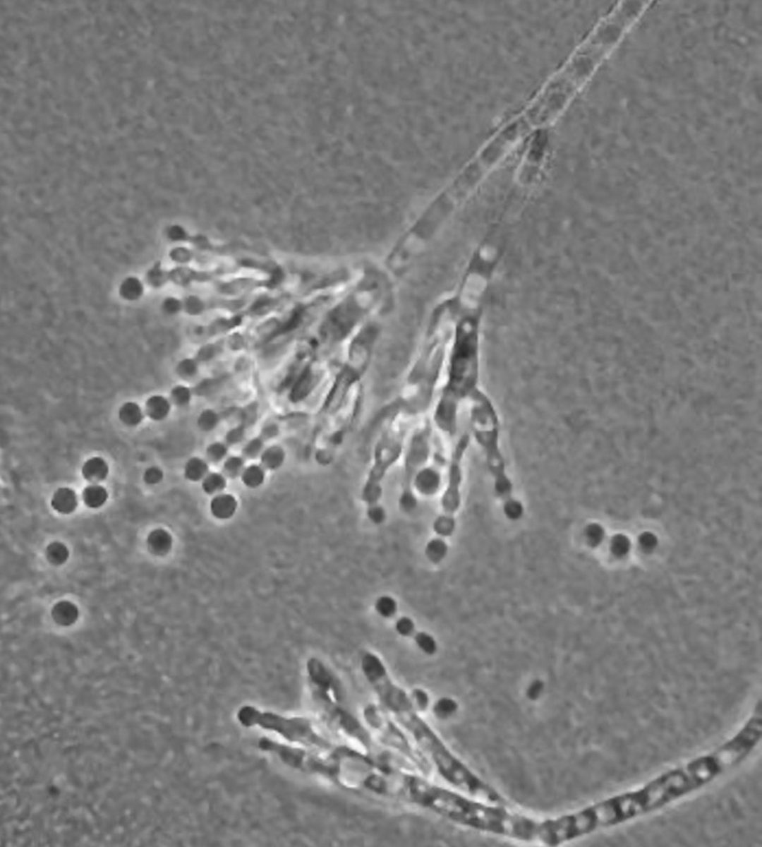
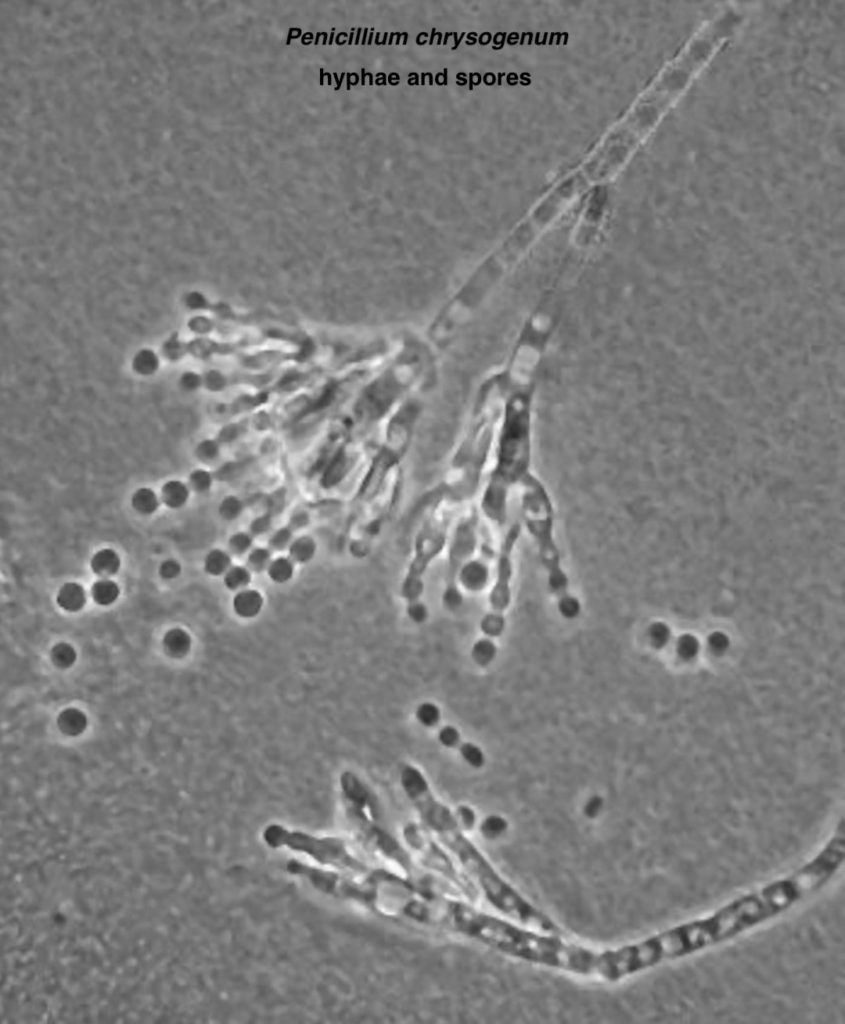
Mold Anatomy: Do molds exhibit anatomical features? Indeed they do! Molds grow by producing a network of transparent, branching tubules known as hyphae which lend molds their typical woolly-appearance. Molds also may have a dusty surface due to the production of spores at the tips of hyphae.

Claire kens that examination of spores and hyphae may help identify the type of mold she seeks; hence, experiments, microscope and Apprentice Marsali!

She seeks to grow a Penicillium strain that will provide a crude antibiotic. Claire, and now Marsali, will search for hyphae that look like wee paintbrushes in the microscope👇🏻! The small round globs are spores.

Let’s wish them the best of luck, although I do fash over Fergus’ recent paper grab; possibly a page of Claire’s lab notes?  😱

Spores act like seeds, a clever strategy to start new colonies. Mold spores released from parent hyphae are widely dispersed by:

    • air currents (can be airborne for long periods and distances)
    • fur
    • feather
    • clothing, etc.

If spores land on organic material (e.g. chunks of homemade bread!) along with moisture, dim light, and warmth, they undergo cell division to produce the fuzzy molds our Sassenach seeks!

Fun Fact: Few molds will grow below 39 °F  (4 °C) which is why refrigerators recommend settings around 38 °F.  But, some hearty molds can survive Antarctica, acids, anti-bacterial soap, and jet fuel! 😳

Molds are a nuisance when they spoil food and damage property. They may also be pathogens causing allergies, invading tissues (e.g. lungs), or generating harmful toxins. On the other hand, molds are extremely useful in producing various foods and beverages, and by providing enzymes, pharmaceuticals, and antibiotics!

Read about Claire’s mold experiments in The Fiery Cross:

I twiddled the quill, rolling it between thumb and forefinger. I had kept a faithful account of my experiments with penicillin—the growing of cultures on media ranging from bread to chewed pawpaw and rotted melon rind, painstaking descriptions of the microscopic and gross identification of the Penicillium molds, the effects of—to this point—very limited applications.

Read the books, folks, wherein Diana faithfully details the mold experiments! 🙏🏻

See Claire’s bread mould/mold in Outlander episode 502, Between Two Fires. Yuck! 😝

The deeply grateful,

Outlander Anatomist

Follow me on:

Photo Credits: Sony/Starz, www.botit.botany.wisc.edu