Mini Anatomy Lesson: Abdominal Aortic Aneurysm (AAA)


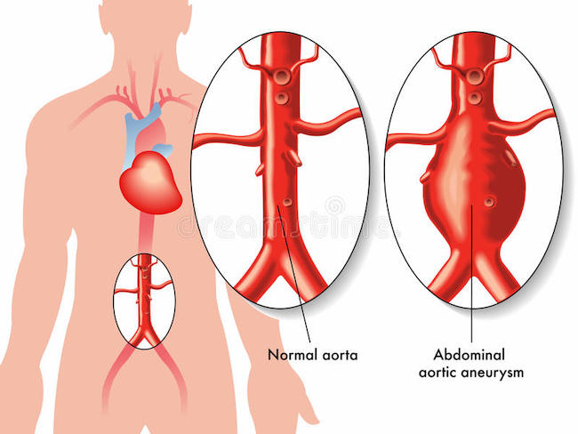
Anatomy Def: An abdominal aortic aneurysm (AAA) is a bulge or swelling of the aorta that occurs in the belly.

Outlander Def: Mrs. Wilson’s untimely #death-resurrection-death wherein “kindly” fisher folk put Claire firmly on her witchy broom!!! 🧹😱

Hello, anatomy students! To date, I have not written a full lesson about the cardiovascular (CV) system. So, the following quick anatomy lesson of the aorta will be useful in understanding AAA. 🤓

And, we’re off!!! 🏇

Aorta: The aorta is the largest blood vessel of the body! 👏🏻👏🏻👏🏻

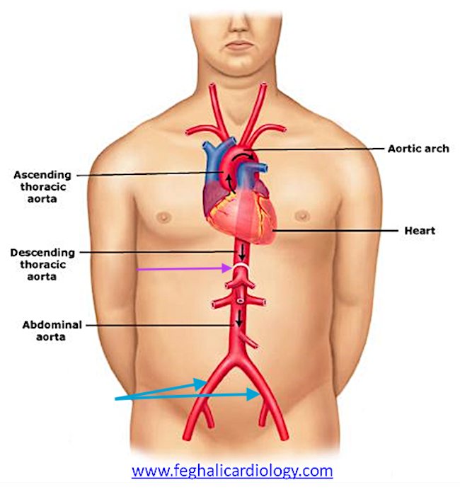
What Does it Do? The aorta is an artery, so it is tasked with carrying oxygen-rich (oxygenated) blood from the heart to general regions of the body. The direction of blood flow is indicated by the small black arrows in the next image.

Also, by convention, arteries are colored red to indicate these vessels carry oxygenated blood away from the heart and veins are blue to show vessels carrying oxygen-poor (deoxygenated) blood toward the heart. (pssst…there are two exceptions to this pattern, but I won’t confuse you by including those. 😜)

Along the entire course of the aorta, various arteries branch off to supply blood to the head and neck, upper limbs, chest and its organs, abdomen and its organs, and lower limbs. These branches are not labeled in the image below.

Divisions: Inside the chest is the thoracic aorta – the part within the belly is the abdominal aorta. The names switch where the aorta traverses the thoracic diaphragm (purple arrow).

The part of the thoracic aorta that rises is the ascending thoracic aorta, the curved part is the aortic arch, and the part that descends is the descending thoracic aorta.

Pathway:  The aorta starts at the left ventricle (chamber) of the heart, rises toward the neck, makes a 180º, descends through the chest, and enters the belly (abdomen) where it terminates at the 4th lumbar (L4) vertebra by dividing into left and right common iliac arteries (turquoise arrows). Whew! 😅

How can a practitioner locate L4 when looking at the belly?

Here’s how: the umbilicus (navel) can be used as a fairly reliable surface landmark for termination of the aorta because it lies slightly above L4. Thus, a finger’s width below the navel will locate L4. If a person is heavy or pregnant, this can distort the position of the navel. 

L4 is important because the practitioner must palpate above that level to detect a AAA.
Like other larger arteries, a pulse in the abdominal aorta can be felt (palpated). Sometimes the belly can be seen to rise and fall with the pulse especially after a meal, during pregnancy, or while lying down. This can be perfectly normal.

However, sometimes a pulse in the abdominal aorta can be a warning sign!🚫 

Aneurysm: If the aortic wall becomes damaged, it can developed a distinct bulge or balloon; this is an aneurysm. (Pssst…🤫 Aneurysms also can develop in smaller arteries and in veins.)

If an AAA is present, a practitioner may be able to detect the enlargement by palpating the abdominal wall. The care giver must press firmly enough to feel the abdominal aorta because it lies very deep, near the vertebral column. 

If a patient has a very taut abdominal wall (next photo) or the waist girth exceeds 100 cm, it is more challenging for the practitioner to accurately feel and identify any aneurysm that may be present. A tight wall or a fairly thick layer of belly fat inhibits the deep pressure required for the exam. Lastly, clothes worn by the patient may also impede effective palpation! 😮
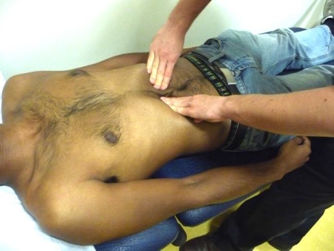
Although the following informative video is designed to train students on how to palpate the abdominal aorta, it is easily understood by a lay person. This will give you an idea of how a pulse is felt and how an aneurysm is detected.

The big problem with AAA is it can enlarge to the point of rupture, allowing blood to seep into the wall of the aorta (dissecting aneurysm) or bleed into the abdominal cavity causing dangerous blood loss or even death. 

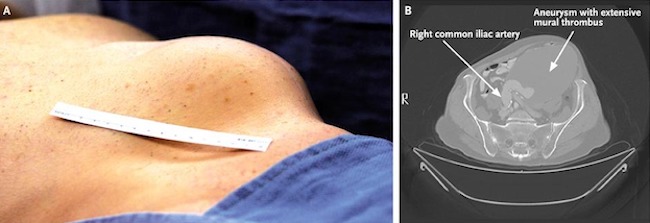
Rarely, an AAA can become so large, it is clearly visible in the abdominal wall (next image)! The left panel shows an huge bulge at the left side of a person’s abdominal wall. The right panel shows a cross-sectional CT scan of the same mass, identified as a huge blood clot (thrombus) in an AAA. 
OK. I hear the bell tolling…… Time to head back to the wee kirk on the Ridge! 

Germain’s “I spy with my little eye,” alerts Granny Fraser that something is amiss with Mrs. Wilson. 👀  

“LOOK!“     

“Stay back!”

Claire rushes to the other granny’s side and palpates her abdomen. Can she feel a possible aneurysm? Very likely, because Mrs. Wilson is fairly trim and her stays appear to be of the type that end near the waist, so Claire should be able to palpate – no problemo. 

Claire slips her fingers between the folds of the skirt and takes a wee feel. Yep! Just as Claire suspected…..the auld lady has an abdominal aortic aneurysm – it has ruptured and she is bleeding internally.
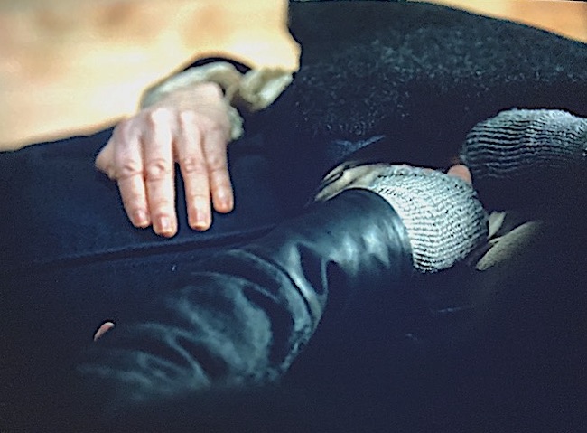
If Mrs. Wilson had been sans clothes, Claire might have witnessed something like this:

She hasn’t got long, Reverend Roger. Just minutes and she stands before her Maker! 

Wait! Wait! What? How could Claire ken the aneurysm had ruptured?

Well, here are some clues that Doc Fraser likely observed..…. 🤓

Signs and symptoms that an AAA has ruptured:

    • Sudden, intense and persistent abdominal or back pain – a tearing sensation
    • Low blood pressure
    • Loss of consciousness
    • Pasty, cold, and clammy skin 

Although Claire does not know if Mrs. Wilson experienced the back pain (no time to take a proper history), all the other symptoms are recognizable. Claire recognizes that given the lady’s advanced age, fainting is likely due to low blood pressure as a result of internal blood loss. Blood loss plus age suggest aneurysm as a likely cause. These coupled with palpation of the enlarged AAA leads Claire to tell Roger that Mrs. Wilson’s death is imminent! (She’s a lot closer to God than you, Roger Mac –  Bree will be grateful for that 🙏🏻)

Mrs. Wilson revives long enough to forgive her SIL – “You’ve been a good lad.” He did provide her with a hame these past 20 years. ♥️

… And she survives long enough to accept the sin-eater’s gift of devouring her sins. 😮

Lastly, you might be interested in what causes aneurysms and any risk factors. 

Common Causes:

    • Hardening of the Arteries (Atherosclerosis): Fat and other substances build up in the wall of the aorta (or other blood vessels) and weaken it.
    • High blood pressure: Prolonged, elevated blood pressure can damage and weaken the aortic walls.
    • Blood Vessel Diseases: Diseases such as Marfan Syndrome damage vascular walls. 
    • Infection: Although rare, a bacterial or fungal infection can cause AAA.
    • Trauma: Abdominal injury (e.g. serious car accident) may cause an AAA due to a tear in the aorta.

Risk factors: And, like many other diseases, there are risk factors for AAA. Most of these we cannot control but there is one clear exception! 🚬🚭

    • Tobacco use: Yep. Smoking is the strongest risk factor for AAA! Why? Because smoking weakens the aortic walls, increasing the risk of aortic aneurysm and the likelihood one might rupture.  The longer and more one smokes or chews tobacco, the greater the chances of developing an aortic aneurysm. (Doctors recommend a one-time abdominal ultrasound to screen for an AAA in men ages 65 to 75 who are current or former cigarette smokers.)
    • Age: AAA occurs most often in people ages 65 and older.
    • Being male: Men are 3-4x more likely to develop AAA as women.
    • Being white: People who are caucasian are at higher risk of AAA.
    • Family history: Having a family history of AAA increases the risk.
    • Other aneurysms: Having an aneurysm in another large blood vessel, such as the artery behind the knee or thoracic aorta can increase the risk of an AAA.

Read about Mrs. Wilson’s walk through the valley of the shadow of death in A Breath of Snow and Ashes, Diana’s longest big book (1,152 pages)! 

“No, Grannie! Look!” 

I followed his outthrust finger, and for a moment, thought he was pointing at his father. But he wasn’t. 

Old Mrs. Wilson had opened her eyes.

There was an instant’s silence, as everyone’s eyes fastened at once on Mrs. Wilson. Then there was a collective gasp, and an instinctive stepping back, with shrieks of dismay and cries of pain as toes were trodden on and people squashed against the unyielding rough logs of the walls.

“Give her a bit of air, please,” I said, raising my voice. The stunned silence was giving way to a rising murmur of excitement, but this quelled as I fumbled to untie the bandage. The room waited in quivering expectation as the corpse worked stiff jaws. 

“Where am I?” she said in a quavering voice. Her gaze passed disbelievingly round the room, settling at last on her daughter’s face.

I, meanwhile, had been doing my best to check the old lady’s vital signs, which were not all that vital, but nonetheless fairly good for someone who had been dead a moment before. Respiration very shallow, labored, a color like week-old oatmeal, cold, clammy skin despite the heat in the room, and I couldn’t find a pulse at all—though plainly she must have one. Mustn’t she?

I put my own hand on her abdomen, and felt it instantly. A pulse, where no pulse should be. It was irregular, stumbling, and bumping—but most assuredly there.

See Hiram Crombie’s MIL and her final moments before leaving her mortal coil in Outlander, Episode 602, Allegiance. A verra poignant scene, indeed!

Gobs of grannies in this episode! 😉

The deeply grateful,

Outlander Anatomist

Follow me on:

Photo Credits: Starz, www.familydoctor.org, www.feghalicardiology.com, www.nejm.org, www.oxfordmedicaleducation.com


Mini Anatomy Lesson: Dupuytren’s Contracture


Anatomy Def: Dupuytren’s Contracture, a disease involving changes to deep tissues of the palm.
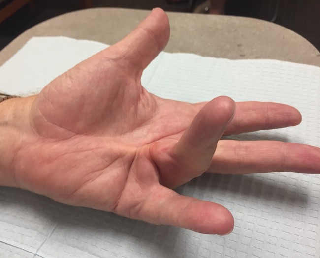
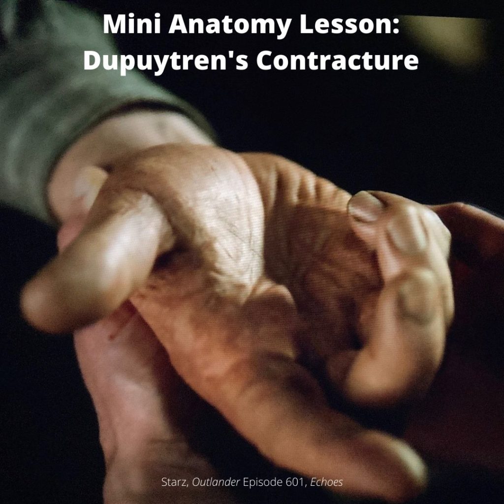
Outlander Def: Tom Christie’s right hand enjoys Claire’s touch waaay too much. Watch out, Jamie! 😉

History: Dupuytren’s Contracture (DC) is named after Baron Guillaume Dupuytren (1777-1835), a French anatomist and military surgeon who gained considerable notoriety treating Napoleon Bonaparte’s hemorrhoids! 😲 Although he was not the first to recognize the condition, he was the first to perform a successful repair on DC and published the results (The Lancet, 1834). Hence, the disease is named for him. 

Also Known As: Like Jamie, Dupuytren’s Contracture (DC) enjoys a number of aliases:

    • Dupuytren’s disease (DD) 
    • Morbus Dupuytren 
    • Viking disease
    • Celtic hand
    • Palmar fibromatosis/familial palmar fibromatosis
    • Palmar fascial fibromatosis.

Symptoms: Tight bands and/or firm lumps (nodules) appear in the tissues deep to the palmar skin. Gradually, the fingers may curl and pull sideways or toward the palm. Overlying skin may pucker. Sufferers may experience inflammation or sensations of tenderness, burning, or itching of the hand. Pressure or tension may be felt when attempting to straighten the fingers. Sometimes both hands and even the feet are affected. More fortunate individuals experience only mild cases that do not require medical intervention. The following image shows an example of DC involving the left ring finger.

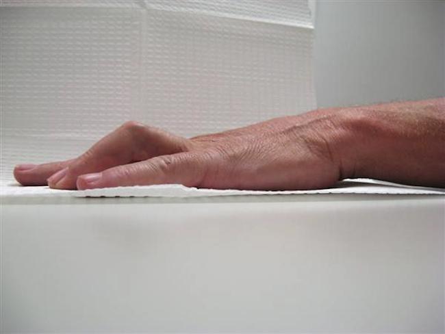
Diagnosis: Care givers use the tabletop test to help diagnose DC: the hand is placed palm-down on a flat surface. If DC is involved, the hand cannot be laid flat.

Now, onto the anatomy. Yay! 🤓

Try This:  Look at your hand. Note that it has two main surfaces. The palm is known as the palmar surface; the back of the hand is its dorsal surface. See that the palmar skin is tightly adherent to underlying tissues, whereas the dorsal skin moves rather easily because it is loosely attached.

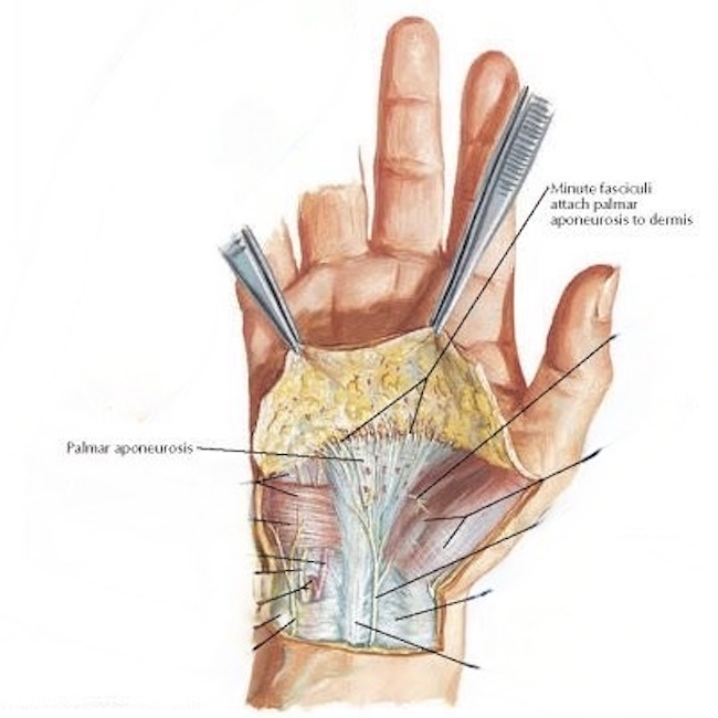
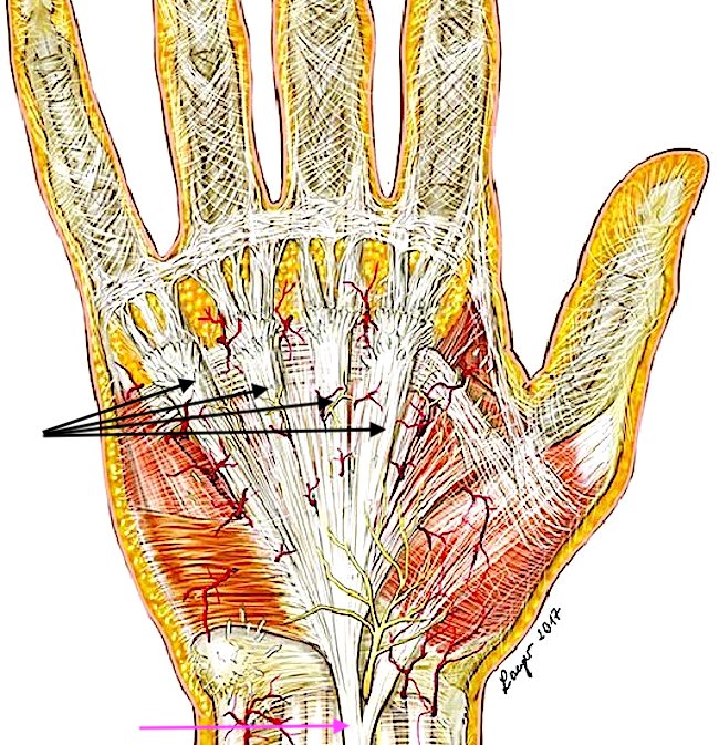
Palmar Aponeurosis: Deep to our palmar skin lies a thick fan-shaped layer of collagen known as the palmar fascia or, more formally, the palmar aponeurosis (next image). Collagen fibers (fasciculi) tightly bind the palmar skin to the underlying palmar aponeurosis. This anatomy enhances our grip, helps cup the palm, and protects underlying flexor tendons.

With the skin removed, the fan-shaped palmar aponeurosis is evident (next image): The point of the fan lies at the wrist (pink arrow), then it widens sending complex collagen bands to each of our four fingers (black arrows). 

In case of DC, the long bands of collagen, along with some cross fibers, thicken, tighten, and may form nodules. Ring and little fingers are most commonly affected; thumb, and index and middle fingers are less often involved. Sometimes DC affects both hands and even the feet. Although some sources claim there is no correlation between occupation and development of this condition, others cite manual labor as a predisposing factor. 

Learn more about the complex anatomy of the hand in Anatomy Lesson #22, Jamie’s Hand Symbol of Sacrifice and Anatomy Lesson #23, Harming Hands, Helping Hands, Healing Hands

Cause: Although poorly understood, several parameters suggest DC is likely is an autoimmune disease caused by a single dominant gene which is unaffiliated with either  X or Y chromosomes (the sex chromosomes). Even if present, the gene doesn’t always express itself. The reason for this is unknown.

Risk Factors: There are interesting risk factors associated with DC. 

    • Middle aged or older
    • Male – much less common in females
    • Scandinavian or Northern European descent (uncommon in Asian and African ethnicities)
    • Family history

Other Associated Factors: 

    • Liver disease
    • Long term use of phenytoin (anti-seizure med)
    • Cigarette smoking
    • Alcoholism
    • Diabetes 
    • Nutritional deficiencies

Treatment of DC: For many years, surgery was the only treatment for DC. In recent times, this has changed and new strategies have appeared.

    • Surgery: The most common treatment. Rarely, finger amputation is needed. 
    • Steroid injection: If a lump is painful, a steroid injection may help ease the pain. Repeated injections may be necessary.
    • Radiation therapy: Low energy X-rays are directed at the nodules to soften them and reduce contractions. Uncommon treatment in the U.S.
    • Enzyme injection: Newer, less invasive procedure. Hand is anesthetized, enzyme is injected. Over several hours, the enzyme breaks down and dissolves the tough bands.
    • Needle aponeurotomy: Newer, less invasive procedure. Hand is anesthetized. Surgeon uses a needle to divide and release the diseased tissue.

Read about Tom’s disease in Diana’s sixth big book, “A Breath of Snow and Ashes.” Dr. Claire describes Tom’s suffering: 🤒

He had been suffering from a condition in the right hand called Dupuytren’s contracture—or at least it would be called that, once Baron Dupuytren got round to describing it in another sixty or seventy years. Caused by a thickening and shortening of the fibrous sheet that kept the hand’s tendons in place when the fingers flexed, the result of it was to draw the ring finger in toward the palm of the hand. In advanced cases, the little finger and sometimes the middle finger as well were involved. Tom Christie’s case had advanced quite a bit since I had last had the chance of a good look at his hand.

See Claire’s assessment of Tom Christie’s Dupuytren’s Contracture in Outlander episode 601, Echoes! Stay tuned! Claire will operate on Tom’s hand in Outlander Episode 603, “Temperance!” 😲

The deeply grateful,

Outlander Anatomist

Follow me on:

Photo Credits: Starz, www.johnericksonmd.com, Netter’s Atlas of Human Anatomy, www.OrthoBullets.com, www.researchgate.net

Mini Anatomy Lesson: IP Joint


Outlander Def:  Wait!  What???  Roger Mac has a joint? But, but….he is a Presbyterian minister (almost)!

No worries. NOT that kind of joint! 😉

Anatomy Def:  IP joints are between bones of each digit.

Let’s take a wee keek at hand anatomy to ken Roger’s bones and joints. The next figure shows bones of the (left) hand.

Collectively, the digits have 14 bones. Every finger  contains three tiny bones, each known as a phalange. The proximal (near) phalange is green, the middle plalange is blue, and the distal (far) phalange is pink.

But, alas, Mr. Thumb only enjoys two phalanges: proximal (green) and distal (pink). 

To identify the digits, anatomists number them from one to five

    • Thumb =  digit #1
    • Index =  digit #2 
    • Middle = digit #3
    • Ring = digit #4
    • Little = digit #5

But, wait for it….drum roll…. 🥁 Anatomists number fingers, from one to four:
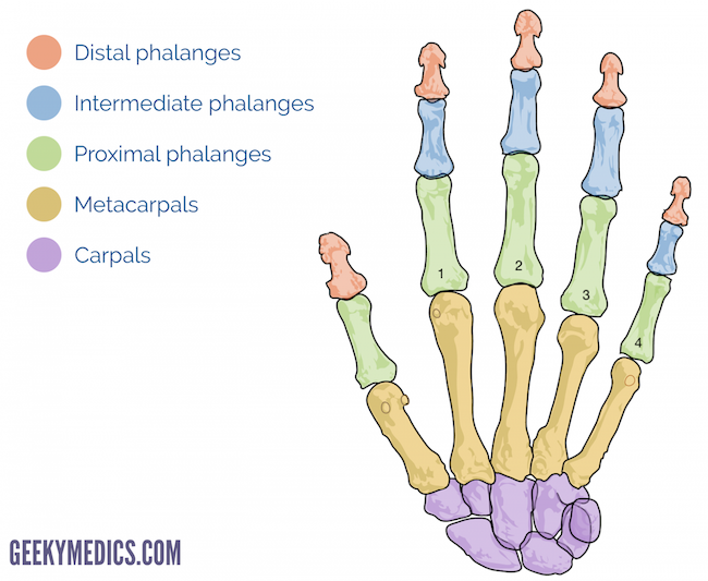
    • Index = finger #1
    • Middle = finger #2
    • Ring = finger #3
    • Little = finger #4

Horror of all horrors…. This means that finger #1 is also digit#2!  😱

You  possible can see how this numbering system could get physicians into trouble. Adding fuel to the fire, anatomists in some countries regard the thumb as a finger and thus describe five digits and five fingers. Bottom line, US hand surgeons often prefer using terms (not numbers): thumb, and index, middle, ring, and little fingers to avoid confusion and mistakes! 🙄

Moving on!

In anatomy, a joint is the site where two or more bones meet and allow for movement –  the greater the number of joints, the greater possible movements. Thus, the many joints between phalanges of our five digits permit greater flexibility allowing digits to flex (bend) or extend (straighten).  Thumb also can oppose (touch) each finger, individually, and little finger also can oppose the thumb. These movements are possible because forearm and hand contain numerous muscles that move the bones.

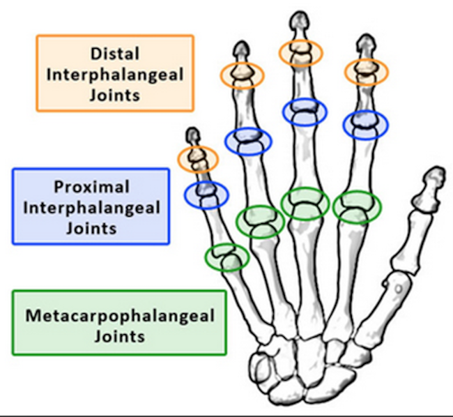
Joints between phalanges are named interphalangeal meaning “between phalanges.” Because each finger has two interphalangeal joints, these are further defined (see next figure of right hand –  ignore metacarpophalangeal joints):

    • PIP (proximal interphalangeal) joint occurs between each proximal and middle phalange.
    • DIP (distal interphalangeal) joint occurs between each middle and distal phalange.

Puir wee thumb only has one joint between proximal and distal phalanges, so it has just a single IP (interphalangeal) joint. But, no tears for Mr. Thumb – he is verra special!

Whew! 😅 Took a bit to explain that!

Now, back to Roger’s anatomy! 😜 (I thought you would be up for that!)

Ergo, manly Roger is flexing the IP joint of his right thumb! 👏🏻👏🏻👏🏻  And, since in most people, the left hand is our shield hand while the right is our sword hand, it is just possible that Mr. Minister is contemplating a stramash! 

Try This: Flex (bend) one of your thumbs. Note it has one joint between its phalanges, the IP joint. Now flex any finger. Note it has two joints, the PIP joint nearest the palm and the DIP joint nearest the fingernail. Well done, student!

The hand and its fingers are elegantly engineered and staggeringly complex. If you wish to read more about them, I have written two long anatomy lessons of the hand.

Learn about  IP, DIP, and PIP joints in Anatomy Lesson #22, “Jamie’s Hand, Symbol of Sacrifice  and Anatomy Lesson #23, “Harming Hands, Helping Hands, Healing Hands.” 

Read about finger and thumb joints in Diana’s first book, Outlander. The following excerpt is from Jamie and Claire’s lovely outing in the countryside, shortly after the wedding : 

“Above one dark speckled pool, Jamie showed me how to tickle trout.

…  “All it is,” he said, “is to pick a good spot, and then wait.” He dipped one hand below the surface, smoothly, no splashing, and let it lie on the sandy bottom, just outside the line of shadow made by the rocky overhang. The long fingers curled delicately toward the palm, distorted by the water so that they seemed to wave gently to and fro in unison, like the leaves of a water plant, though I saw from the still muscling of his forearm that he was not moving his hand at all.

…  “There he is.” Jamie’s voice was low, hardly more than a breath; he had told me that trout have sensitive ears.

…  One finger bent slowly, so slowly it was hard to see the movement. I could tell it moved only by its changing position, relative to the other fingers. Another finger, slowly bent. And after a long, long moment, another. I scarcely dared breathe, and my heart beat against the cold rock with a rhythm faster than the breathing of the fish. Sluggishly the fingers bent back, lying open, one by one, and the slow hypnotic wave began again, one finger, one finger, one finger more, the movement a smooth ripple like the edge of a fish’s fin.”

See Roger’s IP joint in Starz season six outlander promo photo!

The deeply grateful,

Outlander Anatomist

Follow me on:

Photo Credits: Starz, www. geekymedics.com, www.quizlet.com