Anatomy Lesson #24 “Hear, Here – The Ear”

Welcome to summer school Outlander Anatomy students! Today’s topic is Anatomy Lesson #24: The Ear – Part I so listen up! We’ll start with a Starz Outlander scene followed by the anatomy lesson, then a short quiz and end with a Pinna Poll (you’ll soon know what that is grasshopper!). Are you game? Of course you are – it’s summah time and the livin’ is easy!

I’m saying it right up front: the ear is one of the body’s most elegant organs. Yes, I ken I already “handed” that distinction to the hand, but surely there’s room on the winner’s podium for yet another awesome body part? This one truly deserves our collective “Hear, Here!”

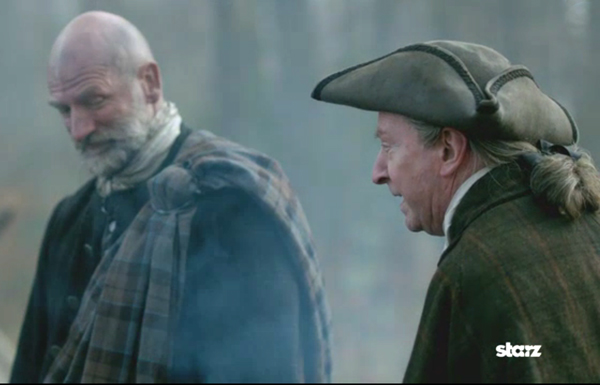
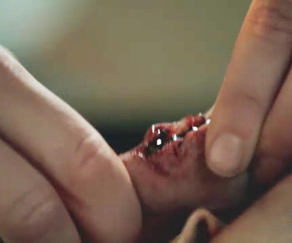
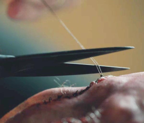
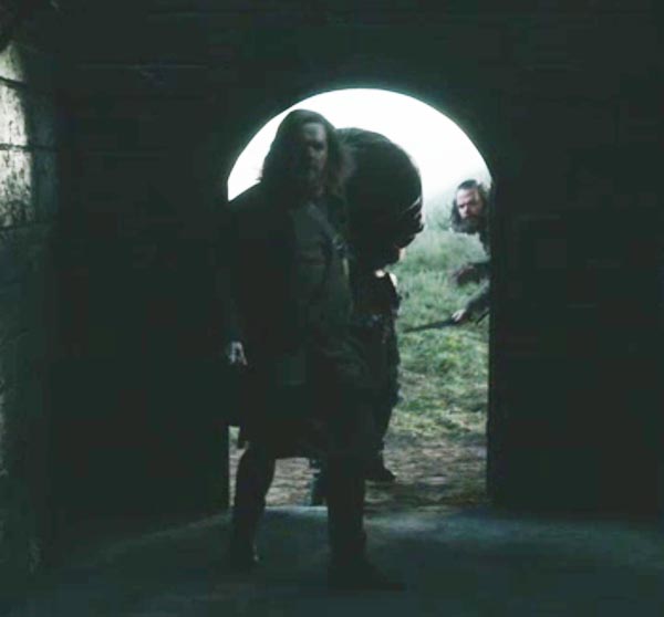
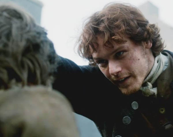
I started planning an ear lesson waaaay back during Starz episode 3, The Way Out. Remember the tanner’s lad whose ear was nailed to the pillory? Herself describes the scene in Outlander book:

He had in his custody a youth, perhaps twelve years old… a tanner’s lad. The priest had the boy gripped… the crowd drew back a bit to allow the locksman free movement for the ear-nailing. The lad… uttered a high, thin scream when the nail was driven in…

ep 103 tanners lad ear 01 KLS edited

Ouch! After an hour nailed to the pillory, the two-bannock thief is free to go but first he must tear his ear free! Cute cagey Claire is no having that! She drags Jamie MacTavish into a 20th century freedom march for the lad; she will feign a faint for crowd control while Jamie has a chit-chat with the wee laddie. Again, from Outlander book:

‘Na then, lad, he said clicking his tongue. Got yourself in a rare swivet, have ye no? Och, laddie… yon’s no job to be making heavy weather of. A wee snatch o’ the head and its over. Here shall I help ye?

ep 103 tanners lad ear 02 KLS edited

And, continuing the quote:

The ear was pinned firmly through the upper flange, close to the edge, and a full two inches of the nail’s square, headless shank was free….

So, Mr. MacTavish grabs the nail with his looong strong fingers (Aye, we all ken the fine work they do. Snort!) and lickety-split out comes the spike! Hie home lucky laddie!

ep 103 tanners lad ear 03 KLS edited

That likely got us in the mood, so let’s get on with the anatomy lesson: the ear is very important for our well-being. Those who have suffered hearing and/or balance loss can attest to the importance of sensory input provided by the ear. Herself writes about such loss in “Written in My Own Heart’s Blood.”

…Artilleryman, from his uniform. So something big had exploded near him— a mortar, a cannon?— and not only burned his face nearly to the bone but also likely burst both his eardrums and disturbed the balance of his inner ear.

Yep, she’s right! The Human ear is the organ that collects, processes and interprets sound waves as well as aids in balance and awareness of head position. The paired ears of vertebrates are symmetrically placed on opposite sides of the head for the purpose of localizing sound sources; if only one ear is operative, the direction and distance from which a sound arises is difficult to pinpoint.

We often refer to these visible flaps of flesh as the ears but this is only the beginning (Photo A – left ear).

Figure0001 ear KLS edited

Photo A

Have you read Diana Gabaldon’s article about story telling and the Rule of Three? Well, you may be surprised to learn that anatomy also has a Rule of Three as many body parts come in trios (like Jamie’s acromion, clavicle and humerus – see Anatomy Lessons #3, #19 and #20) and we will cover several in this lesson.

The human ear actually includes three distinct ears: an outer ear (Photo B – green overlay), a middle ear (Photo B – yellow overlay), and an inner ear (Photo B – blue overlay). Today’s lesson will cover only the outer (external) ear.
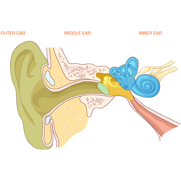
Ear-KLS-edited

Photo B

The outer ear consists of three (yes, three) parts: ear flap, ear canal and outer layer of the ear drum (Photo C). Anatomy has two names for the ear flap and either one works: pinna (Latin meaning feather or wing) or auricle (Latin meaning ear). The ear canal is the external acoustic meatus (Latin meaning passage). The ear drum is the tympanic (Greek meaning to strike) membrane. Let’s examine each of these three parts.
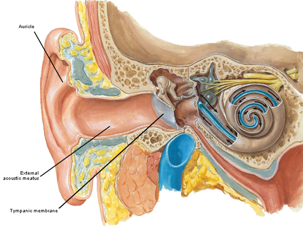
Figure0087 outer ear KLS edited

Photo C

But, wait! First, do you recall from Anatomy Lesson #6 that finger print whorls are formed by dermal ridges and the pattern is unique to each human? Even humans with identical DNA (e.g. identical twins) do not have identical finger prints (Photo D – small dots are openings of sweat gland ducts).
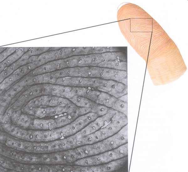
fingerprint KLS edited

Photo D

In a similar vein (ha ha), the venous pattern on the dorsum of hand (Anatomy Lesson #22 & Anatomy Lesson #23) is entirely unique to an individual; so much so, that some board examinations now require test takers to undergo an infrared scan of the hand venous network for identification purposes as it is more reliable and interpretable than a fingerprint (Photo E).
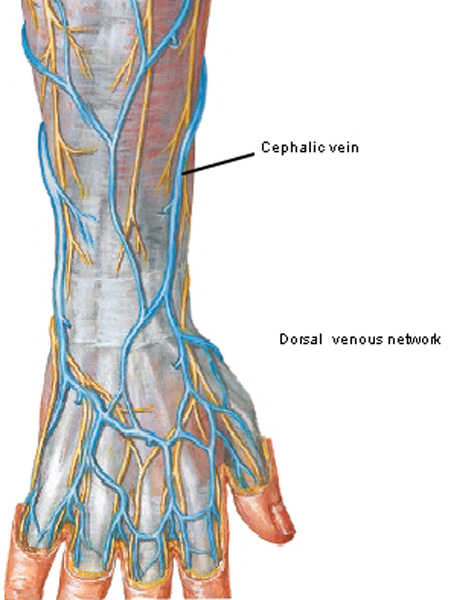
Figure0463B dorsal venous network KLS edited

Photo E

Hmmm, has this prof’s mind gone bye-bye? Why is she writing about fingerprints and hand venous patterns when the lesson is about the ear? Because, I want to emphasize a small cohort of unique body parts which includes the pinna as no two are alike. The pinna bears hills and valleys all of which are named (this is how anatomists make a living) but its overall shape and relationships are unique to its owner.

The pinna includes a fleshy earlobe/auricular lobule (Photo F – blue overlay) and the rest of the ear flap (Photo F – green overlay). The earlobe is soft and flexible because it is made of skin and collagen.
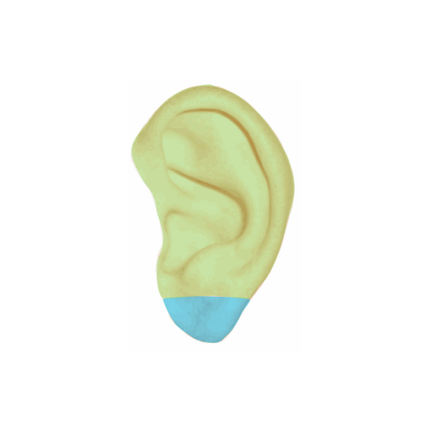
pinna-KLS-edited-copy

Photo F

One can even find references to the earlobe in Diana’s books (I swear anatomy abounds in her written works). This quote from Outlander book:

I wriggled closer and pulled Jamie’s head down as though overcome by amorousness. “What is it?” I whispered in his ear. He seized my earlobe between his teeth and whispered back. “The horses are restless. Someone’s near.

The way earlobes connect to facial skin is an interesting and disputed topic. Common wisdom is that earlobes come in two varieties: attached, wherein the lobe slopes to connect with facial skin (Photo G – red arrow) or free, wherein a distinct cleft separates earlobe from facial skin (Photo G – blue arrow). Science teachers often teach basic Mendelian genetics using earlobes as an example: earlobes are either free or attached and this trait is controlled by a single gene. But family studies have shown that this is not so. There is a broad range of earlobes all the way from “free” to “attached” with many variations in between. This range means that lobe attachment is a multifactorial  trait that is not determined by a single gene.

Try this: check out your earlobes and those of friends and family. Are the lobes attached, free or some variant between the two? Do your earlobes look like those of your father or mother? What about your biological children – do their earlobes look like yours or those of the other parent?
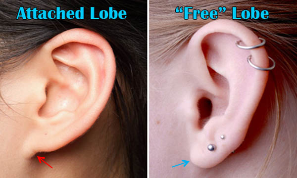
ear-lobes-KLS-edited

Photo G

The remainder of the pinna is well-defined with skin tightly bound to an underlying endoskeleton of cartilage (Photo F – green overlay). The body contains three types (Three, again. I kid you not!) of cartilage: hyaline cartilage covers the surfaces of moveable joints; fibrocartilage forms much of an intervertebral disc and elastic cartilage shapes most of the pinna. (NOTE: the three cartilage types are found in other sites too but these will be covered in later lessons). The curved elastic cartilage of the pinna is complex with elevations and depressions; it also forms the outer 1/3 of the ear canal (Photo H – dissected human auricular cartilage of right ear).

Try this: fold one of your pinnae (pl.) down and release – it immediately springs back to its normal configuration. This is because elastic cartilage contains special elastic fibers that have shape memory (sort of like Spanx!). Now, this next image may be a bit gross (it is gross anatomy, after all) but pretend it’s Play-Doh!
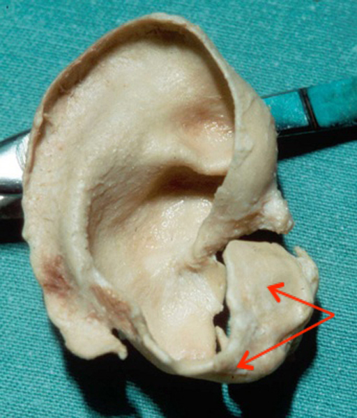
pinna-cartilage-KLS-edited

Photo H

Next, let’s examine details of the pinna. The outer rim of the pinna is the curled helix extending from just above the ear canal to the earlobe (Photo I – yellow arrow); it looks like a bit like a question mark – ? – with the lobe serving as the dot. Roughly 10-11% of people bear a nodule on the upper helix known as “Darwin’s tubercle” (Photo I – black arrow), a bump that may represent the point of the mammalian ear (Photo I – white arrow). First described by Charles Darwin in The Descent of Man (1879) as the Woolnerian tip (sounds like something Angus might find betwixt his toes or Rupert might have in his beard), for decades it has been considered a dominant genetic trait controlled by a single gene. Again, family studies have also shown this is not so.

Stay tuned! Science does stumble, but its general direction is onward and upward!
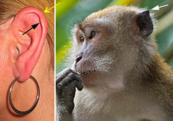
Darwin-s-tubercle-KLS-edited

Photo I

Now moving inward, the next mound (ahh…such a great anatomical word…describes so many…oops, I digress) is the stout Y-shaped antihelix (Photo J). The tragus (Greek meaning goat) is a mound (hee hee, there it is again! Say it with me…m-o-u-n-d) covering the opening of the ear canal, so named for a conspicuous tuft of hair that often graces its surface in men. Opposite the tragus is a small hillock the antitragus (Photo J).

Try this: Feel one of your pinnae (pl.) and note that although the earlobe (auricular lobule) is very flexible, the skin overlying the remainder of the pinna is tightly bound to the underlying cartilage. Palpate the curve of one helix; do you have a Darwin’s tubercle? It may be absent from both pinnae, present in one but not the other, or present in both. Check out Photo J or use a partner (I am a huge fan of playing doctor – extra credit for students who do!) to identify Darwin’s tubercle, helix, antihelix, tragus, and antitragus.
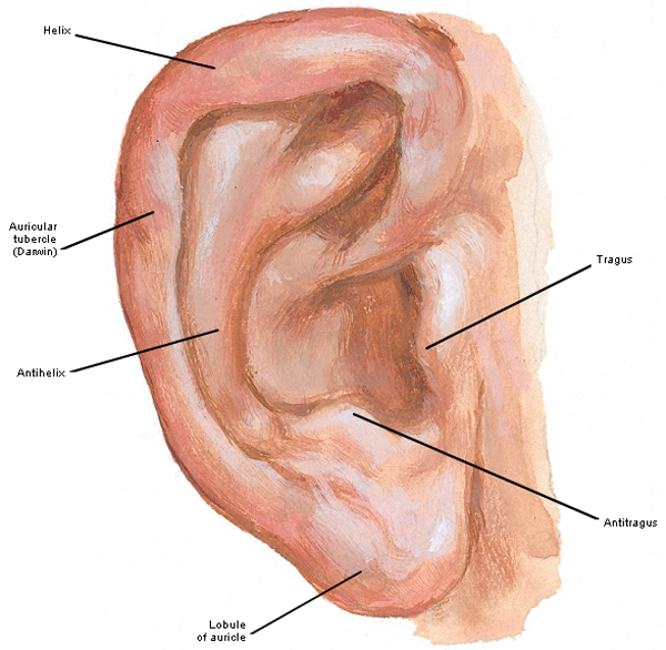
Figure0088A auricle 01 KLS edited

Photo J

Between elevations (another great word…quick! Name three body parts that can elevate!) of the pinna are valleys and depressions. The trough-like scapha separates helix and antihelix; the cymba of the concha is a pit in front of the upper antihelix; the cavity of the concha is the major depression leading to the ear canal. Finally a cleft between tragus and antitragus is the intertragic notch (Photo K).
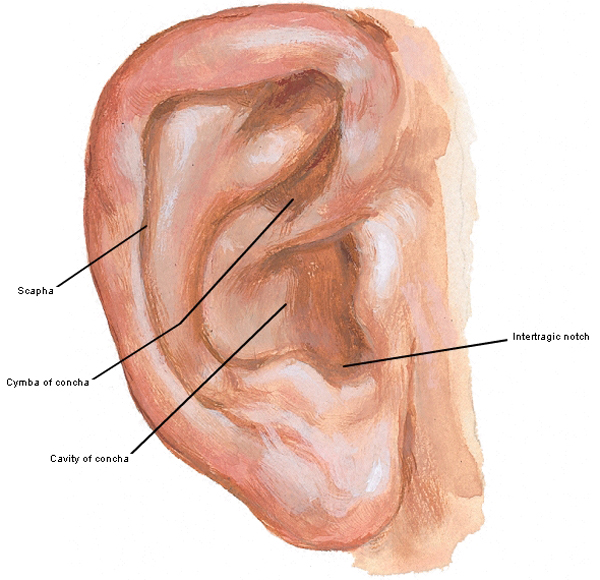
Figure0088 auricle 02 KLS edited

Photo K

So, how do pinnae make a living? The complex array of bumps, depressions and whorls of each pinna is designed to gather sound waves from the environment and transmit them to the ear canal. But, here’s a fine fascinating factoid: the oddly-shaped pinna can also slightly amplify (increase) or attenuate (decrease) sounds of certain frequencies. Yes it can!

I will likely get shot down for this next comment, but you understand that the pinna wasn’t designed for hanging earrings, seating earplugs or hosting earspools (Photo L)? Sure we all do it; just saying!

Piercing earlobes has been done successfully for eons but the now popular ear cartilage piercings are cause for a bit more concern. Why? Cartilage has no blood supply and because it is bloodless, defensive/immune cells or antibiotics are more difficult to deliver when needed to fight infection. Thus, chondritis (inflammation of cartilage) or pericondritis (inflammation of the covering of cartilage) can be challenging to treat and if unchecked, either can seriously deform the pinna.
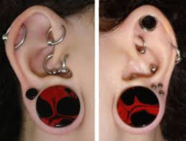
earplugs KLS edited

Photo L

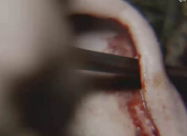
Here’s an interesting Clinical Correlation on the pinna: defects of the pinna are not uncommon and include abnormal folds or location of the pinna, low-set ears, no opening to the ear canal, small pinna or no pinna or ear canal (anotia). Microtia (Latin meaning small ear) is a condition in which the pinna is small and/or distorted. Reconstruction of the pinna is a sophisticated multistep process in which either “harvested” cartilage or synthetic material is skillfully configured to mimic the shape of the pinna cartilage and inserted beneath the pinna skin. The results can truly be remarkable as shown in this series of photos over time (Photo M).
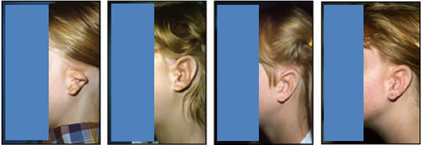
Photo-M

Photo M

Now, we all ken that many mammals turn their pinnae together or even independently. Such creatures have strong auricular muscles that swivel the pinnae to more effectively collect, modify and direct sound waves into the ear canal (Photo N – impala).

Impala KLS edited

Photo N

We humans are equipped with three auricular muscles (anterior, superior & posterior) per pinna. However, these are so dinky they can’t effectively move the pinnae for sound wave collection (Photo O). Most humans I know are proud if they can pull back/wiggle their ears even a wee bit. What this doc wants to know is can Dougal wiggle his ears?
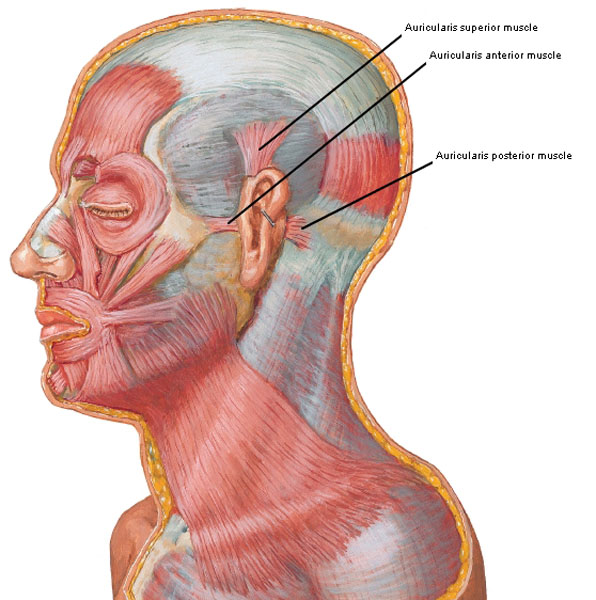
Figure0022 auricularis muscles KLS edited

Photo O

Moving inward, the next structure is the ear canal or external acoustic meatus (EAM). I swear everything anatomical eventually appears in Diana’s books. Claire has just traveled through the stones and meets Jonathan Randall, Esq., Captain of his Majesty’s Eighth Dragoons (Outlander book):

I had by this time recovered my breath, and I used it. I screamed directly into his earhole, and he jerked as though I had run a hot wire into it. I took advantage of the movement to get my knee up, and jabbed it into his exposed side, sending him sprawling into the leaf mold.

Yep. You have to love it even though earhole isn’t a scientific term and yet, very close to another word (may I have an A please, Pat?) I frequently use for the wicked Wolverton wretch. Ha!

EAM is about 2.5 cm (1”) long with a slightly S-shaped path that slopes forward, inward and downward. It is lined with thin skin sporting protective hairs and ceruminous (wax) glands. The first part of the wall contains elastic cartilage (Photo P – black arrow) but bone encircles the rest of the canal (Photo P – red arrow). Throughout the EAM skin tightly adheres to the underlying cartilage or bone; for this reason, a disturbance as innocuous as a blackhead (comedone) can be very painful as there is little room to accommodate swelling and/or inflammation. I’m going to digress again for a moment but can you imagine getting a blackhead in your ear canal in the 18th Century? No wait, don’t answer that. Moving on!
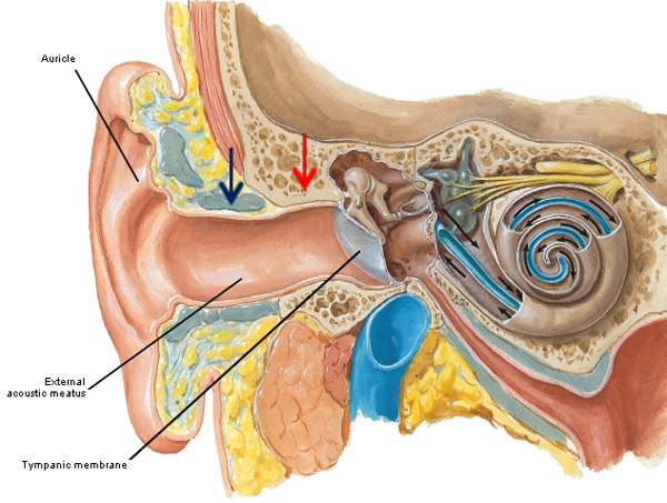
Figure0087-outer-ear-KLS-edited

Photo P

The purpose of the EAM is to modify, direct and deliver sound waves to the ear drum. Although kids will likely disagree, the canal is not for housing beans, peas, BBs, toys, gum, paperclips, etc. (Photo Q)! One does have to wonder though what sorts of items Angus stored in his pinna…

ear objects KLS edited

Photo Q

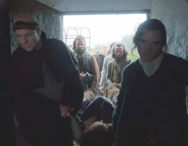
Speak o’ the devil himself…a big shout out to Professor Angus – not only is he good with a sgian dubh (Anatomy Lesson #15), he demos how polite wedding guests should clean the EAM  (Starz episode 107, The Wedding). Och, for safety sake, inserting anything smaller than a pinky finger into the ear canal is unwise!

ep 107 Angus cleaning ear KLS edited

Finally we arrive at the third part of the outer ear, the ear drum or tympanic membrane (TM). TM is a membranous sheet that stretches across EAM and separates outer and middle ears. Photo R shows the external surface of the right TM. Only two of about 10 parts are labelled: the malleus is a small bone of the middle ear and the cone of light is a reflection from the beam of an otoscope.
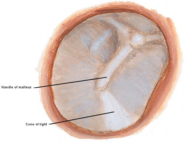
Figure0088B ear drum KLS edited

Photo R

Drawings are great but they may fail to capture subtle details easily revealed in a photograph. If a health care provider examines a normal TM using an otoscope, (s)he sees this image (Photo S – right TM): the red arrow marks the handle of malleus and the blue arrow indicates the cone of light. Infections of the middle ear, perforations, blockages or other disturbances are readily identified using this remarkable instrument. The first time I saw the TM of a volunteer student via otoscope, I squealed (not in her ear, mind you –  was a she); it was an absolutely awesome experience!
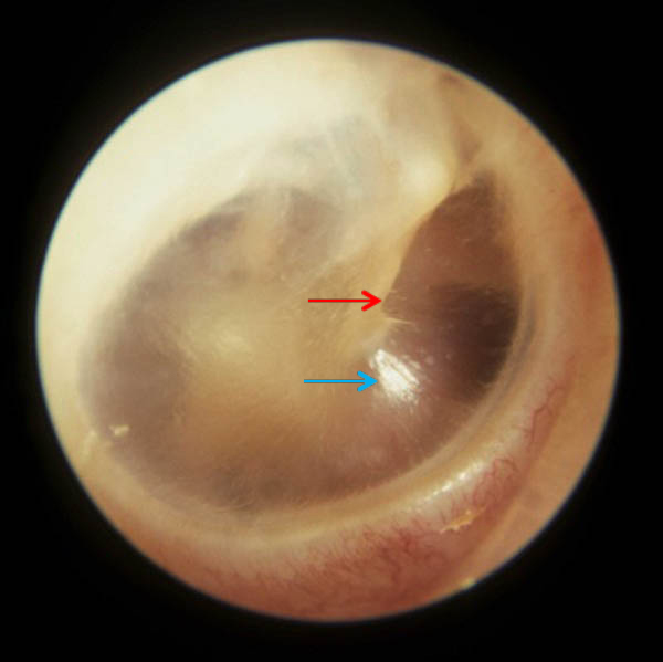
right-TM-KLS-edited

Photo S

OK, you paid attention very well! Pop Quiz time! Pull out paper and pencil and let’s see how you do. There are five questions followed by five answers. Keep score to determine your grade at the end.

Question #1: Name the bump at the tip of the black arrow.

ep-103-Bain-ear--02-KLS-edited

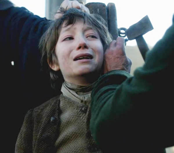
Answer #1: Darwin’s tubercle. Verra guid! Here, Father Bain smells the “vapors of hell” on Claire (Starz episode 103, The Way Out) because her medical meddling endangers Tammas Baxter’s immortal soul!

Question #2: Name the mound at the tip of the red arrow.

ep-116-priest-ear-01-KLS-edited

Answer #2: Antihelix. The good Father hears Claire’s remarkable confession no doubt aided by this Y-shaped mound of cartilage and skin (Starz episode 116, To Ransom A Man’s Soul).

Question #3: Name the mound at the tip of the red arrow.

ep-102-Jamies-ear-01-KLS-edited

Answer #3: Tragus. Excellent! Ye can barely see the tragus because it’s covered by all the long red curls. Jamie messes with Claire’s ears in this scene (Starz episode 102, Castle Leoch) as Herself informs us in Outlander book:

The lad had nice feelings… he sat down, gathered me firmly onto his lap with his good arm and sat rocking me gently, muttering soft Gaelic in my ear and smoothing my hair with one hand.”… No wonder he was good with horses, I thought blearily, feeling his fingers rubbing gently behind my ears, listening to the soothing, incomprehensible speech. If I were a horse, I’d let him ride me anywhere.

Question #4: Name the gap at the tip of the arrow.

ep-103-Tammas-ear-KLS-edited

Answer #4: Intertragic notch. Does this notch really lie between two tragedies? Weel, sort of: puir Tammas Baxter lies ‘tween Claire’s belladonna decoction and Father Bain’s “driving out the demon” approach to universal health care (Starz episode 103, The Way Out)! But, Mrs. Fitz saves the day!

Question #5: Is darling Roger’s wee earlobe (lobule of the auricle) attached or free?

ep-108-Roger-ear-KLS-edited

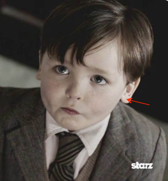
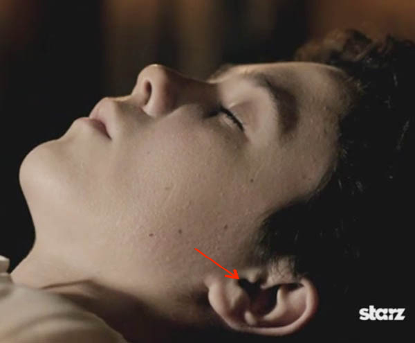
Answer #5: Free. Have ye ever seen a sweeter face (Starz episode 109, Both Sides Now)? What a cutie. May he have another biscuit, please? True story: when one of my grandsons was about wee Roger’s age, he called his earlobes, ear loaves. Butter mine please!

How did you do with the self-graded pop quiz? 5 of 5 is a AAAAA; 4 of 5 is an AAAA; 3 of 5 is an AAA; 2 of 5 is an AA; 1 of 5 is an A; but 0 of 5 is a “please reread the ear lesson!”

Okey dokey, now it’s time for the fun part, the PINNA POLL: which one of the following pinnae do you like BEST? There are six choices and all are awesome so please choose your favorite and leave your vote in the comments! The votes will be tallied and reported at the next anatomy lesson.

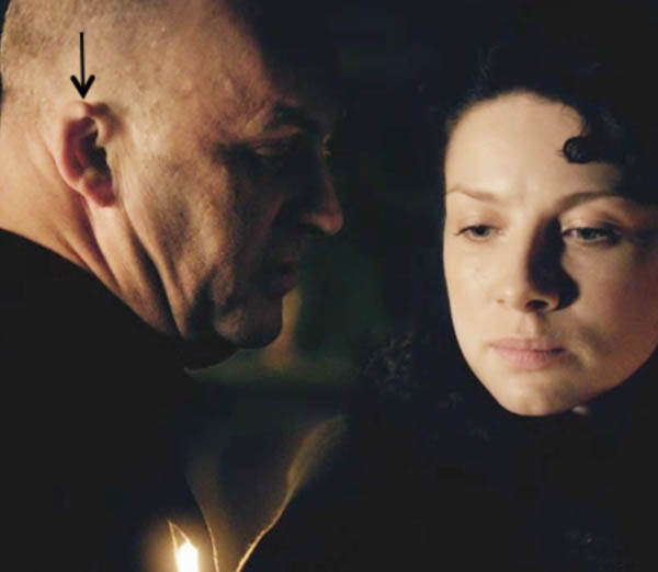
First up is Dougal MacKenzie, Jamie’s favorite Jacobite uncle on his mum’s side (Starz episode 109, The Reckoning). He’s pissed because Colum snags a bag of gold meant for the “bonny Stewart Prince across the water!” Isn’t his helix absolutely fab!

ep 109 Dougal ear 03 KLS edited_1

Next is Geillis, wife of “the man knows no guile” Arthur Duncan, procurator fiscal of Cranesmuir (Starz, episode 103, The Way Out). This wily witch has a one beguiling pinna and plenty of guile to go around!

ep 103 Geillis ear KLS edited

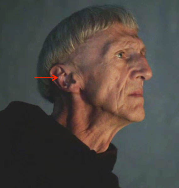
Then, the villain that everyone loves to hate, BJR (Starz episode 115, Wentworth Prison)! Being an officer and a gentleman (yeah, right), he’ll not give into his baser instincts. Vote for his pinna if you like it best.

ep 115 BJR ear 03 KLS edited

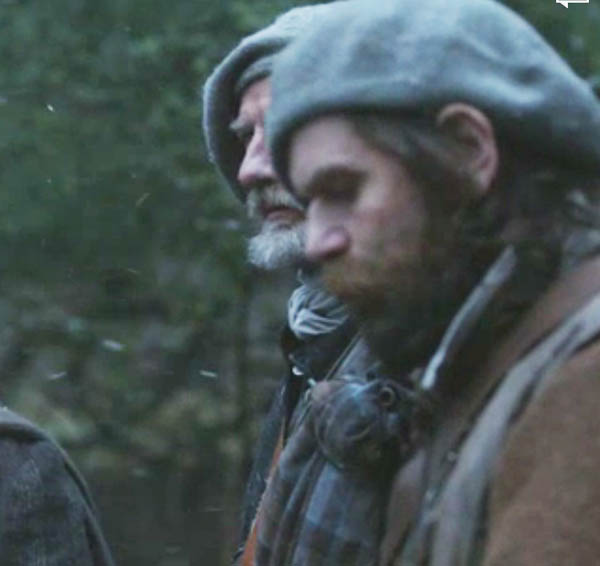
Next we have Murtagh, everyone’s favorite godfather. It’s snowing so his pinna is chilled to the cartilage. He is just about to hock a big ol’ loogie to demo Dougal-disdain (Starz episode 109, The Reckoning). No love lost between those two! Does your vote go for Murtagh’s pinna?

ep 109 Murtagh ear 01 KLS edited

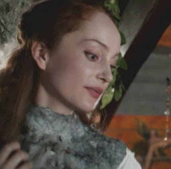
Next up is Claire’s beautiful, wing-like pinna– it looks like porcelain illuminate by the light. NO Jamie, she doesna want to tell her secrets to Geillis (Starz episode 103, The Way Out). Take her outta the fiscal’s house you big, gorgeous strong-fingered hunk!

ep 103 Claire ear KLS edited

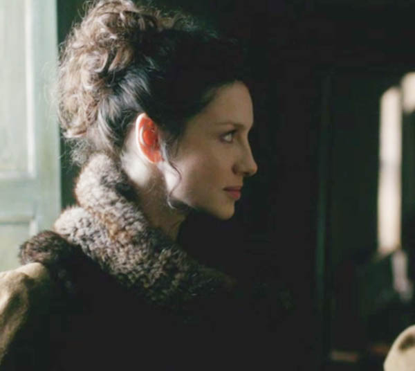
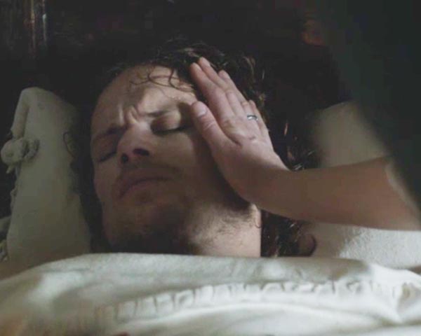
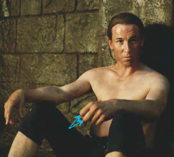
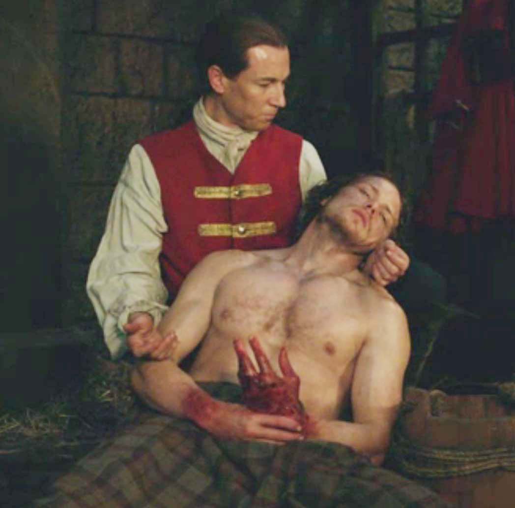
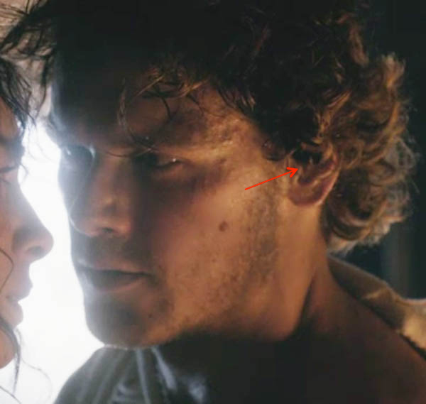
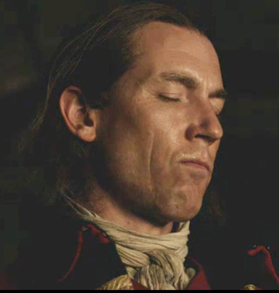
Finally, for the last pinna of the poll! Actually I think this is only time Jamie’s pinna is fully exposed during the entire season one (Starz episode 116, To Ransom a Man’s Soul). Claire’s fingers “ached, wanting to trace the line of his small, neat ear …” (Outlander book). Need an ear rub Jamie? Snuggle up!

ep 116 Jamies ear 02 KLS edited

The middle and inner ears will be covered in our next anatomy lesson, but for now, please understand this: sound waves must travel through gas/air (outer ear), solid (middle ear) and liquid (inner ear) before they are converted into electrical signals that make their way to the brain. The brain translates the electrical signals into a “sound” that we can recognize and understand.

Thanks for playing along! Remember that the outer ear is all about augmenting the hearing function of the ear; it isn’t involved in balance. We’ll learn about hearing and balance in the next lesson. Until then, enjoy stanza III from “The Bells,” a poem by Edgar Allan Poe (1809-1849):

Yet the ear, it fully knows,

By the twanging,

And the clanging,

How the danger ebbs and flows;

Yet, the ear distinctly tells,

In the jangling,

And the wrangling,

How the danger sinks and swells,

By the sinking or the swelling in the anger of the bells–

Of the bells–

Of the bells, bells, bells, bells,

Bells, bells, bells–

In the clamour and the clangour of the bells!

The Deeply Grateful,

Outlander Anatomist

photo creds: Starz, Gray’s Atlas of Anatomy for Students, 2005, Human Anatomy by Martini and Timmons, 1995, Netter’s Atlas of Human Anatomy, 4th ed., Clinically Oriented Anatomy, 5th ed., www.blissbiology.wordpress.com (free & attached earlobes), www.clker.com (pinna), www.nola.com (toys from ear), www.pinna.hawkelibrary.com (elastic cartilage), www.karenscottaudiology.com (three parts of ear), www.microtia.us.com (microtia), www.si.wikipedia.org (Darwin’s tubercle & earplugs), www.wikipedia.org (impala), www.s1.zetaboards.com (attached & free earlobes)

Anatomy Lesson #23: Harming Hands – Helping Hands – Healing Hands

Hallo Outlander Anatomy students! Welcome to Anatomy Lesson #23: The Hand – Part 2. Anatomy Lesson #22 introduced the hand but there is still much more structure and function to learn.

Today’s task is three-fold: to enrich our understanding of hand anatomy, to apply that anatomy using images from Starz episodes and to consider how hands are used. For most of us, our hands are with us throughout life so the way we use them helps define how we work, play and live (Photo A).

So, how do we use our hands? What tasks do we apply them to? Are the tasks helpful, harmful or neither? Outlander books and the Starz series provide abundant examples of hurtful hands, helpful hands and healing hands which are considered in this lesson. Just so we are all on the same page, all Starz images in this lesson are from episode 116, To Ransom a Man’s Soul, unless otherwise stated.

hands of love KLS edited

Photo A

Let’s begin the lesson with a brief review of digit and thumb movements and of the hand skeleton.

Our four fingers (U.S. definition or digits #2-#5) carry out four movements: abduction, adduction, flexion and extension (Photo B). Try all four movements with your own fingers.

finger movements KLS edited

Photo B

Because thumb anatomy differs from that of the fingers, it can perform seven independent movements: abduction, adduction, extension, flexion, opposition, reposition (Photo C) and circumduction (thumb moves in a circle – not shown in Photo C). Try all seven thumb movements.

Reposition (Photo C – right image) is the hand with all digits at rest or repose, a benign anatomical position maintained with little muscular effort.

thumb movements KLS edited

Photo C

In the image below, Claire’s healing hand is held in repose/reposition as she offers comfort to an angry, distraught guilt-ridden Jamie:

“Dinna touch me!” … “You canna save a man that doesna want saving!”

epi 116 good hand in repose KLS edited

Next, consider the crazy creepy crawler’s hand reaching for Jamie, the “object” of his obsession (Starz episode 115, Wentworth Prison). The palm is turned from our view but consider his hand. Is it at rest? Is it neutral? Anatomically, BJR’s hand yearning for Jamie is very nearly in reposition and although it may seem benign, we know it portends great harm. There’s a keen difference between anatomical positions of the hand and the intent behind such positions.

ep 115 BJR hand KLS edited

Next, let’s review bones of the hand (Anatomy Lesson #22). The complex skeleton of the hand contains 27 bones (Photo D – left hand, palm up): 8 carpals in the wrist, 5 metacarpals in the mid-hand and 14 phalanges in the digits. Each finger (digits #2-#5) contains three phalanges but the thumb (digit #1) has only two.

The bones of the hand create many joints (Photo D):

    • blue arrow shows wrist joints (between forearm and carpals)
    • orange arrow shows carpometacarpal joints (CM)
    • green arrow shows metacarpophalangeal joints (MP)
    • turquoise arrow shows proximal interphalangeal joints (PIP)
    • black arrow shows distal interphalangeal joints (DIP)
    • red arrow shows a single interphalangeal joint (IP) of thumb.

hand-bones-KLS-edited

Photo D: ©Stockmedicalart.com 2012

This creative rif on “The Scream” by Edvard Munch (Photo E) warns us that up next is toxic/nuclear waste in the form of handiwork by the bloody black-hearted bastard. In this lesson, I do resort to name calling – I canna help it when it comes to that wicked wretch!

hazard-symbol-munch-scream KLS edited

Photo E

Now for applied anatomy, first of the hand bones: the villainous Jack-Hammer mangled the bones of Jamie’s left hand (right hand in the book) such that Claire is tasked with treating nine fractures. From herself’s own words (Outlander book):

…”A single broken finger is enough to sink a strong man to his knees with nauseated pain…“I carefully picked up the smashed right hand, bringing it into the candlelight for examination. It would have to be set, and as soon as possible. The injured muscles were already clawing the fingers inward.

“It was a long, horrible, nerve-wracking job….. some parts, such as the splinting of the two fingers with simple fractures, went quite easily. Others did not…. I set his middle finger, exerting considerable force necessary to draw the ends of the splintered bone back through the skin.

The next image shows a compound or open fracture of the proximal phalange of Jamie’s middle finger. Compound fractures are characterized by broken bone(s) protruding through the skin and indeed the distal broken fragment is clearly visible (aqua arrow) as Claire rinses it with clean (sterile) water.

ep-116-compound-fracture-KLS-edited

Claire retracts the tip of Jamie’s finger pulling the bony fragment back into the finger. She then approximates the broken ends of the phalange. Retraction could require some effort because following a fracture, attached muscles undergo spasm to form a “natural splint” designed to keep the broken ends of a bone together as much as possible. This explains the quote from Outlander book:

“The injured muscles were already clawing the fingers inward.”

ep 116 compound fracture 01 KLS edited

And a final quote from Outlander book:

“All five fingers eventually lay straight as new pins, stiff as sticks in their bandaged splints… By good luck, only the one joint had been badly damaged…there was nothing I could do about the cracked metacarpal bones or the puncture wound.”

As Claire applies her skills to Jamie’s battered hand, she stitches the lacerations and places his hand on a small wooden pallet. She binds the badly damaged ring finger to the little finger using the latter as a split. All is then secured with a linen strip wound around a wooden peg that is then inserted into a hole in the wood pallet. Wow! This is so impressive and resourceful as no knots are require; the peg can be removed, the strip unwound and the wound attended without messing with knots. Overall, this ingenious device is an external cage which stabilizes the hand in the proper anatomical position and immobilizes it during the healing process. Brilliant!

Finally, did you ken the red, purple, and dusky blue bruises forming on Jamie’s hand? There is more about bruising later in this lesson.

ep 116 hand cage KLS edited

Now for applied anatomy of joints of the hand: in this jarring scene, we see the MP joints (green arrows) of BJR’s right fingers. He tells a traumatized Jamie “I know you can do better than this,” tapping the tip of his nose as one might correct an errant child. The total absence of regard for Jamie’s suffering tells us that the cunning cruel Captain does dwell in darkness and in darkness he shall remain.

ep-116-BJR-I-know-you-can-do-better-KLS-edited

Next, the unholy-one eyes Jamie writhing in agony: “Am I close? Have you reached your limit yet?” Oozing an air of utter boredom and sure knowledge of outcome, the mad menacing maniac sits with flexed left fingers that clearly display his left PIP joints (aqua arrows).

ep-116-BJR-have-you-had-enough-KLS-edited

Soon, BJR instructs Jamie to brand himself with BJ’s glowing wax seal stamp – over the left breast and near the heart. “Are you mine? Show me … do it!” The red arrow shows the flexed IP joint and the green arrow indicates the flexed MP joint of Randall’s right thumb. Remember: the thumb has only two phalanges with one IP joint shared between them.

ep-116-branding-Jamie-02-KLS-edited

Wracked with pain, Jamie brands himself but stubbornly sears his left side; his heart belongs to Claire! The flexed DIP joints (red arrows) are visible on Jamie’s right fingers as he grips the stamp.

ep-116-branding-Jamie-KLS-edited

Remember the anatomical mantra: the more bones in a body part, the more possible joints between the bones; the more joints, the more possible movements at the joints? Bone movements occur when muscles that attach to them undergo contraction. So, to better understand hand movements, we must next consider muscles operating the hands.

In Anatomy Lesson #22 we learned that some forearm muscles move bones of the hand. To review, the forearm is divided into two compartments. The anterior compartment contains eight muscles six of which attach to bones of the palmar (front) hand. The posterior compartment contains 12 muscles nine of which attach to bones of the dorsal (back) hand. Covering all the forearm muscles that move hand bones isn’t feasible in a single lesson, so let’s consider a few.

Three important muscles originate from the anterior forearm and end on bones of the digits; these are the long flexor tendons.

The first muscle is flexor digitorum superficialis or FDS (Latin meaning superficial flexor of digits); this muscle arises from humerus and radius (Photo F). Near the wrist FDS splits into four tendons, one to the intermediate phalange of each finger. Contraction of FDS mainly flexes PIP joints (Anatomy Lesson #22) but continued contraction flexes MP and wrist joints.

Try this: strongly flex (curl) your fingers and wrist joint. You used FDS to accomplish this. Identify wrist, MP and PIP joints. Flexion of these joints is critical for the grip (Anatomy Lesson #22).

Figure0426A FDS KLS edited

Photo F

Please think, if the digits have a superficial flexor will they also have a deep flexor? If you answered yes, then you are getting the idea of anatomy! The next anterior forearm muscle is flexor digitorum profundus or FDP (Latin meaning deep flexor of digits). Deep to FDS, FDP (Photo G) arises from the ulna and breaks into four tendons near the wrist to end on the distal phalange of each finger.

Now consider this: if tendons of FDS insert on the intermediate phalanges how will tendons of the deeper-lying FDP reach the distal phalanges? The body’s answer is ingenious! FDP goes through FDS: all four tendons of FDS split to allow FDP tendons to reach the distal phalanges (Photo G – red arrow). Do you see the splits? The engineering is awesome!

FDP flexes the DIP joints but it also contributes to flexion of PIP, MP and wrist joints.

Try this: Attempt to flex only the DIP joints of your fingers. Can you do it? I know a few folks who can but it is unusual. DIP joints typically flex in conjunction with PIP joints.

Figure0426B-FDP-KLS-edited

Photo G

The third muscle of the anterior forearm is Flexor Pollicis Longus or FPL (Latin meaning long flexor of thumb). FPL originates from the radius and inserts on the distal phalange of digit #1, the thumb (Photo H). Its major action is to flex the IP joint of the thumb but continued contraction also helps flex the MP joint.

Try this: flex your thumb and find the IP joint. Flexion is caused by FPL.

Figure0426B FPL KLS edited

Photo H

Do you recall those eight carpal bones arranged in two rows of four bones each (Anatomy Lesson #22) – aye, the ones with the funny mnemonic? These bridge the gap between radius and ulna and five metacarpals. More than a dozen strong fibrous wrist ligaments bind together carpals, metacarpals, radius and ulna (Photo I –  grey strands).

Figure0437B carpal arch KLS edited

Photo I

Two-dimensional images imply that the carpals lay in a flat plane, but this is not so. Wrist ligaments secure them into a curved carpal arch that is convex on the dorsum and concave on the palmar surface (Photo J – curved red line). The carpal arch is spanned by a fibrous sheet, the flexor retinaculum thus forming a potential space, the carpal tunnel (Photo J). But, the tunnel is NOT empty because it contains four FDS tendons, four FDP tendons, one FPL tendon and the median nerve (Anatomy Lesson #22).

Now for an important clinical correlation: truly a tight squeeze, any swelling of structures within the carpal tunnel compresses the median nerve and affects hand areas it subserves, a condition known as carpal tunnel syndrome. Symptoms may include tingling, numbness and pain with reduced strength of thumb and index, middle and half the ring fingers. Repetitive motions that over-extend the wrist such as keyboard use, piano playing, or prolonged use of vibrating tools may be contributing factors.

Figure0444B-Carpal-tunnel-KLS-edited

Photo J

Next, we consider the muscles of the back of forearm and hand: known as long extensor tendons, several muscles reach from posterior forearm to bones of the hand. We won’t name these but to summarize: three long tendons extend the wrist (Photo K – blue arrows) and 5+ long tendons extend the finger joints (Photo K – red arrows). Three long tendons extend and/or abduct the thumb (Photo K – black arrows). Whew!

Figure0424A-long-extensor-tendons-KLS-edited

Photo K

A closer view at the dorsal hand shows long extensor tendons as they cross the wrist. These tendons are held in check by a fibrous band, the extensor retinaculum (Photo L – black arrow). Note bands of fibrous tissue between the long extensor tendons. Known as intertendinous connections they augment extension of the fingers as a unit; if one extensor muscle contracts, it helps activate its neighbors (Photo L).

Try this: Intertendinous connections are quite variable but let’s try to locate one. Place forearm and hand flat on a surface. Pronate the forearm to expose the back of your hand. Try to lift (extend) just the little finger. Can you do it? Next, lift ring, middle and index fingers one at a time. Are some fingers harder to lift independently? I have greatest difficulty lifting middle and ring fingers independently so I know they are bound by an intertendinous connection. Try to locate one of yours.

Figure0453A-Extensor-tendons-KLS-edited_1

Photo L

Now, let’s consider intrinsic muscles of the hand beginning with the palmar surface. Palmar skin is tightly bound to an underlying fibrous sheet, the palmar aponeurosis, a feature that aids grasping (Photo M – red arrow). Two superficial muscles are apparent: palmaris longus and palmaris brevis. A forearm muscle, the tendon of palmaris longus blends with the palmar aponeurosis and tenses it during contraction. Many anatomists regard palmaris longus to be a vestigial muscle because it is absent in about 14% of people and its absence doesn’t really affect our ability to grip. Palmaris brevis is a tiny postage stamp-sized muscle near the base of the little finger (Photo M); its contraction puckers the palm skin and deepens a cupped palm.

Figure0442A-palmaris-longus-&-brevis

Photo M

Try this: Spread your digits and tap your palm. Feel that taut springy tissue? That’s the palmar aponeurosis. Now, bring pads of thumb and little finger together and strongly flex the wrist. Does a tendon stand out in the midline (Photo N – black arrow)? If so, this is palmaris longus. Repeat with the opposite side. What is the pattern of your forearm? Do you have two palmaris longus muscles, one or neither? Turns out I have one on the left but none on the right.

Palmaris longus KLS edited

Photo N

I love watching Jamie balance and twirl a dirk on the palm of his hand (Starz episode 105, Rent). See the puckered flesh at the base of his palm (red arrow)? This is caused by contraction of palmaris brevis to help cup his palm and support the dirk. Yep, Jamie has one! Is Claire watching his clever performance? Aren’t we all?

ep-105-palmaris-brevis-KLS-edited

The hand contains 19 intrinsic muscles (including palmaris brevis), meaning they arise and insert within the hand itself. These small muscles initiate fine movements of the digits. It would be tedious to name them all so understand that the thumb has four intrinsic muscles concerned with flexion, opposition, adduction and abduction (Photo O – green overlay). The little finger has three muscles which flex, oppose, and abduct (Photo O- purple overlay). Between the long flexor tendons and metacarpal bones are 11 additional muscles (lumbricals and interossei) which help flex, abduct and adduct the fingers (Photo O – black arrows). All totaled a whopping 34 forearm and hand muscles are involved in hand movements!

Figure0443B-intrinsic-hand-muscles-KLS-edited

Photo O

Like most body parts, the hand needs a blood supply. The large ulnar and radial arteries descend through the forearm, cross the wrist to form superficial and deep palmar arches (Photo P). Branches from both arterial arches produce two small arteries (proper palmar digital arteries) for each finger. The radial artery supplies branches to the thumb. The dorsal hand (not shown) has a smaller arterial arch that produces two more small arteries to each finger. Ergo, each finger has four supplying arteries; the thumb has three.

Figure0449B-palmar-arterial-arches-KLS-edited

Photo P

For those who do not understand blood flow, a full lesson on this subject lies in the future. For now, please know that the digital arteries carry blood ladened with oxygen and nutrients to the digits. They divide into smaller arterioles which branch into microscopic capillaries forming capillary beds (Photo Q). In the capillary bed, oxygen and nutrients are exchanged for carbon dioxide and waste products. The capillary bed reforms into venules that unite to form veins draining blood from the tissues. This general plan is followed throughout the hand and in most body tissues.

capillary bed KLS edited

Photo Q

Time for a clinical correlation about blood supply: capillaries have very thin walls. When subjected to trauma, these vessels burst leaking blood into surrounding soft tissues. Extravasated blood presents as a contusion, a reddish-purple-dusky discoloration which doesn’t blanch when pressed: the classic “black and blue” bruise (Photo R).

Bruising KLS edited

Photo R

Several students have inquired about Jamie’s blackened hand and digits following his rescue from Wentworth Prison. They were concerned that his hand exhibited tissue necrosis or gangrene. But no, discoloration is due to the beating administered by that depraved, demented devil. This gut-wrenching scene gives evidence of early bruising (black arrow): a vivid color range from deep red to purple to dusky blue. Very convincing!

ep-116-Jamies-hand-on-Claires-back-KLS-edited

Later on the Cristabel, Jamie’s thumb and fingertips are fully black and blue. The pounding mallet did a number on his hand capillaries and the bruising is fierce!

ep 116 Jamies hand on ship KLS edited

We are done with the real and applied anatomy lesson so let’s finish with examples of how hands are used in Starz episode 116 To Ransom a Man’s Soul.

“Dear God, you are a magnificent creature” utters the crazy cunning cutthroat with Jamie spread across his lap. As I gazed at this imagery, it seemed vaguely familiar. Then, B-I-N-G-O!

ep 116 BJR holding Jamie KLS edited

Intentional or not the position, posture and attitude are very nearly a mirror image of the Pietà, a Renaissance sculpture by Michelangelo Buonarroti (1475-1564) of the Madonna cradling a crucified Christ. This riveting juxtaposition of images reveals universal opposites: dark versus light; evil versus good; cruelty versus mercy, all deeply rooted themes in Diana’s works. Randall even draws the analogy to Jamie’s suffering: “Ah, so that’s your plan; to submit like Christ on the cross?” Helping hands, healing hands, or hurting hands. No brainer, here!

Pieta KLS edited

Later, the foul, festering fiend sensuously rubs lavender oil into Jamie’s painful burn telling him “These are Claire’s hands. Think of your wife.” There are new bruises on Jamie’s left shoulder. We don’t know what caused these – mayhap a mallet blow went astray? We shouldna be deceived by this perceived gentleness because BJ is about to deliver extreme mental pain to Jamie. Hurting hands, helping hands, healing hands?

ep 116 BJR these are Claire's hands KLS edited

After suffering hours of agonizing pain, BJ tells Jamie to:

“Think of Claire. Think of your wife. Wait for me. Claire’s here.”

Sadly, the soulless one said he would have Jamie’s surrender before he left this world and he does. Then BJ delivers his cruel, mocking coup de grâce: “I understand. How could she ever forgive you?” His hands are made for hurting and they are remarkably skilled at their work!

ep 116 how can she forgive you 01 KLS edited

Ugh! Enough of hurting hands…How about some healing hands? We already witnessed Claire’s healing hands as she cleanses and sets Jamie’s battered hand. Here she tidily stitches his skin lacerations. Her competency as a 20th century WWII combat nurse, her ingenuity at using 18th century tools and her brilliant, fertile mind are testimonies of her love and devotion to Jamie. Her hands have come to his rescue over and over throughout the Starz series; a true symbol of healing.

ep 116 stitching KLS edited

We’ve seen Claire reset Jamie’s broken bones and mend his wounds. Picking lavender, grinding buds in a mortar and pestle to make oil of lavender is yet another example of her healing hands as she prepares to enter the darkness with Jamie.

ep 116 lavendar oil KLS edited

As she rests her healing hand on her husband, he struggles with shame and self-loathing. “You belong to no one else but me…Well, Randall had your body but I’ll be damned if he has your soul as well.” Go Claire! Ye make us proud!

ep 116 Claire helping hands KLS edited

Finally, Godfather Murtagh’s healing hands are at work as he cuts JR from Jamie’s skin and casts the scrap into the fiery furnace! Mercifully, healing hands are plentiful in Starz episode 116.

ep 116 removing brand KLS edited

Let’s finish this lesson with examples of helping hands. First is a fantastic image of the three Highland musketeers rescuing Jamie from the “thug under the door.” Murtagh stolidly carries his precious cargo from Wentworth Prison to freedom. Angus furtively peers backward with dirk in hand while Rupert rides shotgun! Yeah, helping hands! Ye made me weep with relief!

ep 116 helping hands 02 KLS edited

So many helping hands at the monastery reach out to the party of Highlanders offering refuge, supplies and care for Jamie, wounded in body and soul.

ep 116 helping hands 03 KLS edited

More helping hands as the goodly Father comforts Claire after her confession: a far cry from Father Bain, no?

ep 116 priest helping hands KLS edited

Finally, we catch Angus’ au revoir kiss but did ye ken that the wee laddie’s helping hand helps itself to some of Claire’s personal real estate? Naughty Angus Mhor!

ep 116 Angus & Claire KLS edited

Helping hands, healing hands, hurting hands… how shall we use our own? Mine will continue to write lessons throughout the summer although most will be shorter and less complex. Thanks to all who love anatomy and Outlander!

Oh! Last week, I was invited by Jennifer Barnes, Ryan Hooks and Keetin Marchi (they are such great fun!) to give a mini anatomy lesson about the hand on the Outlander Aftershow at TheStream.TV. You can see it below around 16:30. Now that we face another bout of withoutlander, you can join our friendly hosts for the Outlander Book Club on Monday nights where they compare and contrast the book and the show: http://bit.ly/1dqHXxQ. Happy Summer!

The deeply grateful,

Outlander Anatomist

Follow me on:

Photo creds: Sony/Starz, Netter’s Atlas of Human Anatomy, 4th ed Gray’s Atlas of Anatomy for Students, 2005, ClinicallyOriented Anatomy 5th ed, www.artsology.com, www.imgbuddy.com, www.medicalartlibrary.com, www.parlmreadingperspectives.wordpress.com, www.wikipedia.org, www.wikiradiography.com, www.shutterstock.com