Anatomy Lesson #53: Dr. Abernathy Meets “Pretty Lady”

Greetings, anatomy students! Several of you have asked for an exploration of the “skeleton scene” shown in Outlander episode 305, Whiskey and Freedom. Although Anatomy Lesson #39, Dem Bones – Human Skeleton, examined the entire human skeleton, this lesson focuses on skeletal analysis performed by Dr. Abernathy with input from Dr. Claire. 

 The “skeleton scene” is a great adaptation of Chapter 20, “Diagnosis,” from Voyager book, by Diana Gabaldon. So, put on your forensic cap and let’s begin with a summary of that scene.

Here, the anthropology office at Harvard seeks Dr. Abernathy’s expertise to determine cause of death using skeletal remains. In the book, the skeleton arrives at his office in a box labelled “PICT-SWEET CORN.” I remember that brand!  

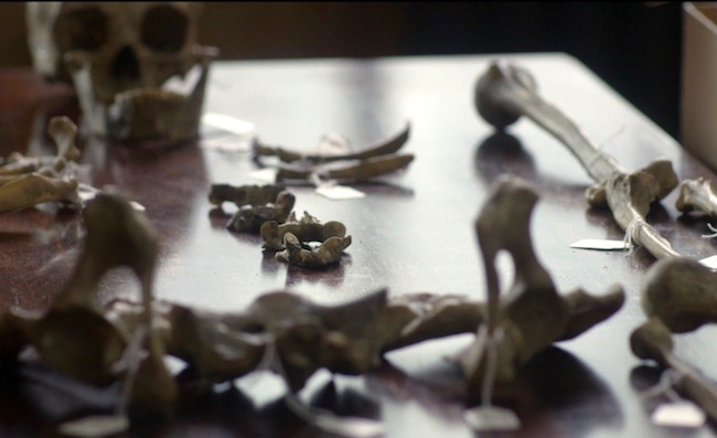
In the episode, bones are laid out on a desk top (Image A). From Voyager:

 Horace Thompson was probably someone from the coroner’s office, I thought. Sometimes they brought bodies to Joe that had been found in the countryside, badly deteriorated, for an expert opinion as to the cause of death. This one looked considerably deteriorated.“

…from the anthropology department at Harvard,” he said… “asked me would I have a look at this skeleton, to tell them what I could about it.”
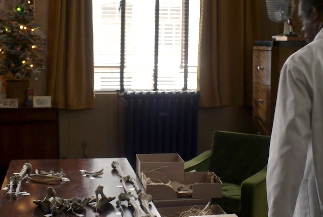
Image A

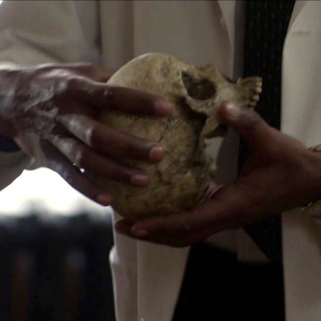
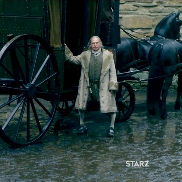
Dr. Abernathy reaches into the box and removes the skull (Image B).
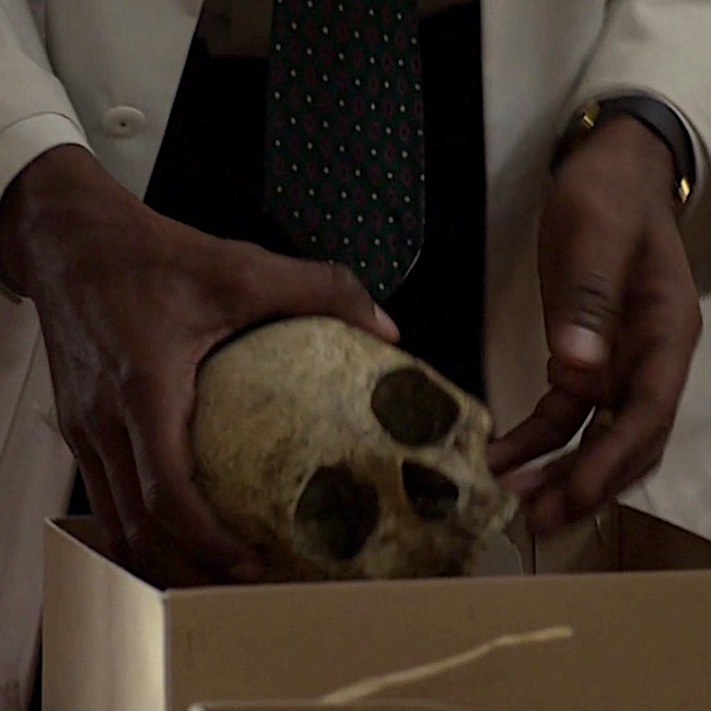
Image B

 Examining the skull, the good doctor concludes it belonged to a pretty lady who was mature and middle-aged (Image C). And, from Voyager:

As the saying goes, “Pretty is as pretty does.” The owner of this skull definitely did not do pretty things! 😳

“Oh, pretty,” he said in delight, turning the object gently to and fro. “Pretty” was not the first adjective that struck me; the skull was stained and greatly discolored, the bone gone a deep streaky brown. Joe carried it to the window and held it in the light, his thumbs gently stroking the small bony ridges over the eye sockets. “Pretty lady,” he said softly, … “Full-grown, mature. Maybe late forties, middle fifties.”

Image C

Clairvoyant Claire picks up the skull, which speaks to her (Image D).  Well, not really –  that would be weird – though Hallowe’en does draw nigh! Another quote from Voyager:

Then I held it close against my stomach, eyes closed, and felt the shifting sadness, filling the cavity of the skull like running water. And an odd faint sense—of surprise?

“Someone killed her,” I said. “She didn’t want to die.”
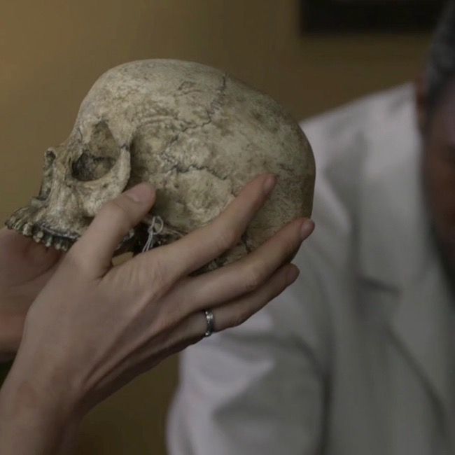
Image D

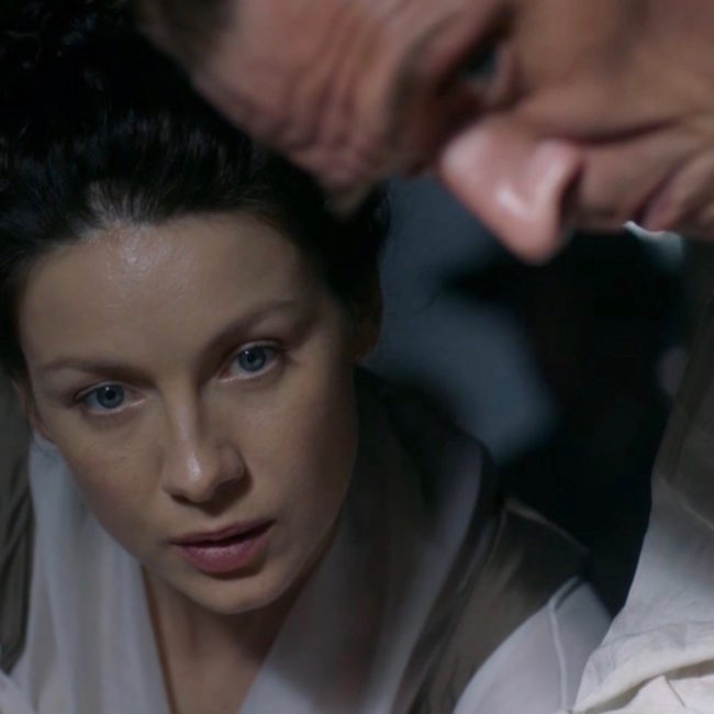
Dr. Abernathy fixes Dr. Claire with a gimlet eye. Lady Jane, have you lost your scalpels???  After all, how could she possibly conjure such details using a touchy-feely method of scientific inquiry? Well, the lass does harbor some awesome powers that seem to grow with time (Image E)? Again, from Voyager:

… “Where did you find her?” I asked….

“She’s from a cave in the Caribbean,” he said. “There were a lot of artifacts with her. We think she’s maybe between a hundred-fifty and two hundred years old.”
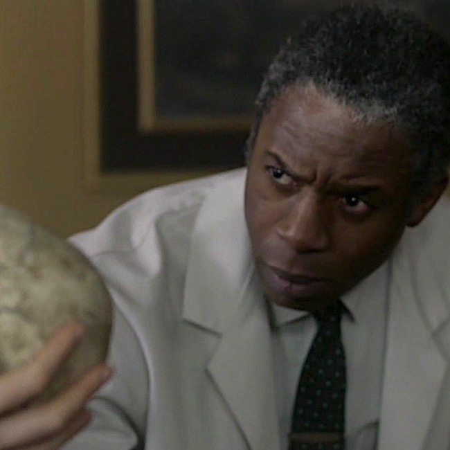
Image E

Next, Dr. Abernathy plucks two pieces of bone from the corny box (Image F). “You were right,” he says. Looking at them, he observes that the fracture plane runs through the centrum, although he doesn’t identify which bone has been fractured. 

Voyager book reveals the fractured bone as a vertebra (bone of spine) – more specifically, it is the axis, aka the second cervical vertebra (C2).

The wide body of the axis had a deep gouge; the posterior zygapophysis had broken clean off, and the fracture plane went completely through the centrum of the bone.

Joe’s finger moved over the line of the fracture plane. “See here? The bone’s not just cracked, it’s gone right there. Somebody tried to cut this lady’s head clean off. With a dull blade,” he concluded with relish.
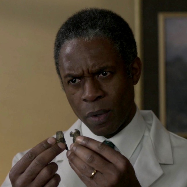
Image F

Then, Dr. Abernathy notes that although the burial site was a cave filled with slave artifacts, this lady was not a slave! He points to two leg bones (Image G), the tibia (Anatomy Lesson #9, The Gathering or Boar Gore) and the femur (Anatomy Lesson #7, Jamie’s Thighs or Ode to Joy!).  Ahhh, Claire sagely nods, “the crural index.” Back to Voyager, again:

“Not a slave,” he said… “No,” Joe said flatly. He tapped the long femur, where it rested on his desk. His fingernail clicked on the dry bone. “She wasn’t black.”

“Take a look at this,” Joe invited. “You can see the differences in a lot of bones, but especially in the leg bones. Blacks have a completely different femur-to-tibia ratio than whites do. And that lady”—he pointed to the skeleton on his desk—“ was white. Caucasian. No question about it.”

…“If you want to think blacks and whites are equal under the skin, be my guest, but it ain’t scientifically so.” He turned and pulled a book from the shelf behind him. Tables of Skeletal Variance, the title read.
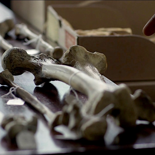
Image G

 OK, that pretty much summarizes the salient scientific points of this scene, although I see three issues that warrant comment:

      • Can bones reveal sex, age and/or beauty of the owner? 
      • Does the fractured bone match with “pretty lady’s” death? 
      • Can crural index determine race? 

Issue #1: Can bones reveal sex, beauty and/or age of the owner?

The answer is a qualified yes

When Claire arrives at the office, Dr. Abernathy has already laid out most of the skeleton and presumably, has already examined each of these bones. It’s pretty iffy to hazard a reliable guess with one measurement. But, if a series of measurements tend to fall within a given range, a forensic scientist can venture an educated guess. So, assuming he examined the skull and pelvis, then sex can be surmised with a resonable degree of confidence. Here’s why.

Sex & Skull: “Typical” female and male skulls exhibit differences (Image H). A female skull is usually smaller, with rounder orbits (red dot), prominent frontal eminences (yellow dot), smaller mandible (pink dot), less prominent temporal lines (green dot), smaller brow ridges (dark blue dot), and less pronounced glabella (turquoise dot), and the brow ridges are sharper. There are also other, more subtle differences, but understand that not all skulls show all structural differences, regardless of genetic sex.
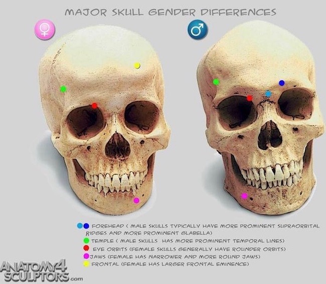
Image H

Sex & Pelvis: The bony pelvis also supplies important clues to sex. “Pretty lady’s” bony pelvis lies in three pieces (two hip bones plus sacrum). The pelvic width, shape of sacrum, sub-pubic angle and shape of obturator foramen (two front holes) are consistent with a female pelvis (Image I – obturator foramen not labeled).  Pelvic inlet and outlet are difficult to demonstrate in a dissembled bony pelvis but, assembled they would be similar to those shown in Image I, lower figure. Ergo, pretty lady’s pelvic features are consistent with those of a female.

Age:  Age can be estimated if cranial sutures (sites where cranial bones meet) are thin, bony ends are fused to bone shafts (growth plates are ossified), teeth are mature and bones are hard, along with the presence of wear-and-tear diseases, etc. So, yes, with careful analysis, general age can be estmated. Presumably, Dr. Abernathy considered these before Claire’s arrival. So far, so good!

Beauty:  As we all ken, beauty is in the eye of the beholder. Dr. Abernathy expresses an opinion when he dubs the owner a “pretty lady.” Of course, he cannot know how flesh draped those bones, but he considers the skull to be delicate and beautiful to his practiced eye. This is a subjective response on his part, but it is arguably consistent with the appearance of the skull which is delicate with good teeth. Nowadays, forensic scientists can reconstruct a face using computer programs, or the older clay sculpting technique

Thus, sex and age can be assessed with a fair degree of confidence if and only if multiple measurements and observations are considered, collectively. But, as beauty remains in the eye of the beholder, this issue receives a qualified yes.
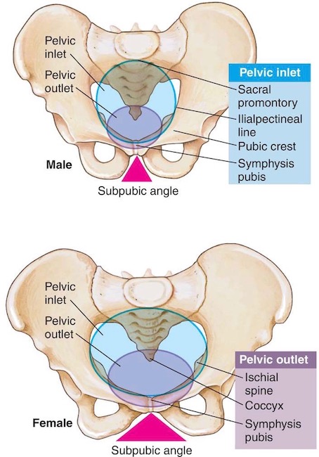
Image I

Issue #2: Does the fractured bone match with “pretty lady’s” death? 

Because I have some issues with this issue, the answer is mostly yes for the book, but no for the episode. Here’s why. 🤓

Dr. Abernathy holds up a wee bone, which is broken vertically into two nearly symmetrical pieces. Voyager identifies these fragments as belonging to the axis. The axis is one of seven neck bones (numbered 1-7 from skull downwards); it is also designated as C2, meaning it is second of the seven cervical vertebrae. The purpose of cervical vertebrae is to support the head and supply flexibility to the neck, augmenting movements of the head and shoulders.

Voyager book states:

The wide body of the axis had a deep gouge; the posterior zygapophysis had broken clean off, and the fracture plane went completely through the centrum of the bone.

Although the bone fragments of shown in episode 305 are supposed to represent a vertebra, the fragments do not  appear to be an axis. The axis is an atypical, weird-looking bone (Image J – gif):
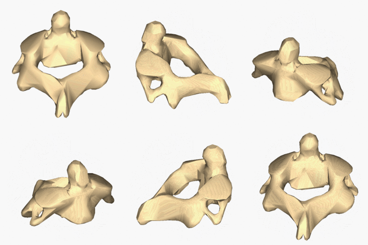
Image J

However, please appreciate that the axis is a splendid bone, allowing us to rotate our head from side-to-side in a “no-no” gesture (Anatomy Lesson #12 Claire’s Neckthe Ivory Tower!). 

Try this: Cervical vertebrae are buried rather deeply in the neck and difficult to demonstrate. But, if you able, sit up straight with chin level, place a finger in the groove of your neck just below the skull. You may feel a small bulge under your fingers. This is the spine of the axis. 

Let’s examine the parts of the axis (Image K – front view) to help us understand the quote from Voyager.

  • The axis has a robust body, located at its front surface.
  • Inside the body is a centrum, a remnant of embryonic development. You cannot see the centrum from the surface.
  • Dens is a tooth-like structure perched atop the body.
  • Superior articular facets (aka superior zygapophyseal processes) are flat surfaces where C2 forms joints with C1 above.
  • Inferior articular facets (aka inferior zygapophyseal processes) are flat surfaces where C2 forms joints with C3 below.
  • Joints are sites where movement occurs between bones – between vertebral facets these are called zygapophyseal joints.

To reiterate, Voyager book states:

The wide body of the axis had a deep gouge; the posterior zygapophysis had broken clean off, and the fracture plane went completely through the centrum of the bone.

Analysis: The book statement is a wee bit awkward because if the fracture plane passed through the centrum, it must necessarily cleave the body, so a deep gouge would not have been left to discern.

Also our vertebrae don’t have posterior zygapophyses – I suspect Diana intended inferior rather than posterior. Assuming this is correct, then: The dull blade cut horizontally (as in cutting off a head), breaking off the inferior zygapophyses (forming zygapophyseal joints with C3), and passing through the axial body and its centrum. Otherwise, the description makes sense.

One may also conclude, the blade must have passed through the lower part of the axis. Had the blade passed through its upper part, the stroke would have sheared off dens and superior zygapophyses but completely missed the centrum! Make sense? Yay! 👏🏻👏🏻👏🏻
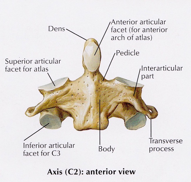
Image K

Conclusion: Dr. Abernathy holds two bony fragments of the fractured bone and I think, hum… that ring of bone appears to have been cleaved in halves by a vertical blow (Image L)! The only way a vertical fracture could occur is if the blade sliced downward, vertically cleaving the skull and its supporting cervical vertebrae. Gah! I think you will agree, this is pretty unlikely plus, the skull remains intact. So, no vertical swipe of the blade!

Ergo, although dramatic and interesting, the bone fragments do not reflect the axis damage as described in Voyager or by episodic Dr. Abernathy. Now, does all this keep me up at night? Hardly – I loved this scene despite its wee anatomical issues!
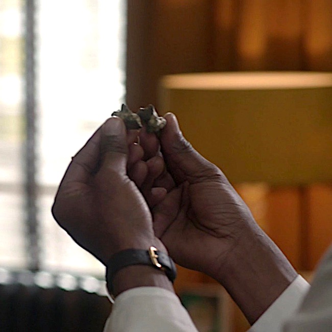
Image L

Issue #3: Can crural index determine race? 

The answer is a qualified no.

This topic is a super sticky-wicket but very important to consider, so bear with me.

Best start with a definition: the crural index, established in 1933, is the ratio of tibia-to-femur (not femur-to-tibia, as this yields different results). The formula to determine crural index is:

 (length of tibia x 100) / length of femur  OR  

(length of tibia/length of femur) x 100

Using this ratio, many studies showed that individuals of African descent had higher crural indices than those of European descent. Image M is a simplified, summary version of such findings.

In 1968, Dr. Abernathy was positive that a low crural index meant “pretty lady” was white….“No doubt about it.”

(BTW, I am pretty sure Claire pronounces this “cruel index;” but, it should be crural <kru-ral>, from the Latin crus meaning “leg”)
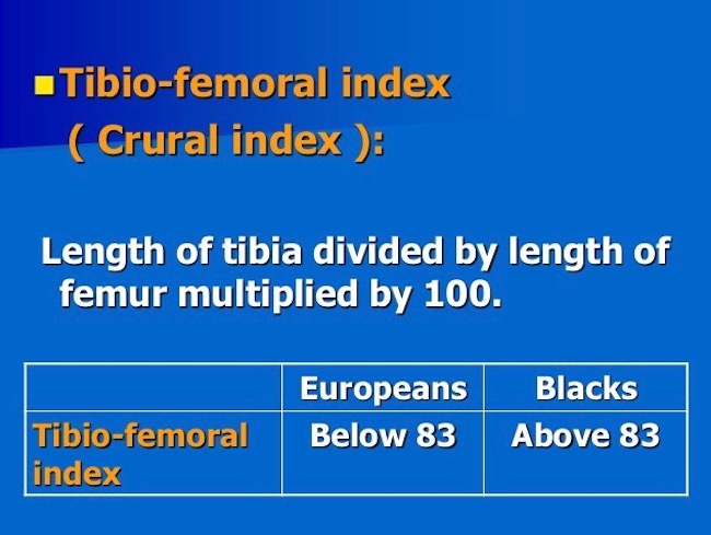
Image M

So, can race be determined from the crural index?

When I started graduate school in 1965, we were taught there were three human races: negroid, caucasoid, mongoloid. Fast forward 52 years and much has changed! Today, many biologists say race cannot be determined from bones because there is no such thing as race. These scientists posit that all living humans belong to one species (Image N): Homo sapiens sapiens (the second sapiens denotes the subspecies – that would be us).

Many designate the term ancestry, because race and even ethnicity have confusing connotations and definitions. Furthermore, they point out, more genetic variations can occur within “racial” groups than between them, meaning findings are limited by the sample studied. What a conundrum!

Just to clarify, some bony physical traits are characteristic of ancestry and can be traced to a particular global location. But, bear in mind, people of mixed ancestry may present features which do not fall neatly into any category. Also, humans are so similar that all bone morphologies are present in all  groups, just at varying rates. Despite such variations, skeletal analysis remains part and parcel of human identification especially when numerous skeletal measurements are obtained. Today, using calipers, x-rays, microscopy, DNA, and a mess of other tools, some of which were unavailable in 1968, forensic researchers can make reasoned guesses as to a person’s ancestry based on skeletal remains.

Summary: Nowadays, before a scientist suggests ancestry based on skeletal remains, (s)he makes multiple measurements, never relying on just one. And, prudent scientists avoid stating “we are sure” (even if they are). Instead, they posit, the data suggests or indicates or is consistent with or is likely. Verra prudent!

Hence the qualified “no” regarding the crural index; it is only one skeletal measurement and insufficient to make a judgement if a person was white or a not. However, Voyager accurately expresses views prevalent in the 1960’s. Make sense? Ta da!
Image N 

Bottom line:

    • Today’s competent (my word) scientist cannot/will not declare ancestry using a single skeletal measurement such as crural index. 👎🏻
    • Cutting (hah) Dr. Abernathy some slack, his surety about the skeleton’s “race” was suitable and justifiable for his time (1968), but untenable for ours (Image O)!

Image O

So, this concludes a brief analysis of the “skeleton scene.”  Much more could be added, but would likely be too technical for most students. Hopefully, this summary was enlightening and will generate some thoughtful discussion and consideration in our ever expanding Outlander world. Buh-bye, pretty lady!

Ode to Pretty Lady

Your bones tell a tale.

Who are you?

Were you well?

Were you pretty?

Were you witty?

Were you sweet?

Did you cheat?

Were you bad?

Were you sad?

Or, were you mad?

Your bones tell a tale.

No Spoilers! Who are you “pretty lady?” Mayhap, we will find out during Outlander S.3!

The deeply grateful,

Outlander Anatomist

Follow me on:

Photo Credits: Sony/Starz (Images A-G, L, O), Clinically Oriented Anatomy by Moore and Dalley, 5th edition (Image K), www.pinterest.com (Image H), www.medical-dictionary.thefreedictionary.com (Image I), www.premiersourceshopping.com (Image O), www.slideshare.net (Image M), www.wikipedia.com (Image J – gif)

Anatomy Lesson #40 “Snap, Crackle, Pop! or How Bones Heal”

Welcome Outlander Anatomy readers! Our last lesson covered the human skeleton (Anatomy Lesson #39 “Dem Bones – The Human Skeleton”). Today’s Anatomy Lesson #40, “Snap, Crackle, Pop! or How Bones Heal” expands on bone anatomy but also addresses bone fractures and how they mend. As always, Starz Outlander images and quotes from Diana Gabaldon’s marvelous books are generously sprinkled throughout the lesson.

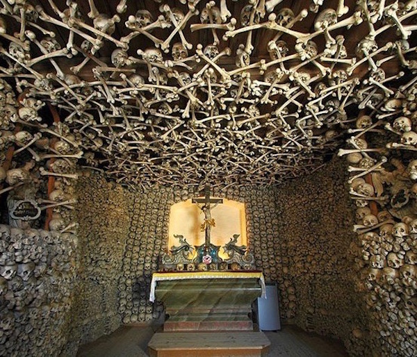
Last lesson began with spectacular images from Master Raymond’s hidden ossuary. This lesson, we start with images of a world famous ossuary located in Kutná Hora, Czech Republic. Known world-wide as the Bone Church, it is an ossuary (‘Kostnice’ in Czech) that houses the bones of an estimated 40,000 – 70,000 people (Image A).

bone church 01 KLS edited

Image A

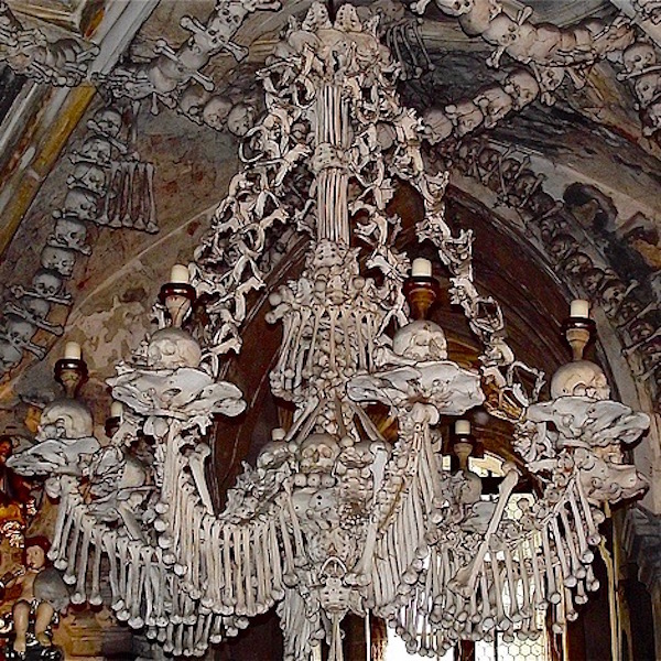
Here, bones are imaginatively and artistically arranged throughout the edifice. One of the most famous displays is a chandelier, purportedly containing every bone of the adult human body (Image B).

During the thirty years’ war, aristocracy of Central Europe wished to be buried in the hallowed graveyard at this site. As the number of burials outgrew available space, remains were exhumed and stored in the chapel, then assembled into artistic presentations.

The ruffled collars beneath the candle-bearing skulls are hip bones! At the tip-top are inverted sacra (pl.). Dangling bones are humeri (pl.). Quite the room-lighter! Makes Master Raymond’s animal skulls seem almost quaint!

bone church 02 KLS edited

Image B

Surely, these images put us in the mood to learn more about bones, although this lesson covers  bones of the living. Today’s lesson will review and present more bone anatomy and types of bone cells. Then, we will cover events that ensue when bones go snap, crackle, or pop, as well as mechanisms of healing. Let’s go!

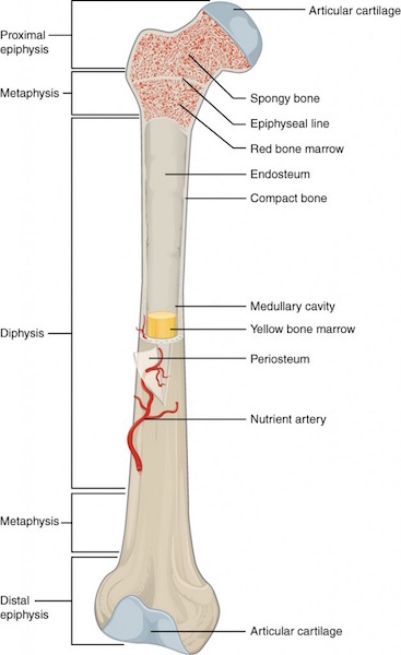
Review of Long Bone Anatomy: Lesson #39 presented long bone anatomy, but let’s take a moment to review. Once again, the femur, is our model (Image C). The shaft of a long bone is the diaphysis. The ends that form joints with other bones are the epiphyses (pl.); these are covered with articular cartilage (ball-shaped epiphysis is head of femur). The flared region between diaphysis and epiphysis is the metaphysis. Cortical bone forms a hard outer rind enclosing the marrow (medullary) cavity. Spongy bone fills epiphyses and metaphyses and is also scattered in the marrow cavity. Depending on the bone and age of the individual, the marrow cavity is filled with either fat cells or hematopoietic (blood-forming) cells, neither of which are elements of bone. Think of them as tenants not owners!

long bone parts 01 KLS edited

Image C

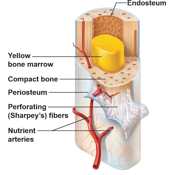
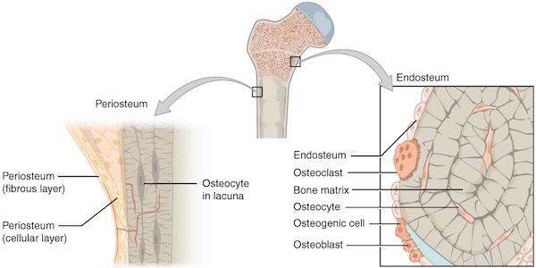
Periosteum and Endosteum: Periosteum is a thick fibrous layer covering the diaphyseal surface (Image D). Endosteum is a thin connective tissue layer lining the bone marrow cavity and covering all exposed surfaces of spongy bone. Both layers contain blood vessels and several types of bone cells as discussed below.

periosteum & endosteum KLS edited

Image D

Vascular Supply: As we learned in Anatomy Lesson #39 “Dem Bones – The Human Skeleton,” bone is living tissue and thus requires a constant blood supply such that the human skeleton receives 5-10% of the entire cardiac output (amount of blood the heart pumps per minute).

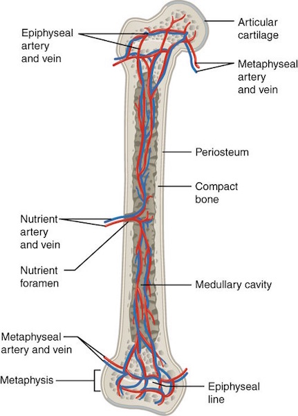
To meet this demand, large bones receive several arteries (Image E). Periosteal arteries supply periosteum and outer compact bone. Epiphyseal arteries supply blood to epiphyses (pl.). Nutrient arteries supply inner cortical bone, endosteum, and help supply epiphyses and metaphyses.

These arteries access a bone via foramina (channels) that traverse compact bone to reach their respective turfs, then break into capillary beds where oxygen and nutrients are exchanged for carbon dioxide and waste products. Corresponding veins form and blood flows back to the heart. Round and round and round it goes…..

Nutrient artery femur KLS edited

Image E

Nerve Supply: You may be surprised to learn that mineralized bone (organic matrix plus inorganic minerals – see Lesson #39) does not have pain receptors. Hum…if this is true, then why does a broken bone hurt so badly? Because periosteum and endosteum as well as articular surfaces are richly supplied with pain receptors (nociceptors). If these structures are compromised as with a fracture, then pain is severe!

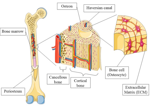
Image F nicely illustrates nerve and blood vessel distribution in cortical bone and margin of the marrow cavity. Anatomy typically color codes arteries red, veins blue, and nerves yellow. The tiny yellow lines indicate that nerves follow blood vessels through cortical bone to the endosteum. Although not shown in Image D, nerves also follow periosteal arteries to innervate the periosteum.

bone nerves KLS edited

Image F

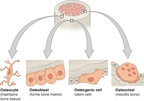
Bone Cells: As mentioned above, bone contains several cell types including osteogenic cells, osteoblasts, osteocytes, and osteoclasts (Image G). These cells are located in periosteum, endosteum, or within mineralized bone. Again, although hematopoietic cells or fat cells fill spaces between spongy bone and within marrow cavities, these are not bone cells.

Osteogenic cells are stem or parent cells giving rise to osteoblasts (Image G); they reside in periosteum and endosteum.

Osteoblasts: From Greek osteo- meaning bone + blastanō meaning to germinate, osteoblasts produce collagen and other proteins as part of the organic matrix. These proteins are released into the extra-cellular environment where minerals deposit around them. Together, the organic matrix provides tensile strength and inorganic minerals provide compressive strength. As to their bulk, organic proteins represent 10% of bone mass with the remaining 90% being inorganic mineral. Osteoblasts reside in periosteum and endosteum.

Osteocytes: In the course of production and secretion of organic matrix and its subsequent mineralization, osteoblasts become “imprisoned” within bone and are renamed osteocytes. Located in compact and spongy bone, osteocytes help maintain the mineralized bone. Further, the mineralized matrix with imprisoned osteocytes is organized into cylinders of bone known as osteons (Haversian systems). A bone such as the femur contains millions of osteons but details about these units must await a future lesson.

Osteoclasts: Giant, multi-nucleated cells, the osteoclasts, are formed by the fusion of macrophages (Anatomy Lesson # 37, “Outlander Owies Part 3 – Mars and Scars”). Found in endosteum, their duty is to dissolve bone.

Print

Image G

As you might imagine, a good deal of coordination among the various bone cells is required to ensure homeostasis (balance) between bone production, maintenance, and resorption. For example, as a bone grows in circumference due to periosteal osteoblast activity, osteoclasts of the endosteum remove bone from the inside so the thickness remains fairly constant, a highly regulated process.

Image H shows in detail the distribution of bone cells in periosteum, endosteum and in compact bone.

Print

Image H

Next, let’s do something really daring and look at a microscopic image of bone cells as viewed through a light microscope (Anatomy Lesson #34 – “The Amazing Saga of Human Anatomy”). Please understand that microscopic anatomy, better known as histology (Greek histo- meaning “tissue” + ology meaning “to know”), is a challenge for most students. Watch this fun video by Harvard medical students as they lament about studying “histo!”

https://www.instagram.com/p/BEYogsIuTPz/

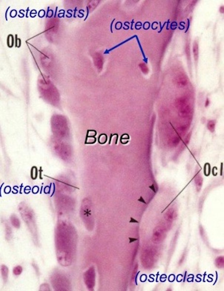
Ha ha! Very clever, medical students! But, you readers might respond the same way looking at a magnified image of a shard of spongy bone (Image I). This image is of a very thin slice of bone that has been stained blue and pink with H&E (hematoxylin = blue + eosin = pink). The so-called H&E stain is the most commonly-used histo stain in the US.

So, what do we see in Image I? The dense pink vertical band is a spicule of ossified spongy bone. The oval cells with pale nuclei along the left border of the shard are osteoblasts laying down a pale layer of unmineralized organic matrix (osteoid). The right border of the spicule show two large multi-nucleated osteoclasts busily dissolving bone – one such interface is a distinct divot indicated by the black arrowheads. Such divots are termed Howship’s lacunae (named for John Howship, a British anatomist). Several irregularly-shaped nuclei are scattered within the bony spicule – these are osteocytes encased in ossified bone.

That’s pretty much it! Being competent at recognizing cells and extracellular substance is based on pattern recognition. Looking at many examples of a given tissue is very useful in obtaining said competence. If you think this is impossible, understand that anatomical pathologists make their living looking at and diagnosing diseases from thousands of such tissue slides.

Osteoblast 02 KLS edited

Image I

Bone Fractures: Time for snap, crackle, and pop, and I dinna mean Rice Krispies! Compressive strength from mineral deposition and tensile strength from organic matrix give normal bones the ability to behave elastically. If trauma overcomes this elasticity, then bones fracture!

Bone fractures fall into two major categories: mechanical bone fractures caused by high force impact or stress, and pathological fractures caused by disease.

Pathological Fractures: Diana presents a fabulous case of fracture caused by disease in Colum. The Laird of clan MacKenzie is haunted by the aftermath of a pathologic fracture and accompanying deformities due to Toulouse-Lautrec syndrome (Starz episode 208, The Fox’s Lair).

Outlander book informs readers that at 18 y.o., Colum took a bad fall breaking the long bone (femur) of his thigh that subsequently mended poorly. Other skeletal anomalies quickly ensued. Diana explains in Dragonfly in Amber:

Legs crippled and twisted by a deforming disease, Colum no longer led his clan into battle…

…He glanced dispassionately down at the bowed and twisted legs. In a hundred years’ time, they would call this disease after its most famous sufferer—the Toulouse-Lautrec syndrome.

Read Anatomy Lesson #27, “Colum’s Legs and Other Things too!” for details about this rare syndrome (1.7 per million births). Of course, there are also many other diseases leading to pathological fractures. Nowadays, the most common in the US is osteoporosis.

Colum legs 01KLS edited

Mechanical fractures: High force impact or stress cause bones to snap. Different types of bones (e.g. short vs. long) often exhibit characteristic fractures. This lesson will consider fractures of long bones using the femur again as our example. Fractures are generalized into six different categories:

  • Closed fracture – bone is broken but overlying skin is unbroken
  • Open fracture – better known as a compound fracture, broken bone pierces the skin
  • Simple fracture – bone is broken but no other tissues damaged
  • Complex fracture – sharp fracture edges damage surrounding soft tissues
  • Complete fracture – bone breaks completely across the shaft
  • Incomplete fracture – bone is partly broken but remains in one piece

Then, fractures are named according to features of the break (Image J).

  • Normal – left image is the normal femur
  • Transverse – break crosses the shaft horizontally
  • Oblique – break crosses shaft at an angle
  • Spiral – torque on the broken halves twists these in opposite directions
  • Comminuated – bone is broken into four or more pieces (including main body of the bone)
  • Avulsion – a piece of bone is torn away usually by pull of a muscle tendon
  • Impacted – bone parts are driven together, also known as a buckle, or compression fracture
  • Fissure – crack along one side of the shaft
  • Greenstick – incomplete break through the shaft. Analogous to breaking a stick of green wood.

bone fractures 01 KLS edited

Image J

Fracture Healing: Fracture healing is the process by which the body repairs a broken bone and what a process it is! Several overlapping steps occur, different descriptors are used, and times may vary depending on the bone, but the general sequence is (Image K):

  1. Hematoma Formation (days 1-5): Bone fractures tear blood vessels, and periosteum and endosteum. Blood pours from the damaged vessels forming a hematoma (blood clot) between the fractured surfaces. Blood flow in intact vessels increases such that an army of white cells are delivered for defense and repair.
  1. Soft Callus Formation (days 3-40): Gradually, a bridge of cartilage and collagen replaces the hematoma and unites the broken ends. The cartilage bridge is made by chondroblasts (cartilage-generating cells) and is termed a soft callus.
  2. Hard Callus Formation (days 30-80): During this stage, cartilage of the soft callus is gradually replaced with new, woven bone (cartilage does not change into bone) and the site is renamed a hard callus. But, woven bone is immature and not as strong as mature compact bone. Mild exercise is often resumed during this phase.
  3. Bone Remodeling (days 80-100): If all goes well, the hard callus is gradually replaced with normal, mineralized compact bone that remodels to align with muscle pull and mechanical stress. Exercise promotes this process which is typically 80% complete within three months. But, depending on the bone and the fracture, step four can take up to 18 months.

Fracture healing 01 KLS edited

Image K

Normal fracture healing requires the following:

  • Adequate blood supply: Oxygen and nutrients must be delivered to the repair site and carbon dioxide and waste products removed; these processes require blood vessels. Thus, re-vascularization occurs at the fracture site.
  • Fracture stabilization: Excessive movement of the fractured bones interrupts callus formation so fractures are immobilized with a variety of ingenious devices such as casts, rods, plates, screws, and external fixators.
  • Adequate nutrition: On average, a fracture heals by 12  weeks. However, healing times depend on which bones are broken, severity of the breaks, age (younger folks usually heal faster), and nutrition. A vitamin sandwich (Image L) looks funny but animal and human studies show that fracture healing improves with supplements of Vitamins C, D, and E, as well as calcium and amino acids. Details of these experiments exceed our lesson goals, but understand that many average diets are marginal in these substances. So, be sure to consult with your physician about supplements while healing a bone fracture.
  • Rehabilitation: Immobilization of a fracture contributes to muscle wasting. Physical therapy can provide critical support in re-building muscle and encouraging proper bone alignment. This includes weight-bearing on a fracture after it has mostly healed in order to build bone strength.
  • No-No nicotine: Studies are pretty clear that nicotine in any form hinders the process of bone healing. Avoidance behavior!

Vitamin sandwich KLS edited

Image L

Now for some real “fun.” With Starz images, let’s use applied anatomy to consider mechanical bone fractures and healing. Starz Outlander episodes offer two excellent examples of bone fractures, both are compound in type.

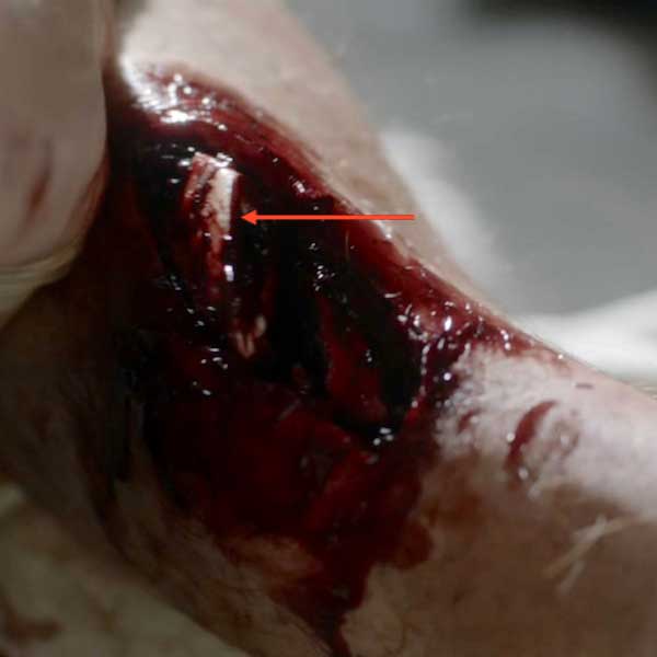
A workman is admitted to L’Hôpital des Anges in Paris (Starz episode 204, La Dame Blanche). He screams in agony because he has suffered a compound fracture of the right tibia (Anatomy Lesson #27, “Colum’s Legs and Other Things too!”). The skin is open and the upper part of the broken tibia is clearly visible at the red arrow.

This injury hurts like holy toothpicks because tears of periosteum and endosteum stimulate nociceptors (pain receptors). Then, muscles spasm as they attempt to stabilize bony fragments, upping the pain meter. And, adding insult to injury, a compound fracture rents the skin and we all ken how sensitive skin is to pain! All around, an agonizing situation.

ep-204-compound-fracture-05

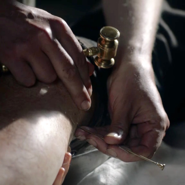
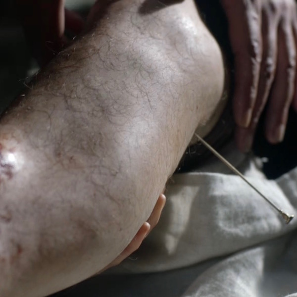
So, what does a good hangman do to ease his patient’s discomfort? Why, he hammers a nail into the side of the poor fellow’s knee, of course! What else? Presumably a type of primitive acupuncture, it momentarily halts the patient’s agony; mayhap from PTSD?

A quote from Dragonfly in Amber:

Monsieur Forez was at work today. The patient, a young workman, lay white-faced and gasping on a pallet. …The leg, though, was something else… Sharp bone fragments protruded through the skin … “Here, ma soeur ,” he directed, taking hold of the patient’s ankle. “Grasp it tightly just behind the heel… Monsieur Forez brought the point of the brass pin to bear …he drove the pin straight into the leg with one blow. The leg twitched violently, then seemed to relax into limpness.

ep 204 compound fracture 03 KLS edited

Monsieur Forez enlightens Claire regarding this startling exhibit of bedside manner (from Dragonfly in Amber):

“There is a large bundle of nerve endings there, Sister, what I have heard the anatomists call a plexus. If you are fortunate enough to pierce it directly, it numbs a great deal of the sensations in the lower extremity.”

Stop! I must pick at a wee bone: During the episode, Mr. Forez explains to Claire that anatomists describe a nerve at the inside of the knee which, if pierced, briefly anesthetizes the leg (Anatomy Lesson #27 – “Colum’s Legs and Other Things too!”). So, the human grease-guy whacks a 4” nail into the side of the workman’s knee for pain relief!

So this is what troubles me: the nail pierces skin near the saphenous nerve (a branch of the femoral) which, unfortunately, supplies only skin of the area and does nothing for bone pain. The correct nerve to pierce would be the tibial nerve, a branch of the sciatic; but, both of these nerves are in the back of the thigh! From the thigh, the tibial nerve sends fibers to the tibia in concert with its nutrient artery where it innervates endosteum and periosteum. Ergo, for this technique to work, the good Monsieur must pierce the tibial nerve in the back of the thigh.

I canna vouch for the efficacy of nail piercing, which seems a wee bit harsh, but I can question Forez’s anatomists and their data base. Soon after the shock of piercing subsides, the patient resumes screaming so I canna think it verra efficacious. Gah! Back to the dissection lab for those anatomists! But, I must say, the special effects are superb!

ep 204 compound fracture 04 KLS edited

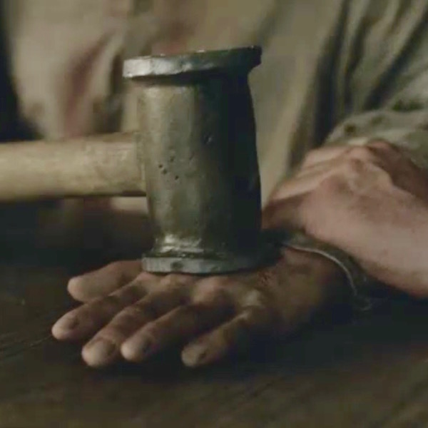
Next, we will consider another compound fracture, arguably the most infamous of the Starz series. The Wentworth Smack-Down delivered high force impact causing mechanical fractures of Jamie’s left hand bones (Starz episode 115, Wentworth Prison). Let’s do a walk through of these fractures.

ep 115 Mallet blow KLS edited

Heading to Outlander book, Diana enlightens us with these quotes:

Luckily the thumb had suffered least; only a simple fracture of the first joint. That would heal clean. The second knuckle on the fourth finger was completely gone; I felt only a pulpy grating of bone chips when I rolled it gently between my own thumb and forefinger…

The compound fracture of the middle finger was the worst to contemplate. The finger would have to be pulled straight, drawing the protruding bone back through the torn flesh. I had seen this done before—under general anesthesia, with the guidance of X rays.

We can easily deduce from Diana’s description that, at a minimum, Jamie’s hand suffered:

  • Simple fracture of the first phalanx of thumb.
  • Communiated fracture of the ring finger (4th finger in English system; 3rd in US system – see Anatomy Lesson #22, “Jamie’s Hand – Symbol of Sacrifice”) – proximal and middle phalanges smashed into bony bits.
  • Compound fracture of middle finger – appears to be middle phalanx of the middle finger.

ep 116 Jamie hand 02 KLS edited

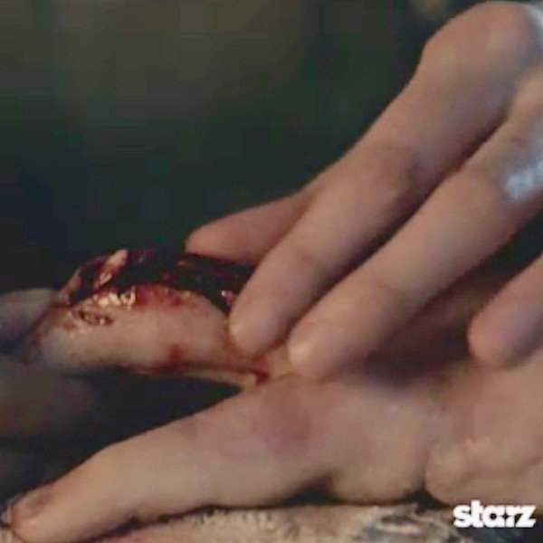
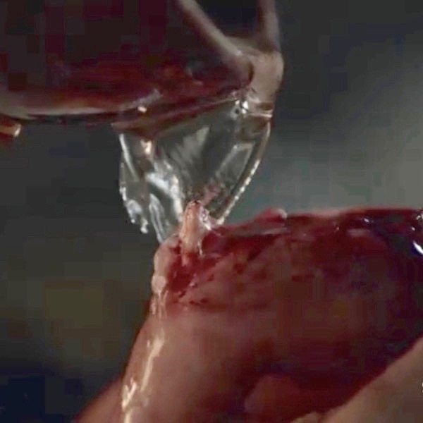
Like any combat nurse worth her salt, Claire cleanses Jamie’s wounds. She has no modern isotonic fluids or antibiotics, so she cleans his compound fracture with (no doubt) sterile water, perhaps containing a wee dram of alcohol for sterilization?

ep 116 Jamie hand 01 KLS edited

Next, Claire ponders the damage and then re-aligns and resets the bony fragments (Starz episode 116, To Ransom a Man’s Soul). And again from Outlander book:

I began to lose myself in the concentration of the job… deciding how best to draw the smashed bones back into alignment.

ep 116 Jamie hand 03 KLS edited

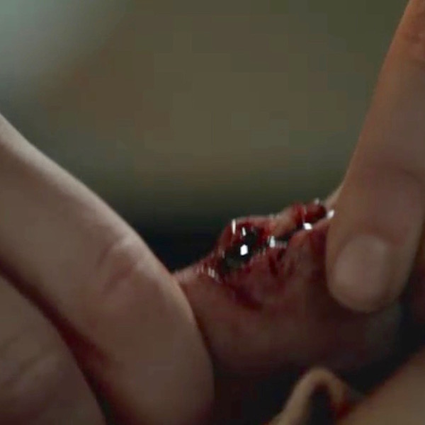
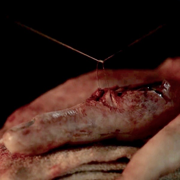
Now, as we learned above, a compound fracture tears surrounding flesh and skin. So Claire carefully closes the wounds with sutures (Starz S.2, introductory image) to exclude pathogens and reduce blood loss.

Opening creds Jamie hand 05 KLS edited

Earlier in this lesson, we read that movement of a new fracture interferes with callus formation. So, various devices are used to immobilize fractures as they heal.

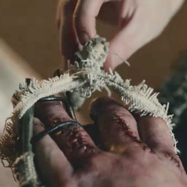
Ever resourceful Claire (or the monks?) devises an external fixator for Jamie’s smashed hand in the form if a very clever wire/leather, linen, and wood device! External fixation is a surgical treatment used to stabilize bone and soft tissues at a distance from the injury. So, check out the amazing invention (Starz episode 116, To Ransom A Man’s Soul) to immobilize Jamie’s hand! The middle finger is reset and stitched. The communiated and most severely wounded ring finger is stitched and splinted against the small finger. Brava, Madam Sassenach!

ep 116 Jamie hand 06 KLS edited

Jamie’s hand care is not yet complete! Claire provides physical therapy in the form a rag ball she made. Squeezing the ball helps Jamie regain hand mobility and strengthen the knitting bones as he and Murtagh plan how best to skewer BJR (Starz episode 203, Useful Occupations and Deceptions)!

In case you forgot from earlier in this lesson, exercising a repairing bony callus augments the ossification process, so Claire’s rehabilitation plan is spot on!

Two quotes from Diana’s Dragonfly in Amber:

It really wasn’t too bad; a couple of fingers set slightly askew, a thick scar down the length of the middle finger. The only major damage had been to the fourth finger, which stuck out stiffly, its second joint so badly crushed that the healing had fused two finger bones together. The hand had been broken in Wentworth Prison, less than four months ago, by Jack Randall.

He had regained an astounding degree of movement, I thought. He still carried the soft ball of rags I had made for him, squeezing it unobtrusively hundreds of times a day as he went about his business. And if the knitting bones hurt him, he never complained.

ep 203 Jamie hand KLS edited

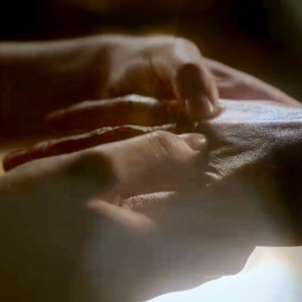
And, pithy quotes from Diana’s Dragonfly in Amber remind us that months later, Jamie’s hand is still not whole (Starz Season 2 opening images), so clever caring Claire applies her personal version of massage therapy… a task that she and he undoubtedly relish! Hee, hee.

I crawled in beside him and took up his right hand, resuming my slow massage of his fingers and palm. He gave a long sigh, almost a groan, as I rubbed a thumb in firm circles over the pads at the base of his fingers.

Opening creds Jamie hand 07 KLS edited

Thus ends Outlander compound fractures and healing. Whether bone breaks or skeletons, people are endlessly fascinated by bones and the stories they tell. Book readers will recognize this poignant commentary from Dragonfly in Amber as Jamie and Claire find the bony inhabitants of an unknown cave in France.

He turned again then to the two skeletons, entwined at our feet. He crouched over them, tracing the line of the bones with a gentle finger, careful not to touch the ivory surface. “See how they lie,” he said. “They didna fall here, and no one laid out their bodies. They lay down themselves.” His hand glided above the long arm bones of the larger skeleton, a dark shadow fluttering like a large moth as it crossed the jackstraw pile of ribs.

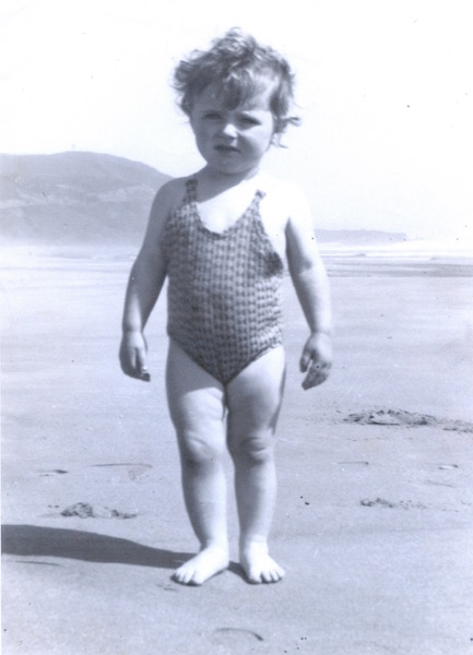
Now, I have my own bone story to share. The following is a true event underscoring the amazing capacity of bones to heal. During WW II, my father was a welder of Liberty Ships in California. When I was 18 months of age, neighborhood kids accidentally ran over me and the trauma snapped off the head (ball-shaped epiphysis) of my right femur. Local physicians were unable to help so they sent us to the Treasure Island Naval Base where many of the best US physicians were stationed to assist with the war effort.

Those docs had never set a child’s femoral head before, but they courageously reset my leg, placed me in a 3/4 body cast, and prepared my parents for the bad news: the blood supply to the femoral head was likely severed, the femoral head would become necrotic (die), and the hip would freeze into an immobile joint. I would not walk normally, if at all.

Well, the good news is that my femoral head did not perish! Somehow and somewhere, an arterial supply survived. Although my colleagues and I share theories about which vessel might have remained intact, no one knows for sure.

Image M was taken in 1944 at Stinson Beach, CA, six months after the injury (like the artery, my appetite clearly remained intact <G>). Observe that the right leg is slightly rotated outward (externally), a classic stance following a femoral head fracture.

Today, the only residual is my right leg is 3/8” shorter than the left. Otherwise, nada! I am able to exercise vigorously and with no noticeable deficit. Blessings to those unknown healers who helped the wee bairn of a common laborer!

Karmen 1944 KLS edited

Image M

Our lesson on how fractures heal has come to an end. After two lessons about the skeleton, there remains much more to be studied, so we may revisit this topic at a later date.

A few last thoughts: Remember, bone is a dynamic tissue which constantly remodels itself throughout life. Individual bones are organs that collectively constitute the skeleton, our internal support system. Bone cells create, maintain, and destroy bone tissue to our benefit.

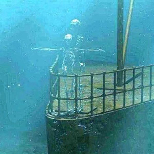
Bones have been prodded, examined, healed, revered, saved, feared, embellished, and cherished. Not all of this lesson is about happy stuff, so let’s end with this slightly irreverent image from the bow of the Titanic.

Titanic KLS edited

Image N

Dem bones, dem bones!

A deeply grateful,

Outlander Anatomist.

Photo creds: Starz, Outlander Anatomy archives (Image M), www.anatomy-medicine.com (Image F), www.askabiologist.edu (Image J), www.apps.carleton.edu (Image B) www.boneandspine.com (Image C), www.geneticliteracyproject.org (Image L), www.medcell.med.yale.edu (Image I), www.reddit.com (Image A), www.en.wikipedia.org (Image E; Image H), www.everythingfunny.org (Image N), www.commons.wikimedia.org (Image G) ,www.studyblue.com (Image D), www.ippe2ools.blogspot.com (Image K)