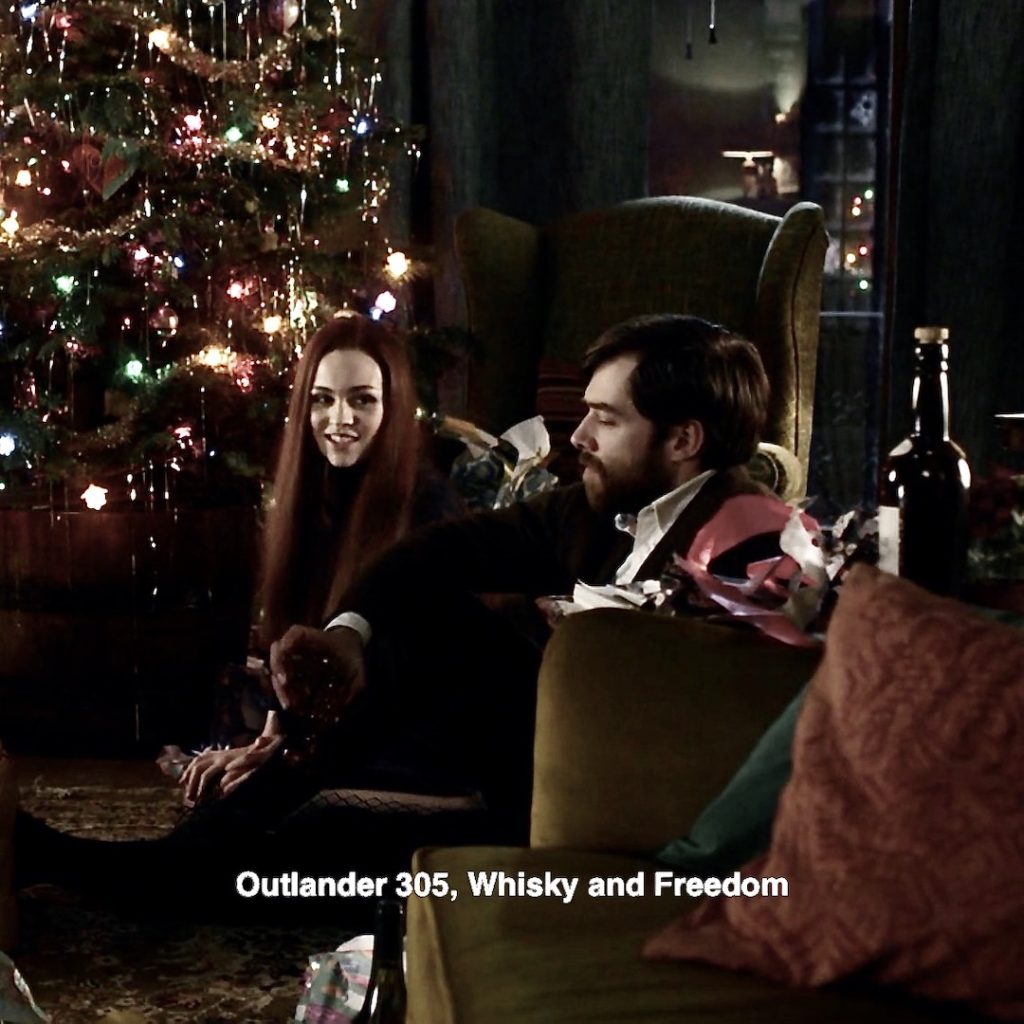
Welcome, Outlander fans and Anatomy students!
This Fun Fact documents my attempt to identify Easter Eggs in Claire’s “dreamscape” from Outlander episode 512, Never My Love. Doubtless, I have missed some, so please chime in with those you have found. And, no problem if you don’t agree with mine.
Each of my eggs is matched with events (mostly) from prior episodes. As nearly as possible, each Easter Egg is followed by an earlier appearance of the thing Claire imagines.
Recall, that during Claire’s horrific abuse, her mind retreats into a dream-like state wherein she cloaks herself with the many loves of her life. Her dreamscape, although distorted, is rooted in past memories.
Here we go!
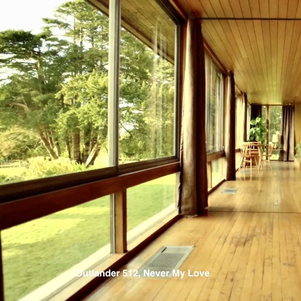
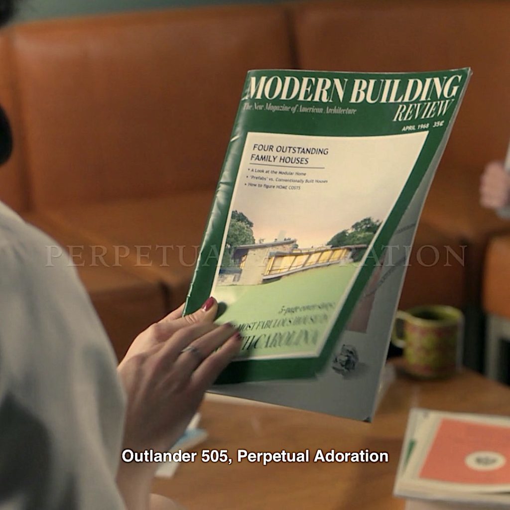
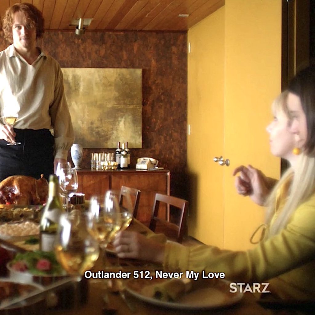
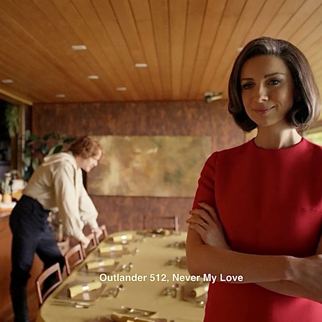
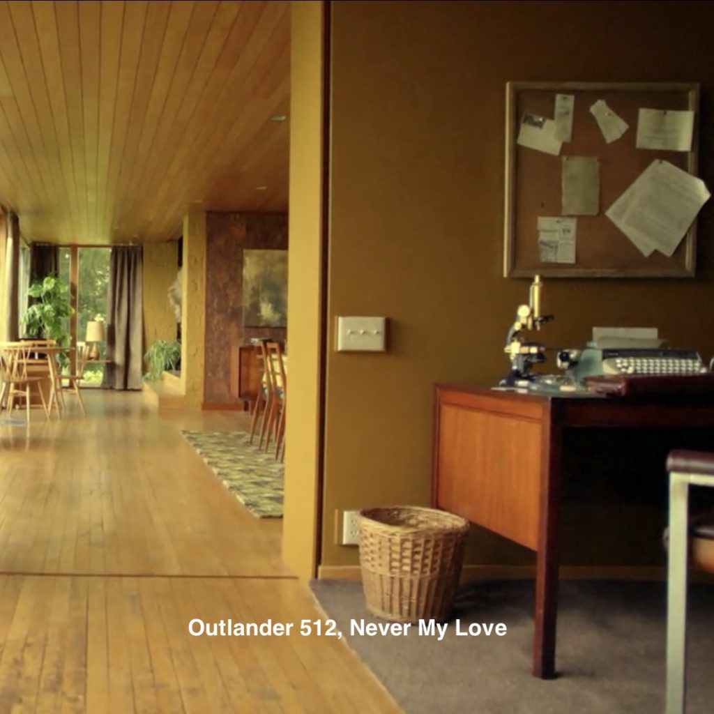
1. Modern House: Claire conjures a picture-perfect home; a real beauty and typical 60’s style, too!
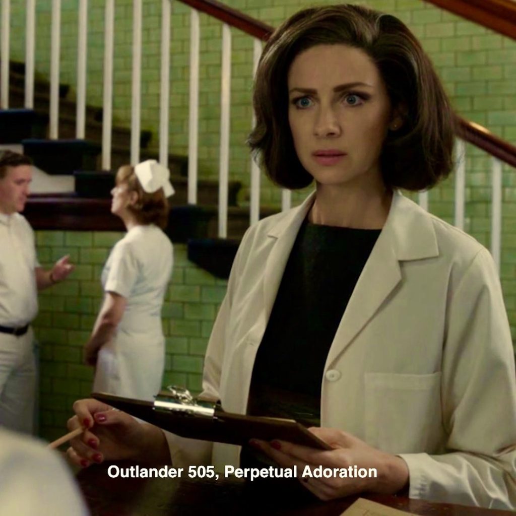
Episode 505: In the doctor’s lounge, Claire picks up Modern Building Review, a magazine featuring four outstanding family houses in North Carolina. Her imagined 60’s home appears on the cover! After a moment, she puts down the magazine in favor of the “Impetuous Pirate,” courtesy of Dr. Joe Abernethy! 😉
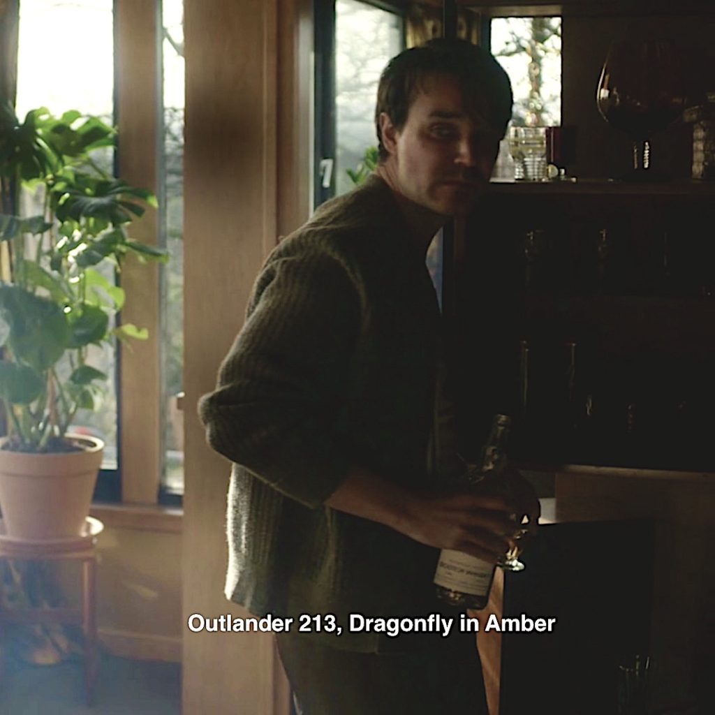
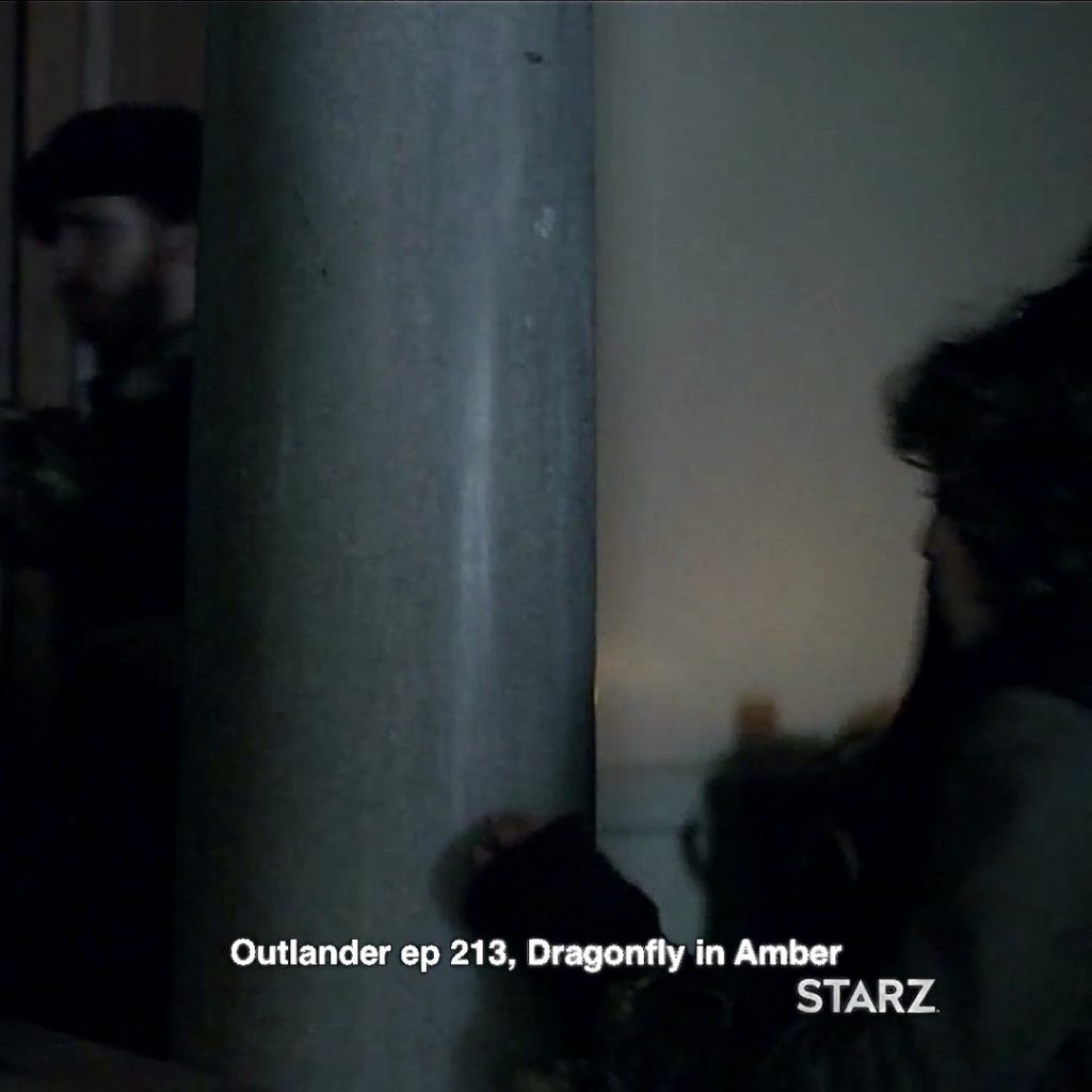
Episode 213: Poor Greg Edgars, Gillian’s (Geillis) ill-fated hubby. In a few hours, he will be toast at the standing stones. A few fans have seen a resemblance between Claire’s imagined 60’s home and the Edgars’ abode: similar wood, windows and plants!
2. Big House: Claire imagines her beloved refuge in the form of an abstract painting. Some fans see flames and smoke rising from the house, a hint to the obituary found by Roger and Bree? You know, the one that sent Bree back to the 18th Century to warn her parents? 🔥
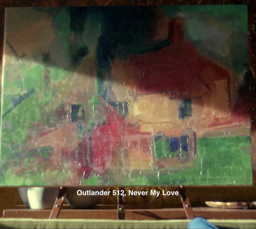
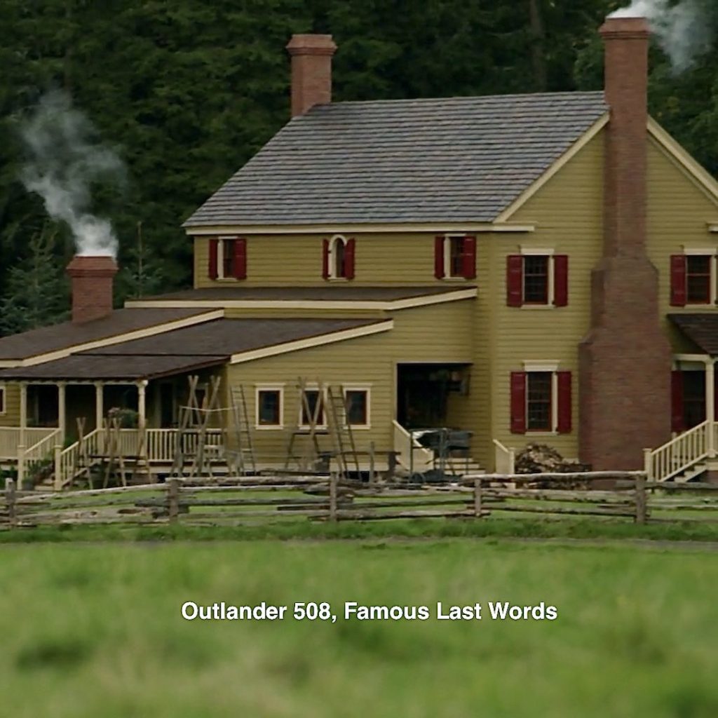
Episode 508: The Big House is safe and sound (for now) on Fraser’s Ridge. Magnificent!
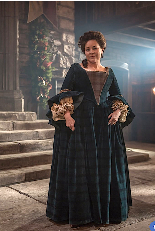
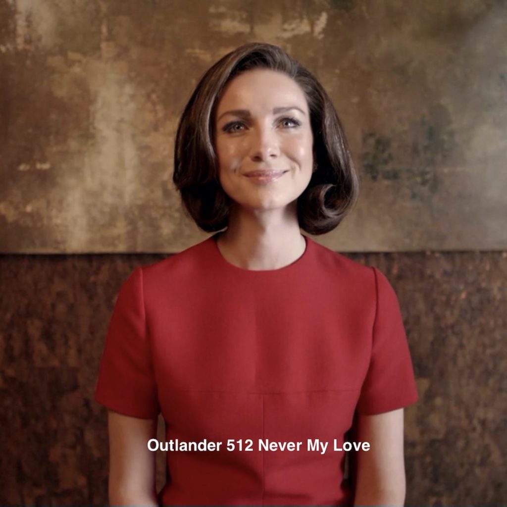
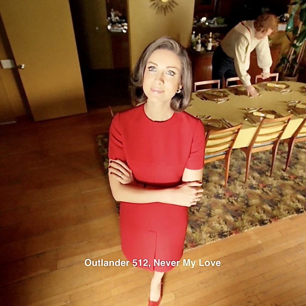
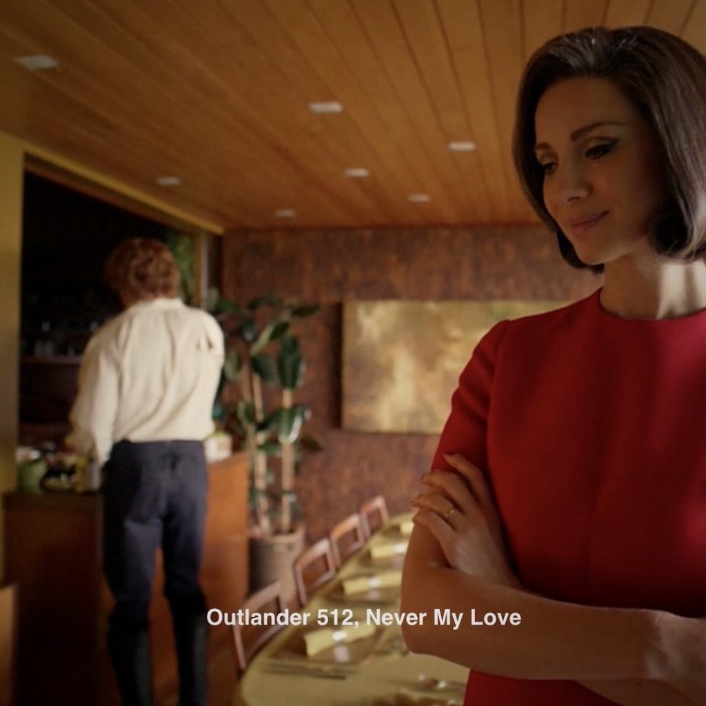
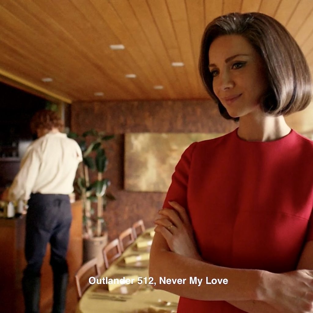
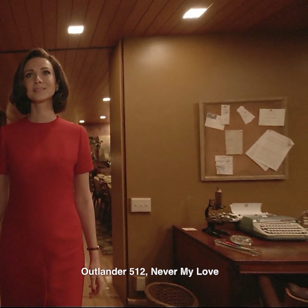
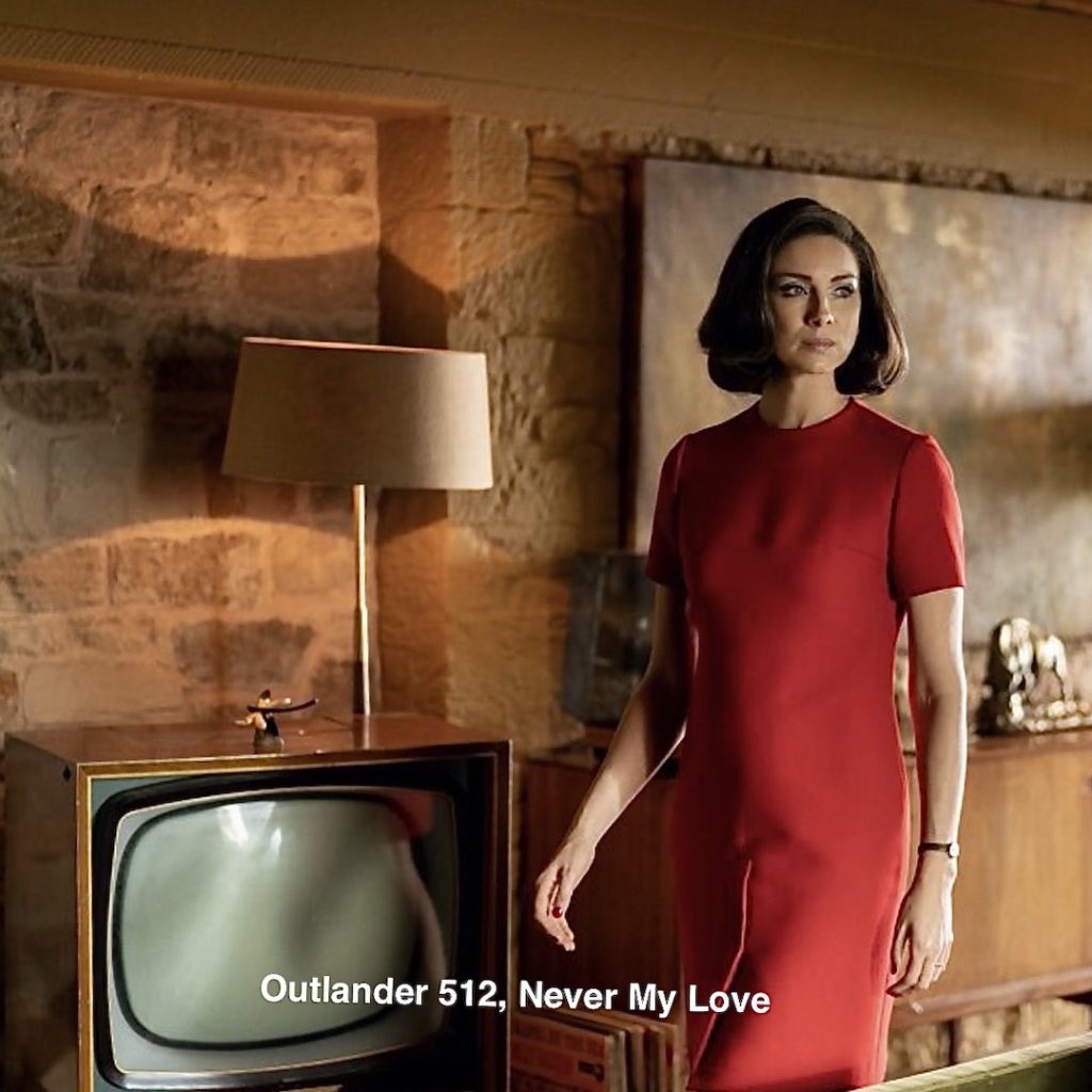
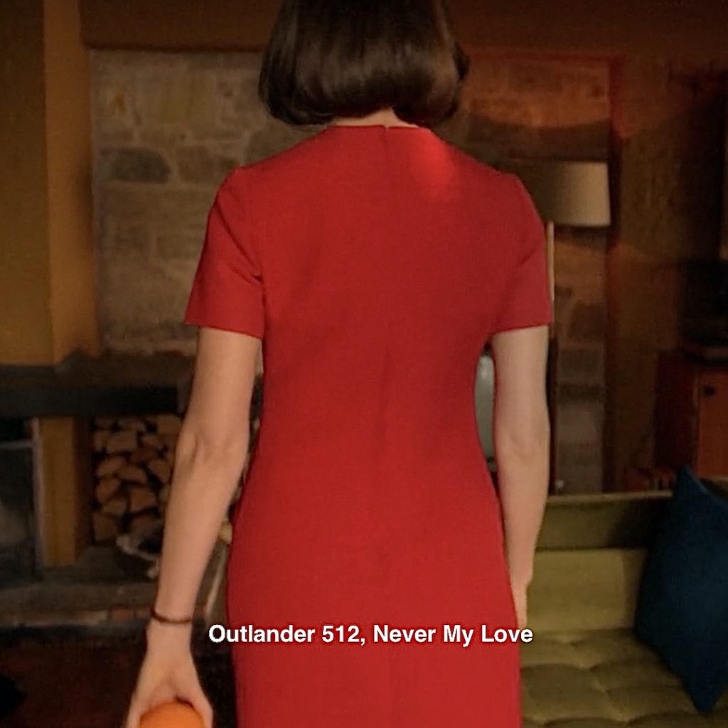
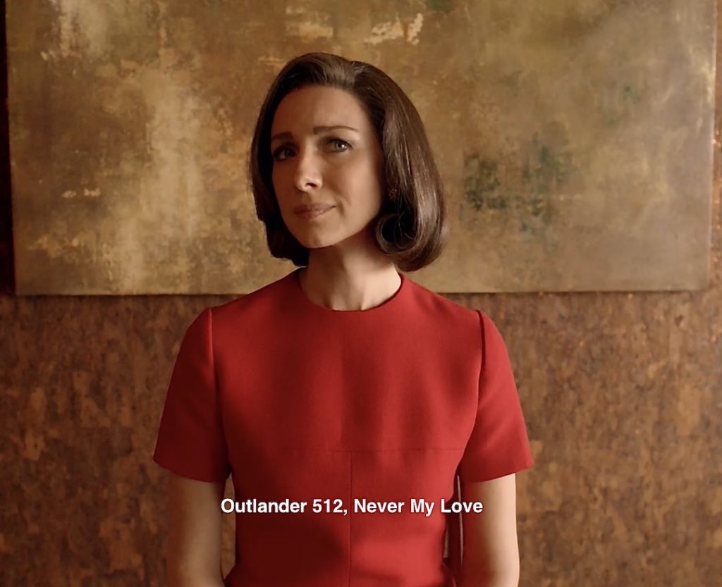
3. Red Dress: Claire outfits herself in a delightful Red Dress for the dreamscape! 😍
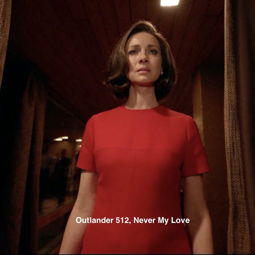
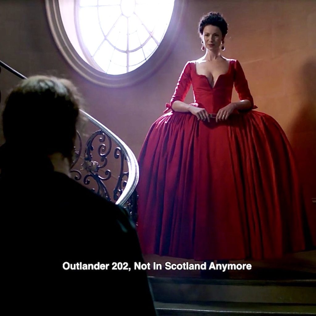
Episode 202: Who can forget Claire’s stunning red dress with plunging neckline? Weil, Jamie asks for a dress that will grab the French King’s attention. And, Claire gives it to him, in spades… or would that be in hearts?
A quote from Diana’s second big book, Dragonfly in Amber! (Louis notices!)
I had forgotten the red dress; His Majesty halted directly in front of me and bowed extravagantly, hand over his waist.
4. Red Shoes: Must have footwear! Claire’s strappy sandals along with Jo’s red boots and Murty’s red loafers. (psst…Who chose Godfather’s pants? Precursor of 70s leisure suits?) 😱
Episode 202: Oooooh! Red shoe porn! ❤️
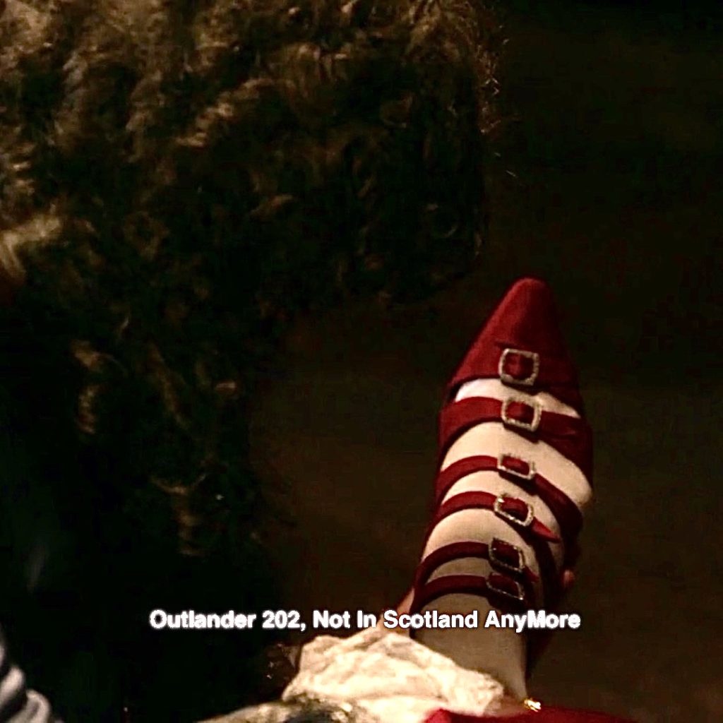
And, another infamous Red Shoe wearer!
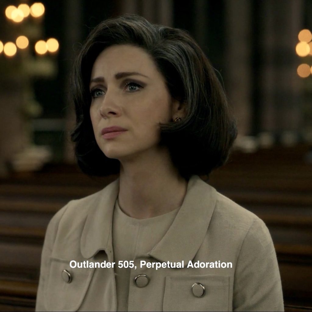
5. Claire’s Hair: Sassenach imagines herself with Jackie Kennedy hair, perfectly coiffed with Claire-ol to the rescue – nary a grey strand in sight! 😉
Episode 505: In reality, Claire has a few silver strands here and there!
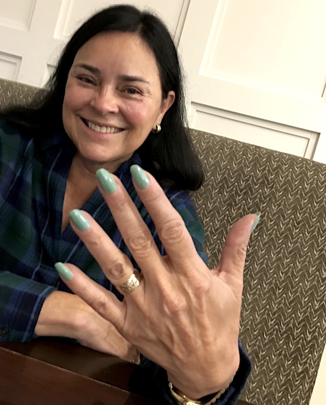
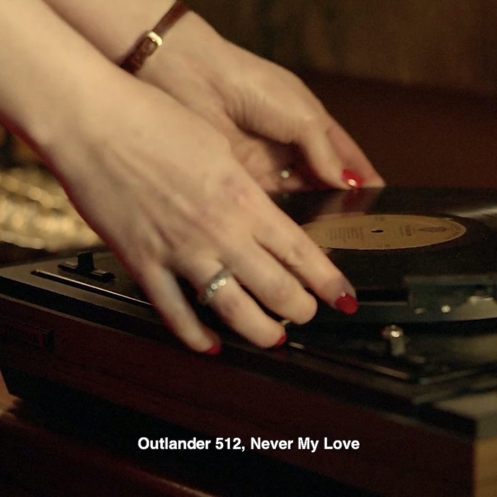
6. Red Nails: Claire grants herself a matching red manicure! 💅🏻
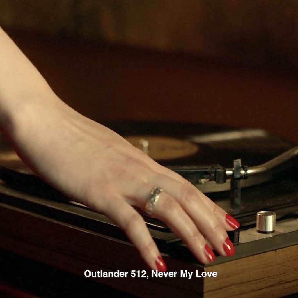
Episode 505: Sometimes Claire wears red nails, even at work? FYI, women in the healthcare field did not wear nail polish in those days, especially a surgeon! 😉
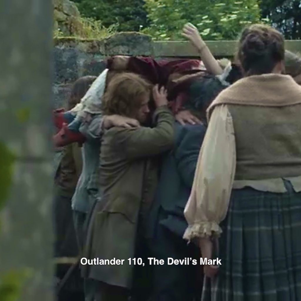
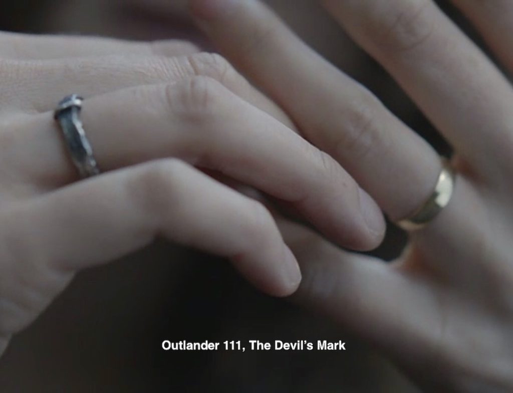
7. Rings: Claire’s dreamscape remains true to her rings; she wears both, just as she does in life. The lass takes her vows seriously! 💍
Episode 111: Claire takes a moment after the witch trial to check her rings. Yep, both are still there!
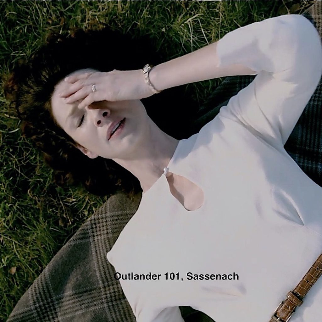
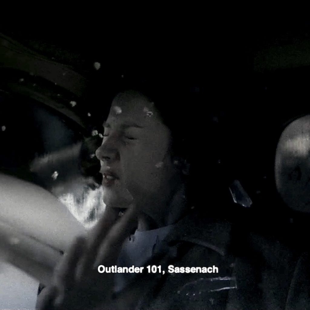
8. Watch: Claire imagines herself at the 60s house wearing a delicate wrist watch.
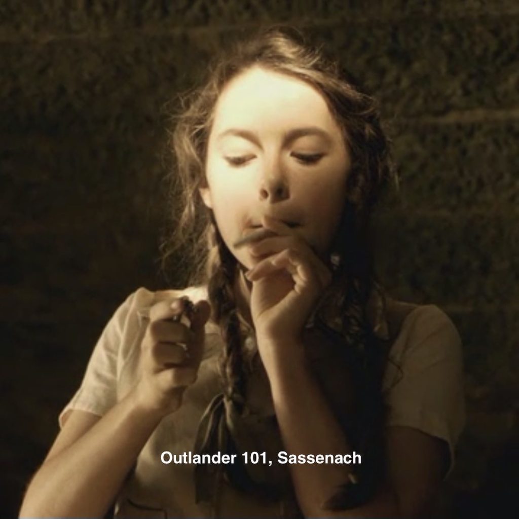
Episode 101: A callback to episode 101, as she rouses after traveling through the stones. The small wrist watch….. why didn’t the jewels inside cause it to burn up?
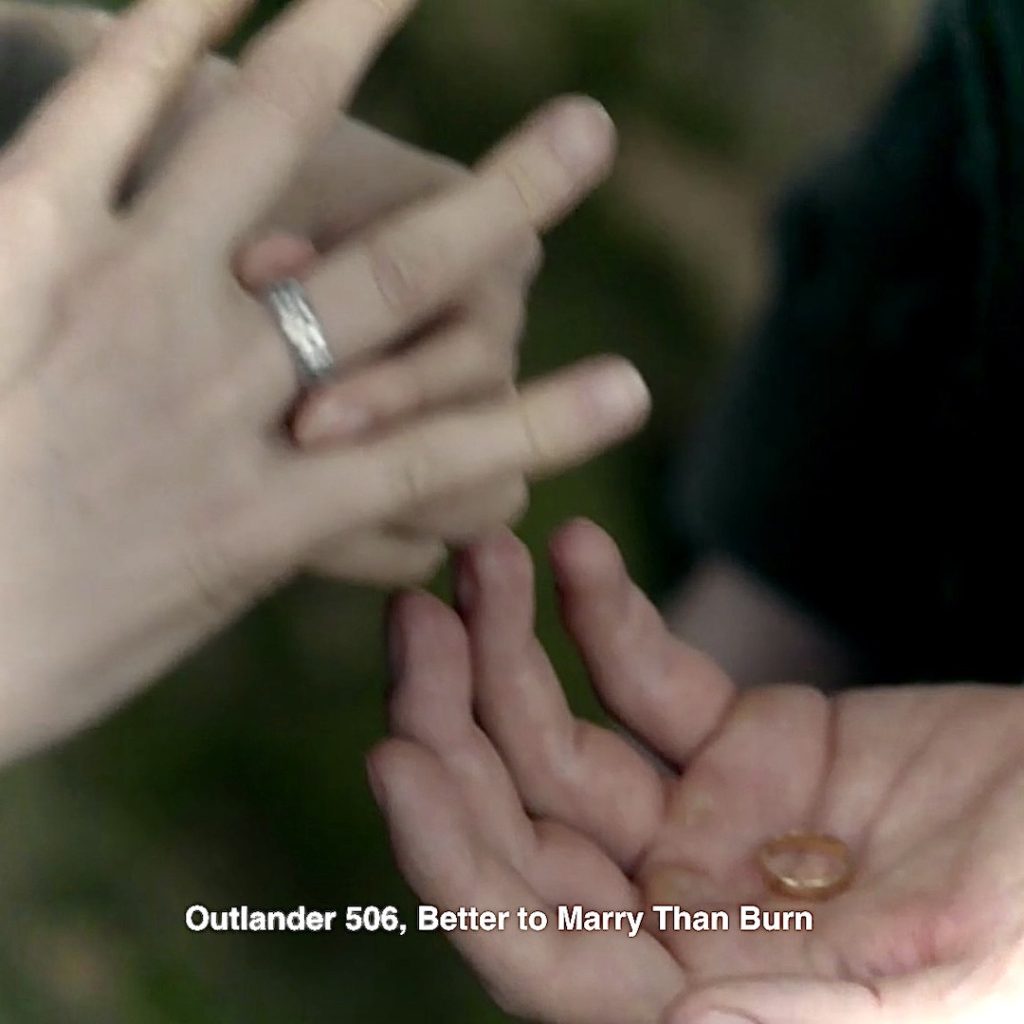
9. Rings: Episode 506: And, another ring callback (there are a bunch of these). This time, Jamie requires Claire’s rings as a stake against card shark, Phillip Wylie!
Herself explains in her fifth big book, The Fiery Cross book:
“The gold is worth more,” he said, and then, after the briefest hesitation, added, “in terms of money.”
“I know that.” I turned round to face him.
… “I meant—hadn’t you better take both of them?” My hands were cold and slippery with sweat; the gold ring came off easily; the silver was tighter, but I twisted it past my knuckle. I took his hand and dropped the two rings clinking into it.
Then I turned and walked away.
OK, Jamie Fraser! Take the rings, but I will never forgive you if you lose them to that handy-dandy fop! 😡
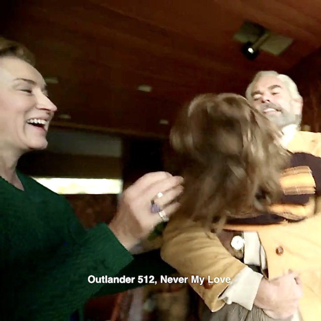
10. More Rings: At the turkey-table, Claire adds a wedding ring on Jocasta’s finger! 😃
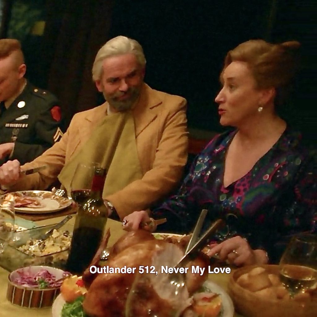
And, a ring for Godfather Murtagh. Seems she imagined a Murcasta wedding!
Episode 506: No doubt, Claire’s grey matter recalls Murtagh’s proposal of marriage to Jocasta on the day she is to wed Duncan Innes! 😢
Switching gears, how does Claire remember Himself!
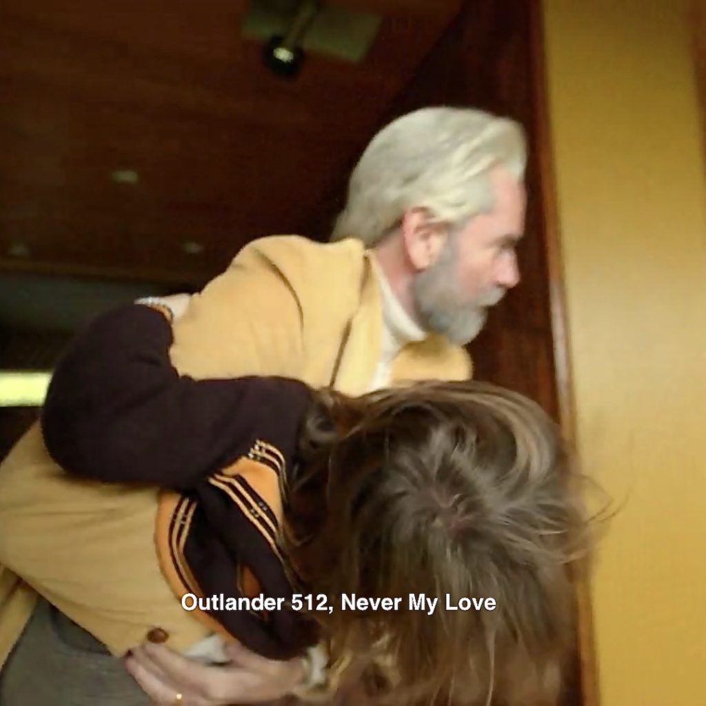
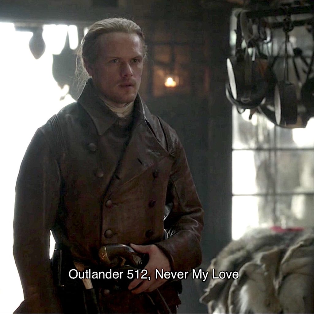
11. Jacket: Jamie arrives wearing a yummy belted, brown leather jacket! The lad looks fab in leather (or not 😉)!
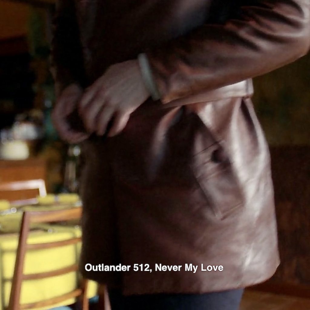
Episode 512: In the same episode, Jamie drops Lionel’s body before older bro, Richard Brown. Talk about a smackdown! 🤼♂️
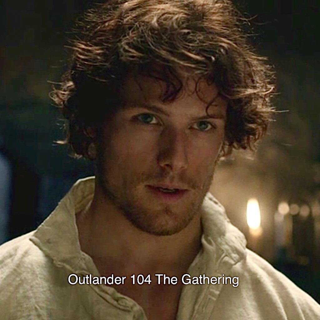
12. Jamie’s Hair: He arrives at their 60’s home sans brush or comb ! 😜
Episode 104: No doubt, Claire remembers Jamie’s “Je suis prest” hair. The lad cleans up verra well. We all love this one! 🥰
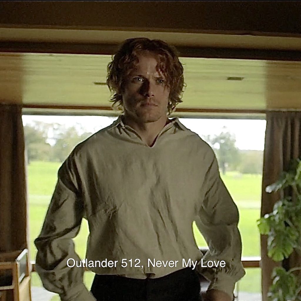
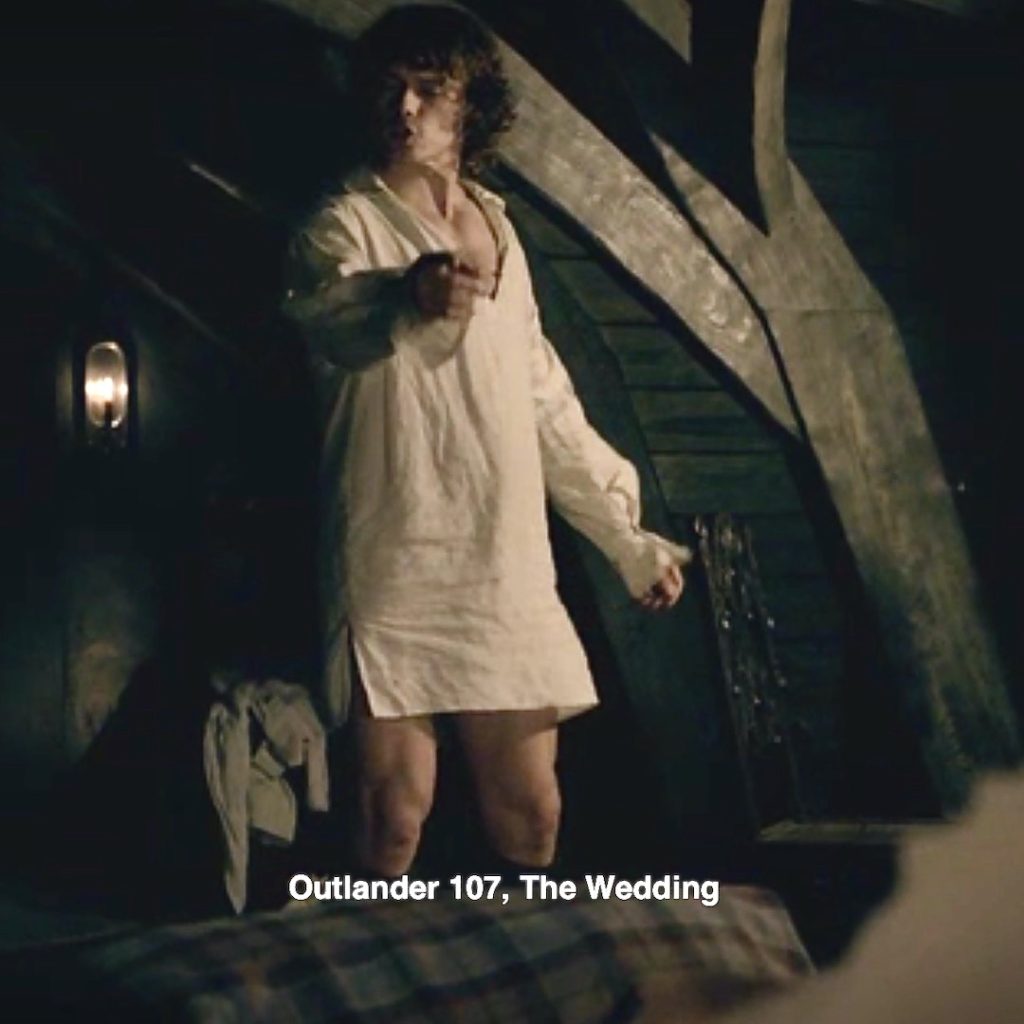
13. Jamie’s Sark: For non-book readers, Claire remembers Jamie’s shirt.
Episode 107: Behold, the sark, in all its glory! A quote from Voyager as Claire reflects on this remarkable garment: ❤️
He would not have worn his kilt—the wearing of all tartans had been outlawed after Culloden. Breeks, then, likely, and a linen shirt. I had made such sarks for him; I could feel the softness of the fabric in memory, the billowing length of the three full yards it took to make one, the long tails and full sleeves that let the Highland men drop their plaids and sleep or fight with a sark their only garment.
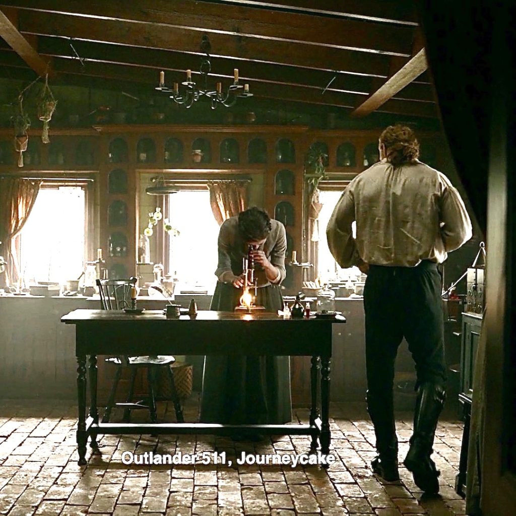
14. Jamie’s Breeks: Claire includes Jamie’s trousers, throw back to the 18th century, for sure!
Episode 511: Jamie’s 18th century breeks sport a wee tie and poofy rear-room, mayhap ease for riding a horse?
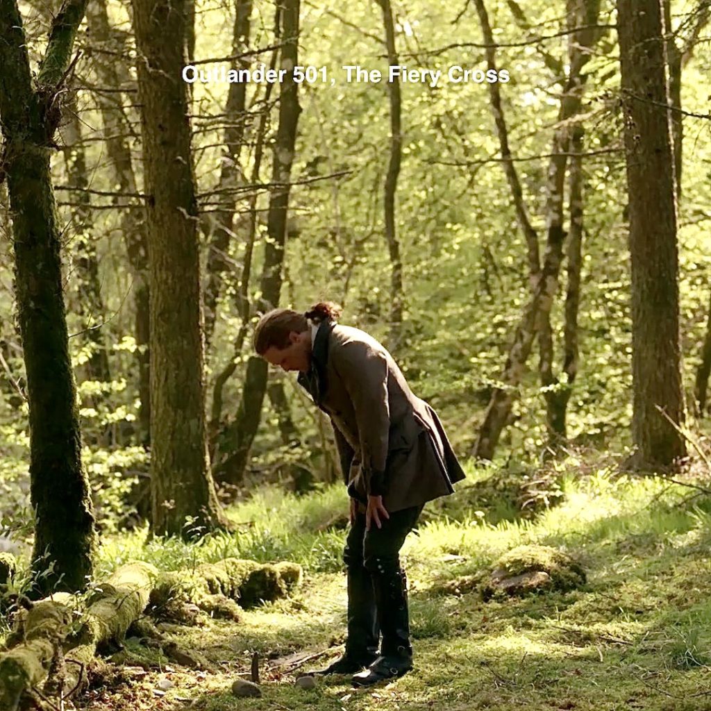
15. Jamie’s Boots: Claire approves of his footwear (As do we! 🥾)
Episode 501: Jamie boots are everywhere – here they are bathed with tears after telling Godfather, “be hard to find!” 😢
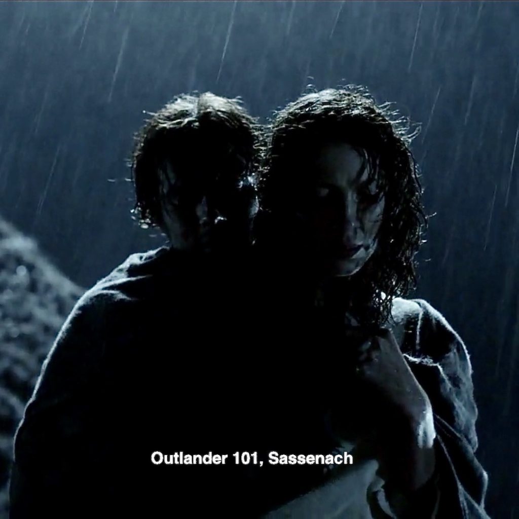
16. Plaid Wrap: Claire dreams she is swathed in the warmth and safety of Jamie’s plaid. She’s shaking so hard it’s rattling his molars! 🦷
Her first vision of the plaid imagines it a light brown, changing until finally it assumes the Full-Fraser plaid as her mind focuses!
Episode 101: We heard that waaaay back in the beginning, after Claire reduces Jamie’s dislocated shoulder. Brrrrr! 🌧🌧🌧
“You’re shaking so hard ye are making my teeth rattle!”
She doesn’t forget Ian!
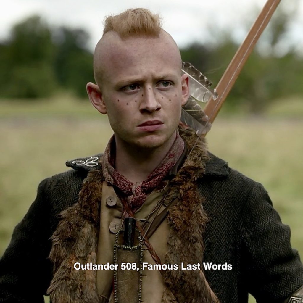
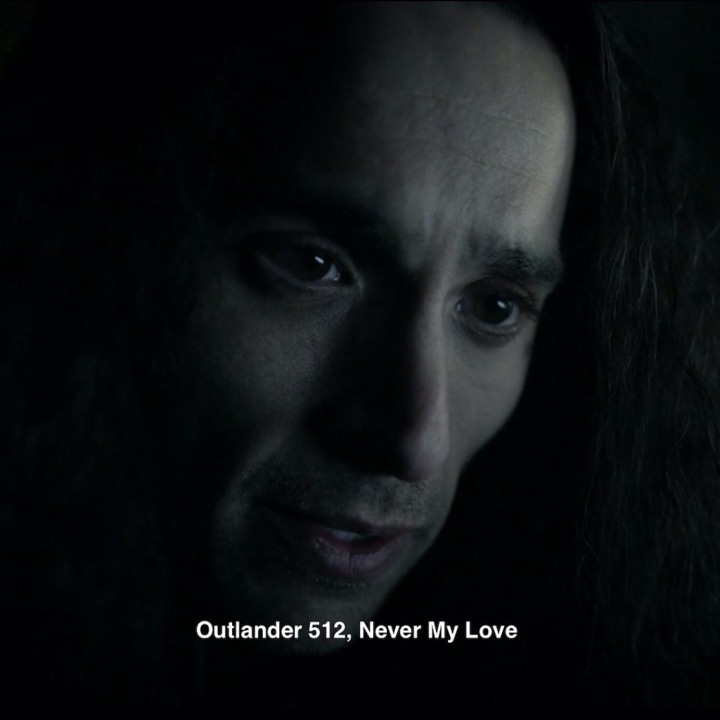
17. No Tattoos! Claire’s dreamscape stripped Ian of his Mohawk tattoos! She also returns his original sunny disposition!
Episode 508: When he first returned from the Mohawk, a line of tattoos covered his cheeks and nose. Claire describes them in Drums of Autumn:
Jamie laughed in gratified surprise, and Ian grinned broadly back. His face was weathered to a deep brown, and the dotted lines of his Mohawk tattoos ran in fierce crescents from nose to cheekbones—but for a moment, I saw his hazel eyes dance with mischief, and saw again the lad we had known.
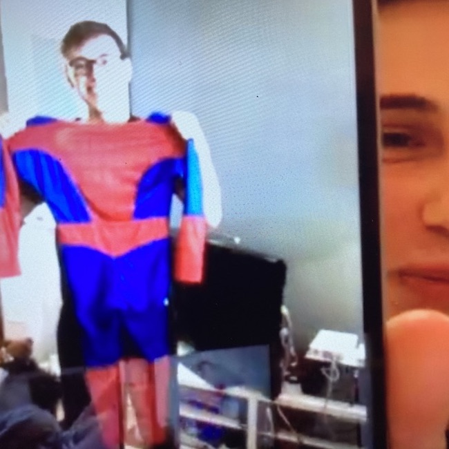
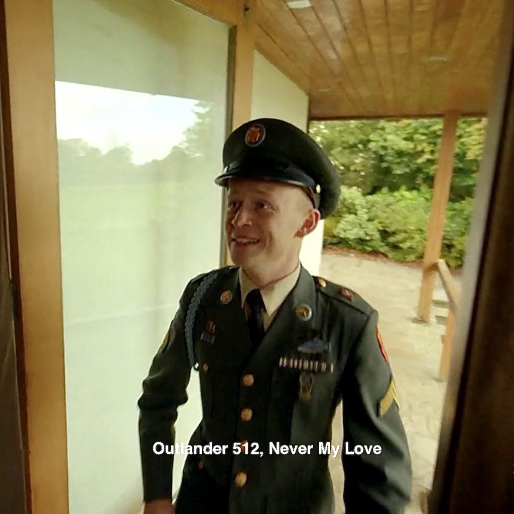
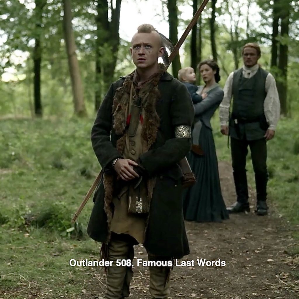
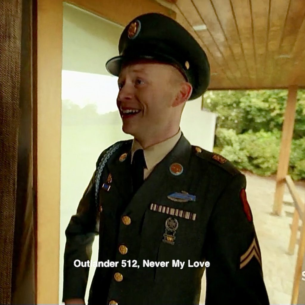
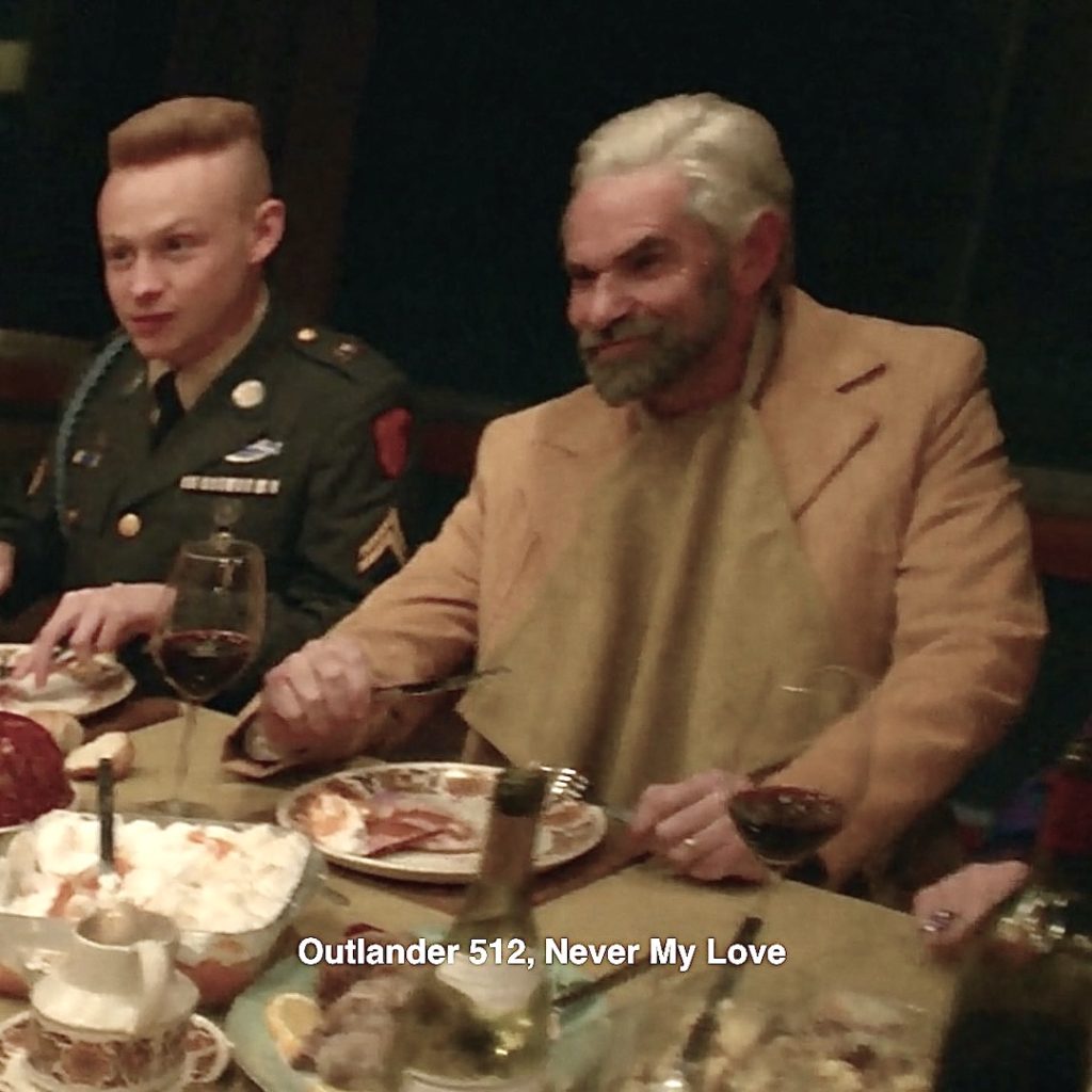
18. Warrior Ian: Claire reimagines Ian as a decorated Infantryman of the US Army! 🎖

Episode 508: In reality, Ian returns from the Mohawk, a warrior in every way!
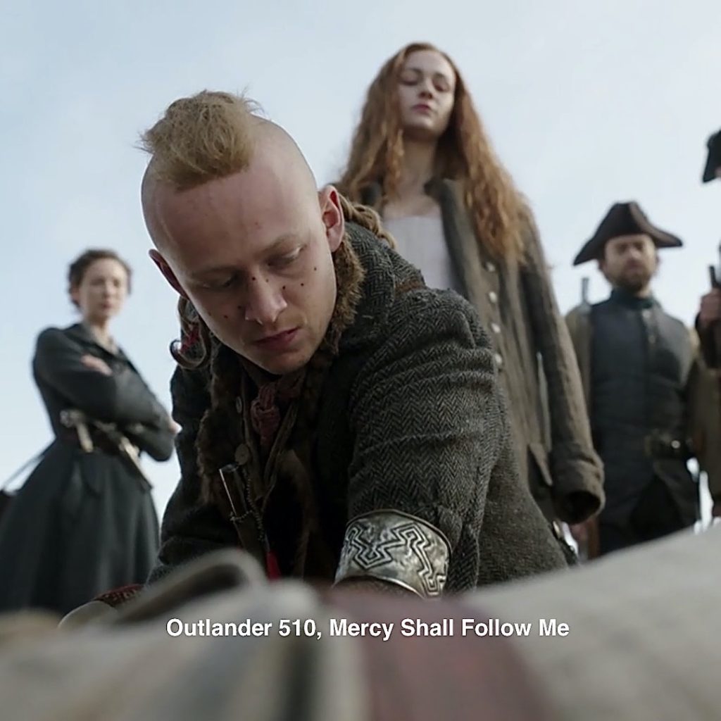
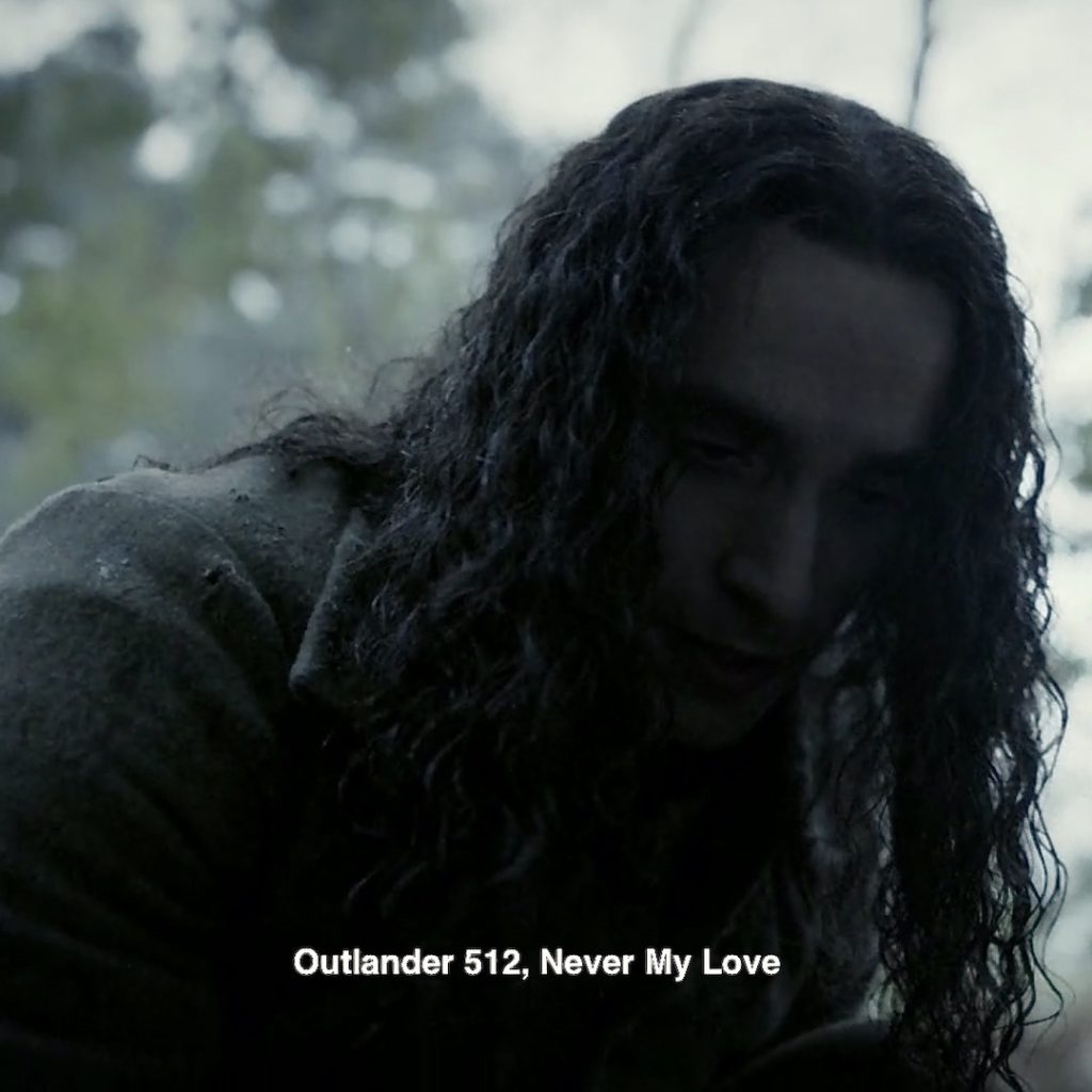
19. Ian’s Hair: Claire’s dreamscape gives Ian a Mohawk-style haircut! ✂︎
Episode 510: Ian’s hair is striking as he muses about removing Steven Bonnet’s! 😳
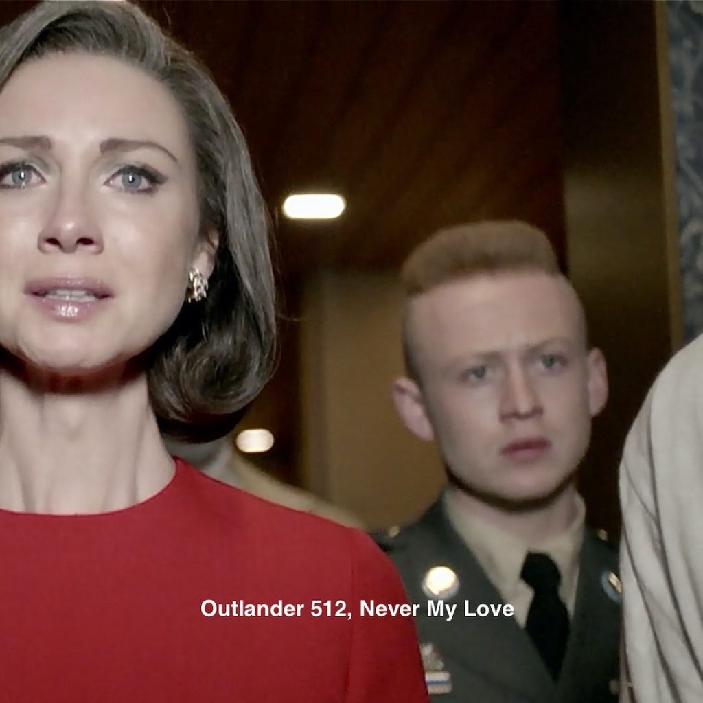
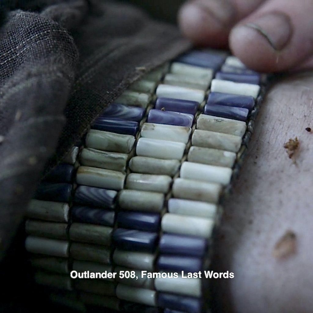
20. Ian’s Awards: Claire imagines Ian with a Decorations and Awards Ribbon above his left breast. Also known as “fruit salad” or “Travel Ribbons,” these are awarded for outstanding service in the US Army. 🎖
Episode 508: So cool because Ian’s Awards Ribbon is composed of his Mohawk beads – likely made by “Works With Her Hands!”
21. Native American Patch: Claire conjures a splendid red and gold Native American patch on Ian’s left sleeve! The patch indicates his Unit of Assignment, the Mohawk, in this instance.
Learn about all the insignia and patches of Ian’s uniform in my last Fun Fact: Anatomy of Ian’s Uniform. Thanks to LTC Edward Maloney for his expertise!
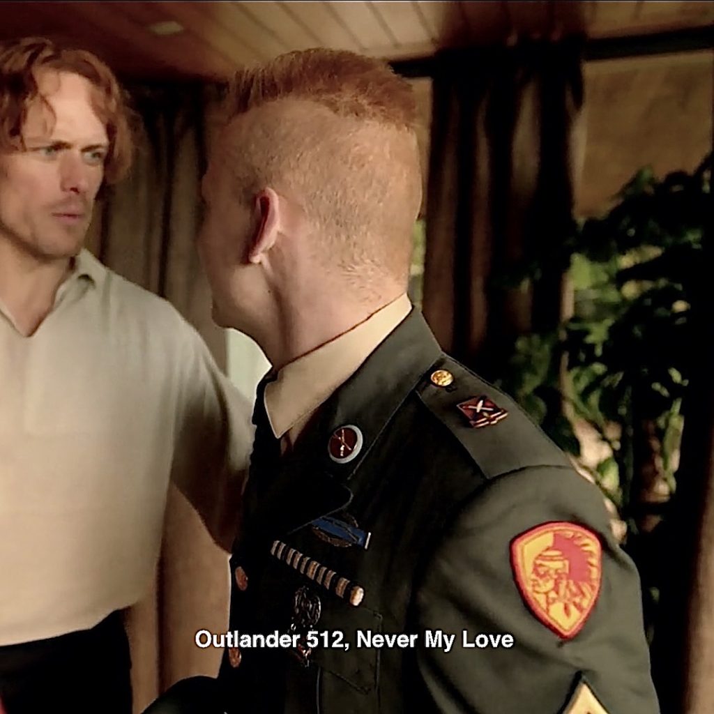
Episode 513: Ian begins his Unit of Assignment when he is adopted into the Mohawk tribe along with his new name! From The Fiery Cross:
“That would be the proper thing to do,” Ian said softly. He had a new, strange stillness to him, and I was forcibly reminded that he had been a Mohawk for the last two years—washed free of his white blood, renamed Wolf’s Brother—one of the Kahnyen’kehaka, the Guardians of the Western Gate.
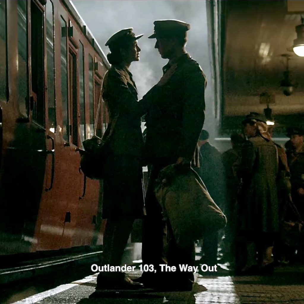
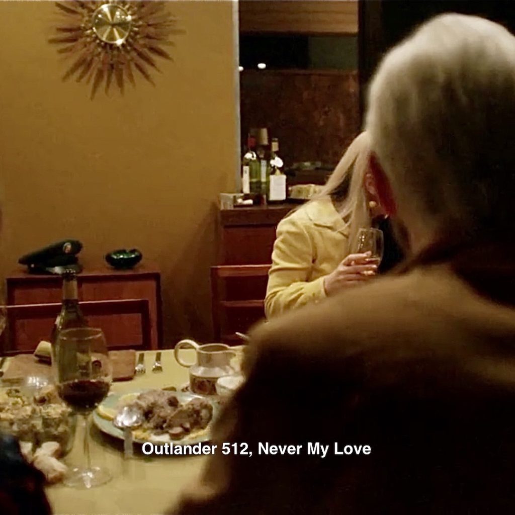
22. Service Cap: Claire’s escape places Ian’s service cap on a side table. Being a Heiland gentleman, he doesn’t wear it to the turkey-table.
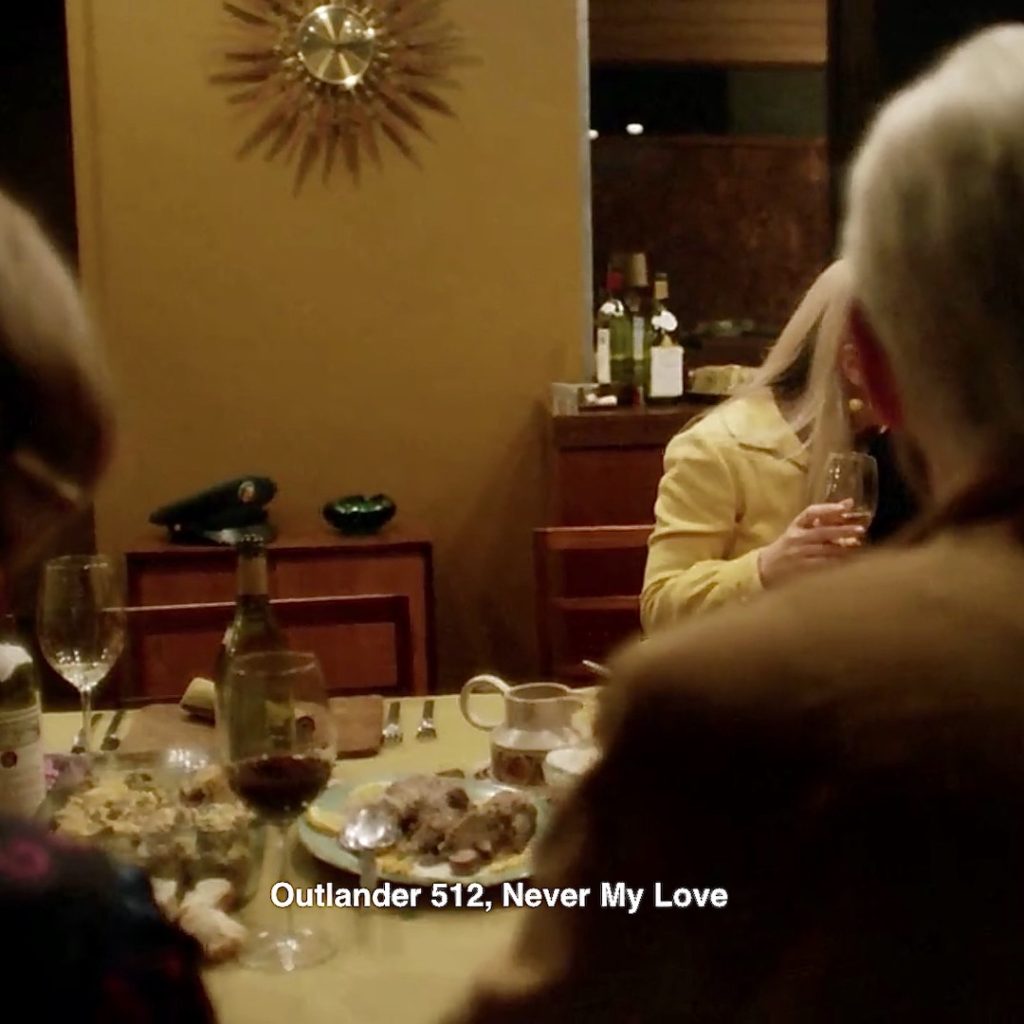
Episode 103: Remember Frank’s and Claire’s service caps, waaaaay back in S1? Claire does!
Let’s see, what else has Claire given to her dinner guests? 🤔
Oh, yes, Murtagh!
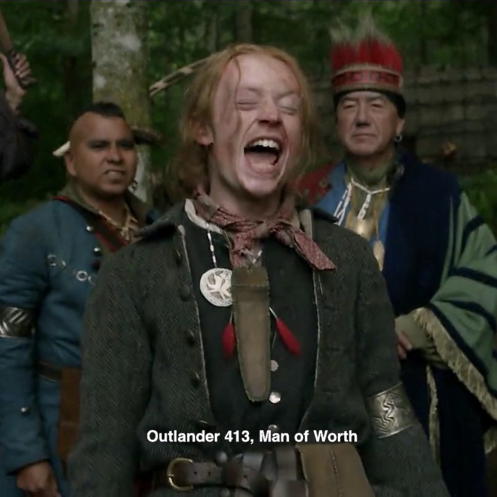
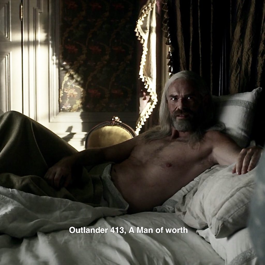
23. Murty’s Hair: Godfather gets a haircut in Claire’s dreamscape, but his hair remains silver and plentiful!
Episode 412, Yeah. That hair! 👇🏻
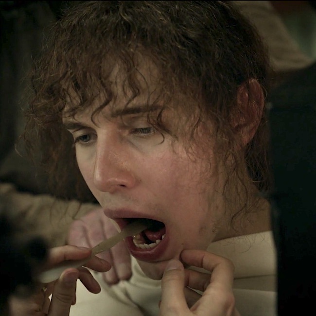
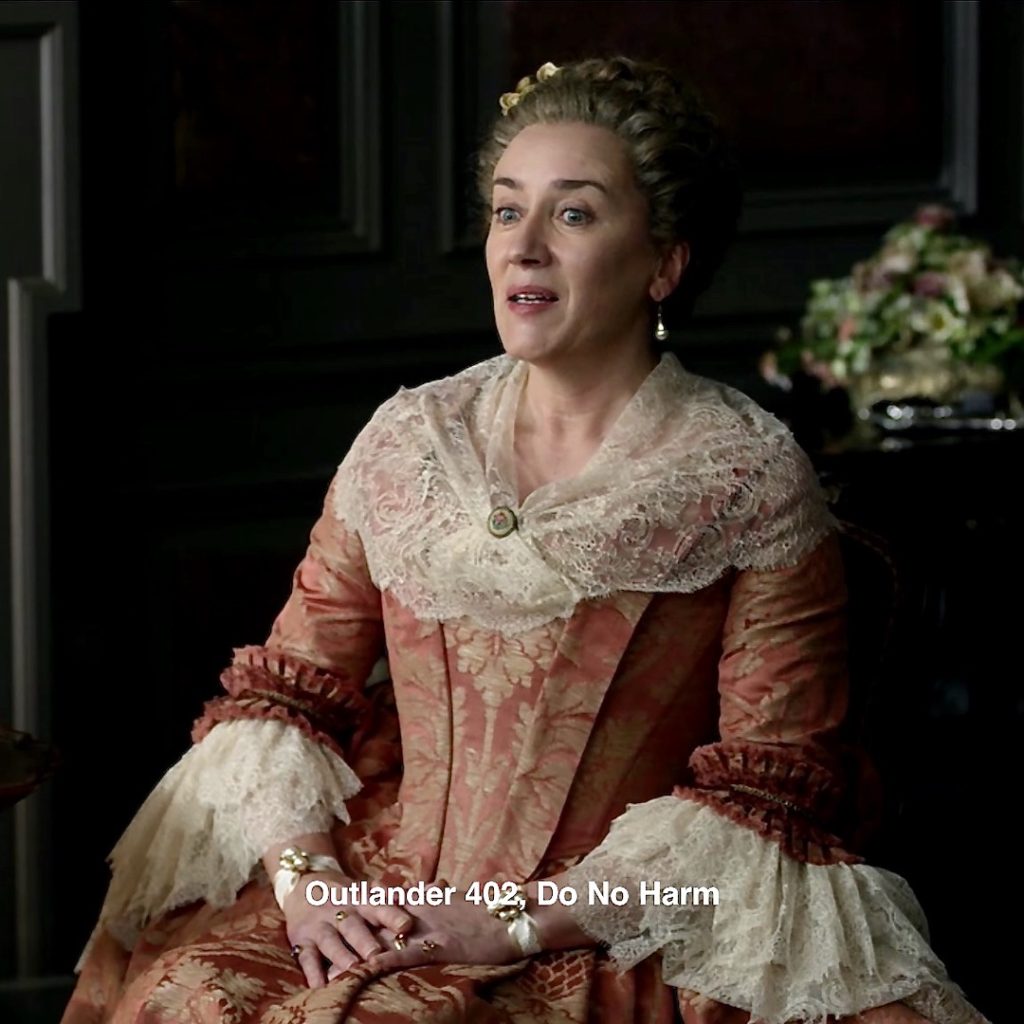
24. Jo’s sight: Claire, ever the healer, chooses to restore Jocasta’s eyesight as when she was a younger woman. So wonderful! 👀
Episode 402: Sadly, from the first we meet, Jocasta is not sighted (ITB, she retains some limited light perception).
Here, when Claire first detects of Jo’s condition, from the Drums of Autumn:
Suddenly, the truth dawned on me: her hand on the butler’s arm, her touching Jamie’s face in greeting, the glass put ready for her grasp, and the shadow on her face when Ian talked of her painting. Jocasta Cameron was blind.
Now, we move to some odds and ends!
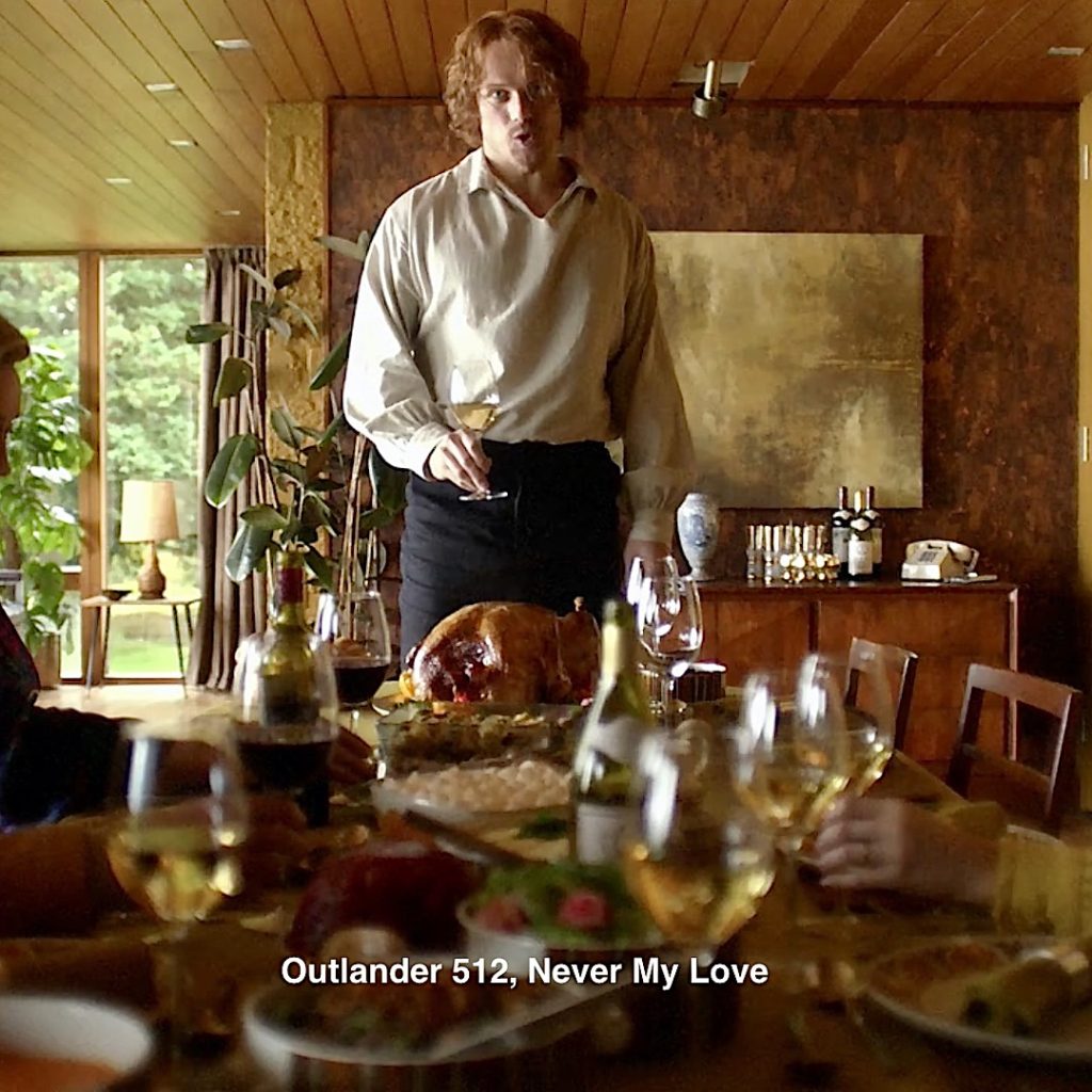
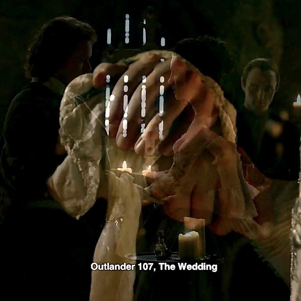
25. Blood Vow: At the 60’s house, Claire graciously accepts Jamie’s turkey-day toast! 🍾
‘Ye are Blood of my Blood, and Bone of my Bone.”
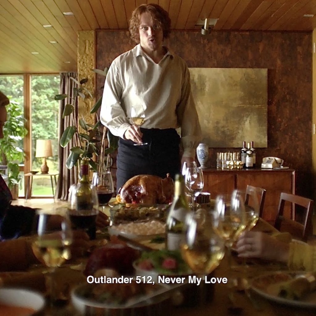
Episode 107, The full blood vow from Outlander book:
“Ye are Blood of my Blood, and Bone of my Bone.
I give ye my Body, that we Two might be One.
I give ye my Spirit, ’til our Life shall be Done.”
🥰
 full
full
26. Telephone! Speaking of phones… OK, we weren’t but, Claire inserts a 60’s style push button phone near the wine bottles! ☎︎
Episode 201: Phones are scattered in a few episodes but this is a good one. Heading back to S2, a rotary phone sits on an officers desk at the constabulary where Frank verbally abuses the chief of police!
“My wife is NOT with another man! “
Erm….. 🙄
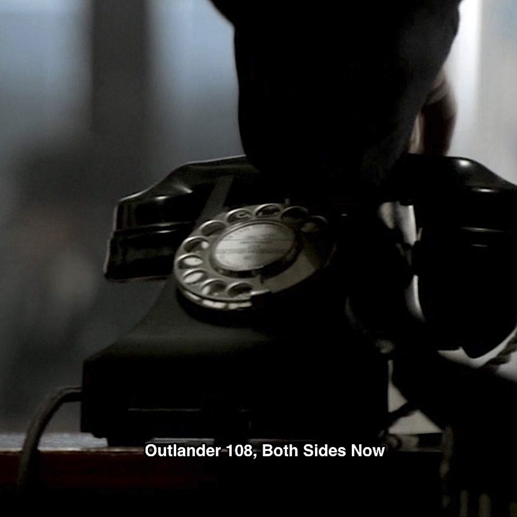
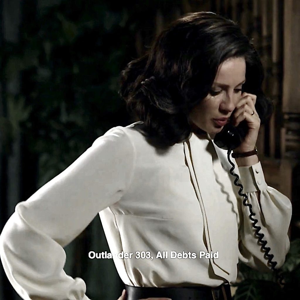
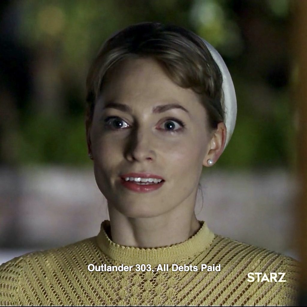
Episode 303: Frank storms out. Claire receives a call summoning her back to the hospital. Such a fateful moment and it is the last time she sees Frank alive! 😔
27. Fergus Left Hand: Bless Claire’s heart! She restores Fergus’ lost hand in her dreamscape. Both hands enjoy turkey, sweet potatoes with marshmallows, and sweet-and-spicy, Marsali! 🙌🏻
Episode 213: The last time Claire saw Fergus’ dear wee left hand was in the final hours before Culloden! 🤚🏻
Jamie signs a “Deed of Sasine” conveying title of Broch Tuarach (Lallybroch) to James Jacob Fraser Murray (young Jamie). And who will take the deed to Janet and Ian? Herself clarifies, in Dragonfly in Amber book:
“Will ye have me take it to your sister?” Murtagh asked as I shook the paper carefully to dry it.
Jamie shook his head…
“No. Fergus will take it.”
28. Ringo: Speaking of Fergus and Marsali, Claire hears them suggest a new name for their unborn babe. Ringo? Please, no! 🚫
Episode 512, In the same episode Wendigo Donner talks to Claire about moon, stars, and Ringo Star. Yep, she kens the drummer!
This Fun Fact has steadfastly held the line to Easter eggs in Claire’s dreamscape, but here is one exception….
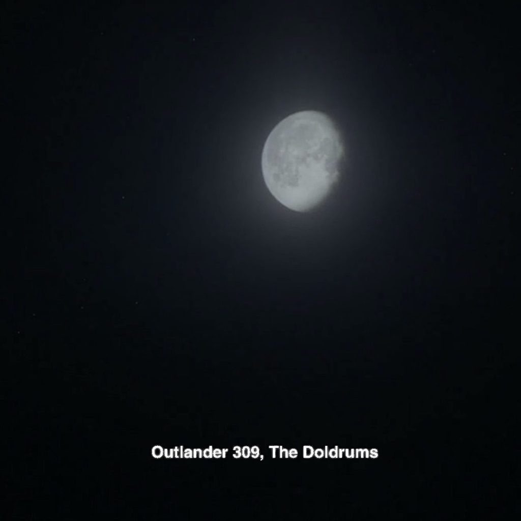
29. Man in the Moon: Speaking of Wendigo Donner, early in the episode, he says to Claire:
“The Moon — That’s the same wherever you go. Looks like a man on the moon”!
Hum…. Actually, Wendigo, the moon doesn’t look the same wherever you go. The moon appears upside down in the Southern Hemisphere compared to what is seen in the Northern Hemisphere! 🌗
Episode 309: Claire tells Jamie about the man in the moon aboard the Artemis while gazing at the night sky.
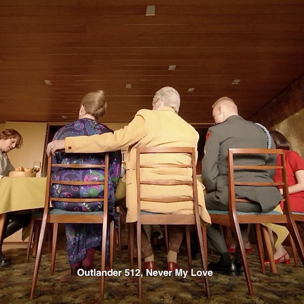
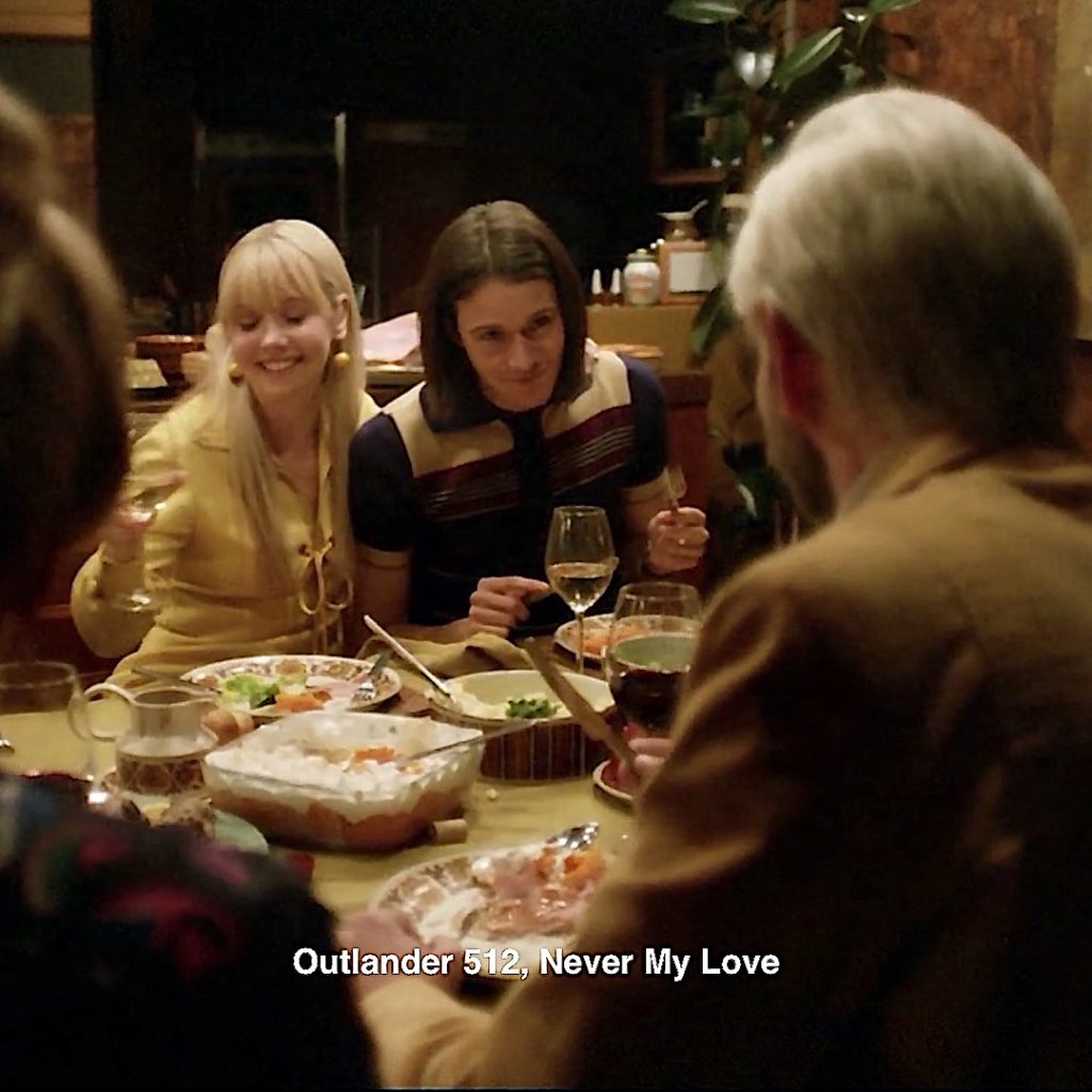
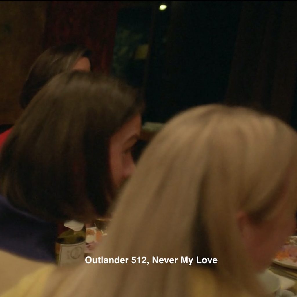
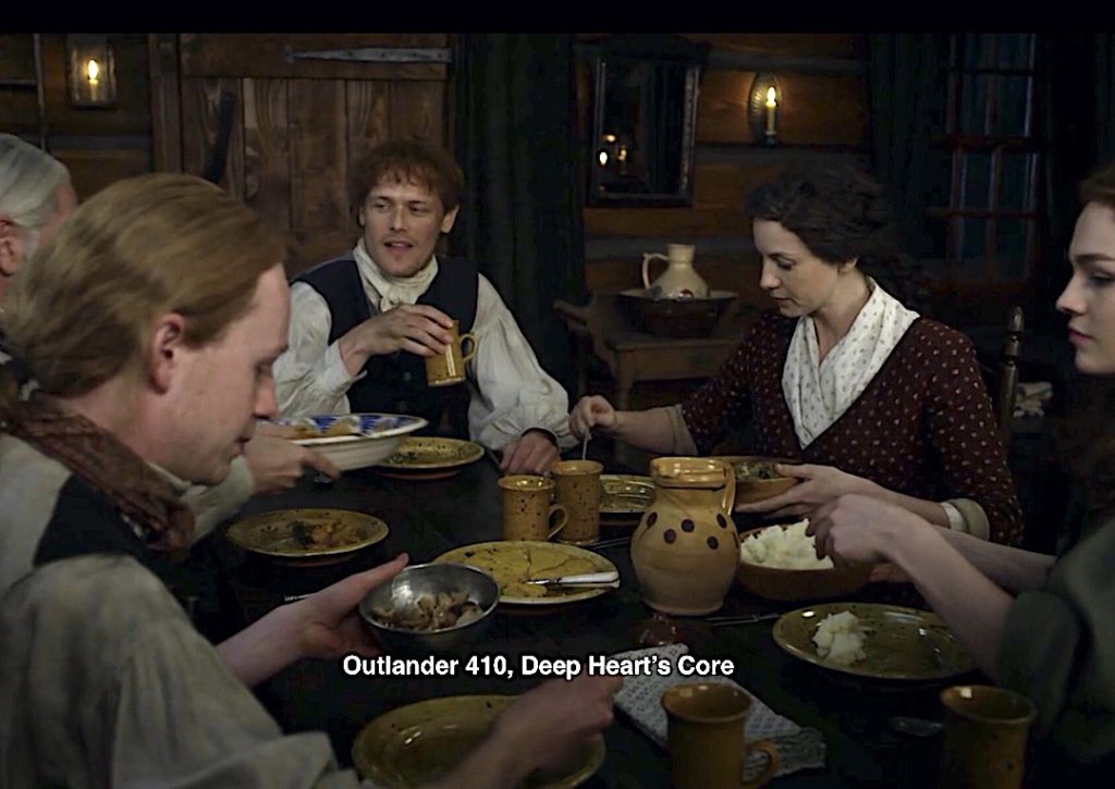
30. Family Dinner: A blessed time for family to gather and share fun, food, laughter, and memories. Any wonder Claire’s mind latches onto such an event? 😋
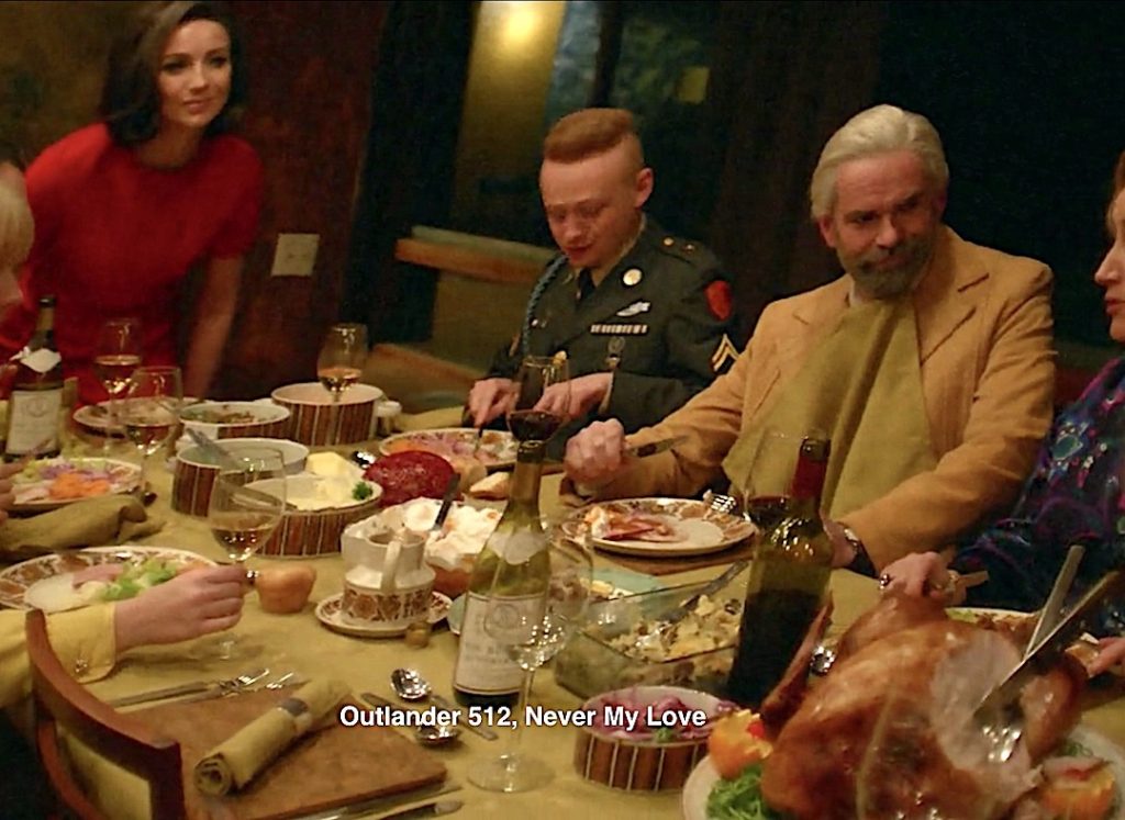
Episode 410: there are other family meals through the seasons, but this one, after Bree arrives at Fraser’s Ridge, is awesome sauce!
31. Wine: Claire imagines a good hostess offers libations; few Thanksgiving meals are complete without wine!
Episode 201: Claire mind harkens back to Uncle Jared’s wine business after sailing to France. 🍷
…Instead of proceeding directly to Paris, though, we had come down the coast from Ste. Anne to Le Havre, to meet first with Jamie’s cousin, Jared Fraser.
A prosperous Scottish émigré, Jared was an importer of wines and spirits, with a small warehouse and large town house in Paris, and a very large warehouse indeed here in Le Havre, where he had asked Jamie to meet him, when Jamie had written
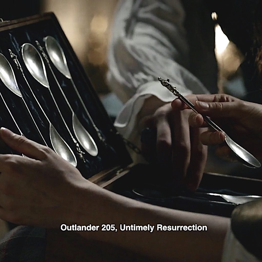
32. Spoons: Recall the table setting prior to guests? Each setting is adorned with a pair of large spoons. 🥄🥄
Episode 205: Possibly intended, possibly not, but these remind some of the Apostle spoon set Jamie gave to Claire in honor of their unborn child….way back in Paris!
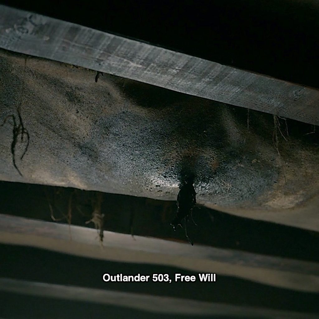
33. Ceiling Leak: A leak in the ceiling! What does this mean? I side with the theory it represents Claire’s attempts to keep at bay the horrific abuse she suffers. But, the limits of her carefully crafted mental barriers are tested. 💦
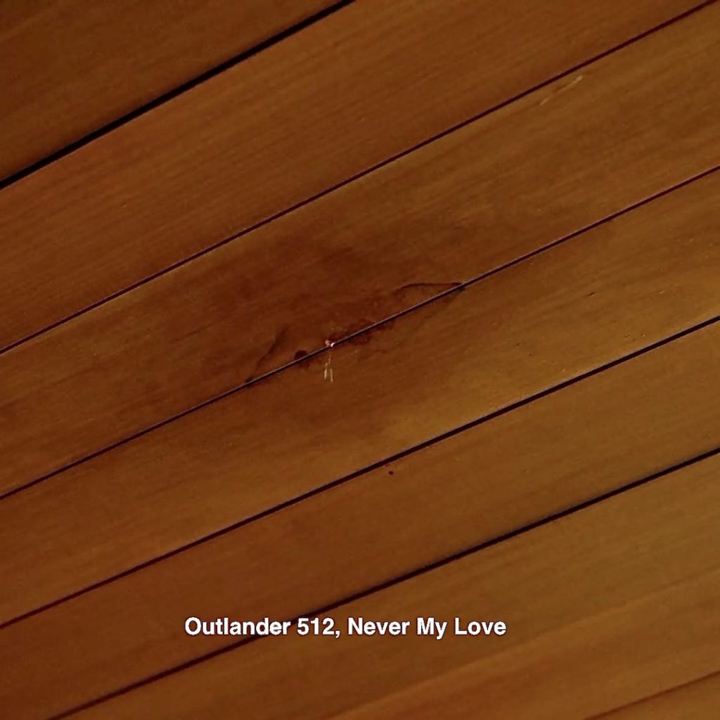
The ceiling leak could also represent that ghastly leak at the Beardsley’s cabin in S5! 😳
And, three other apropos leaks appear in the books.
The first, from Drums of Autumn, deals with a leak in the roof of the new cabin on Fraser’s Ridge!
“Another one!” Jamie sat bolt upright in bed beside me.
“Another what?” I asked sleepily, opening one eye….
“Another bloody leak! It hit me in the ear, damn it!” …
I shifted my glassy gaze from the hairy ankles in front of my nose, to the roof overhead. Sure enough, the torchlight revealed the black line of a split in one shingle, with a spreading dark patch of dampness on the underside…
He halted by the door, glared briefly at me, then, with the rebuking expression of an early Christian martyr, laid down his tools, stripped off the shirt, dropped it on the floor, picked up the tools, and strode majestically out to deal with the leak, buttocks clenched with determined zeal.
The second is from A Breath of Snow and Ashes. This is the book wherein Claire is abducted (in the TV series, it was moved forward to S5) Here, Jamie rescues Claire and brings her back to the Ridge. Not a light -hearted moment… 😭
Another drop struck him in the back of his neck, and he hunched his shoulders in surprise. Caught by his movement, she looked up, and he met her eyes with shock. She shared it; the shock of strangers meeting one another naked. Her eyes flicked away from his, up toward the ceiling.
“The roof’s leaking,” she whispered. “There’s a wet patch.”
And, a third from Voyager book:
The cloak behind me dripped water on the floor, a slow, arrhythmic patting. It reminded me unpleasantly of an old Highland superstition—the “death-drop.” Just before a death occurs, the story goes, the sound of water dripping is heard in the house, by those sensitive to such things.
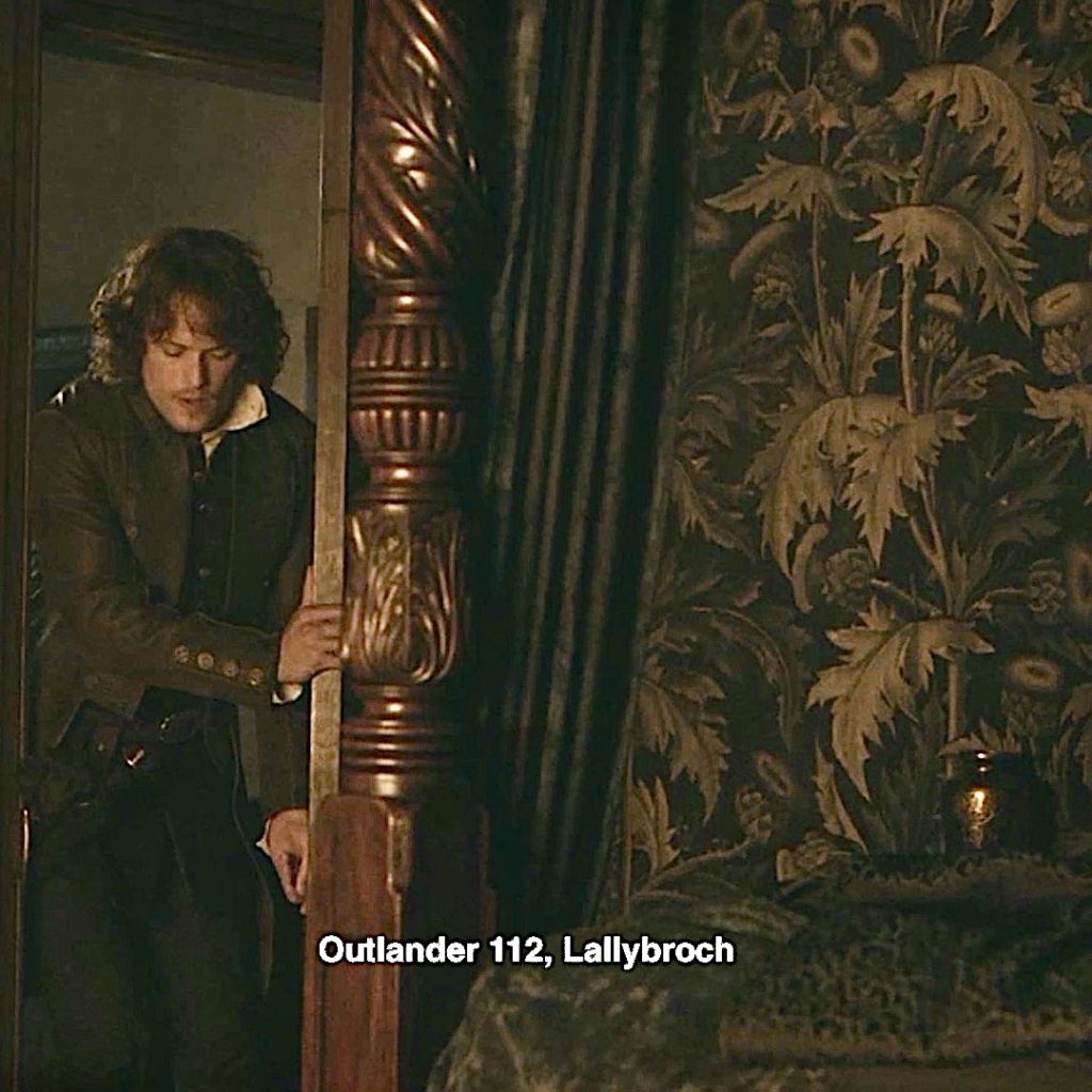
34. Wallpaper: A wall in Claire’s 60’s house is covered with the most interesting print featuring Scottish thistles! Have ye seen it before?
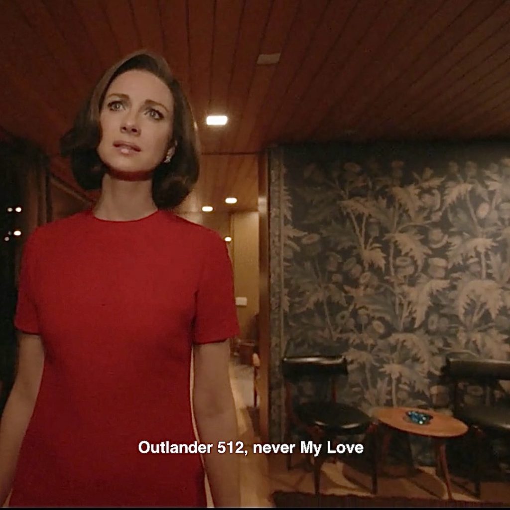
Episode 112: Aye, we have! Same wall covering in the master bedroom at Lallybroch!
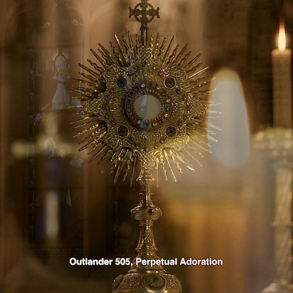
35. Sunburst Clock: Right out of the 60’s! It appears to read 3:15 – was that time significant in Outlander? Not to my knowledge. 🕰
Episode 505: A similar sunburst of the Monstrance at Claire’s house of worship!
Let’s turn to science/medicine. I spy two candidates! 🤓
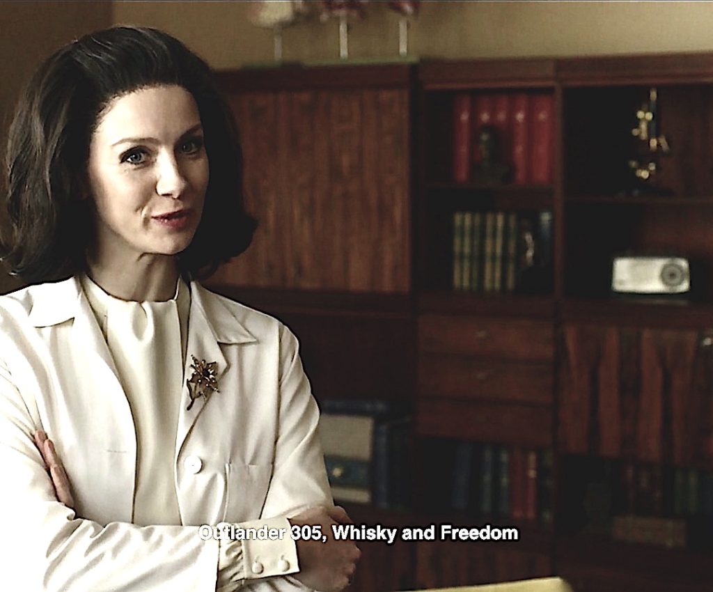
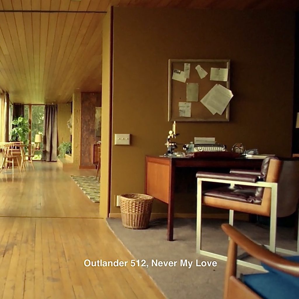
36. Microscope: Claire’s dreamscape includes her beloved microscope, a 60’s style similar to the one she used before returning through the stones! 🔬
If you are interested in a brief history of microscopy, check out Anatomy Lesson #34, The Amazing Saga of Human Anatomy! (Current light microscopes are much more sophisticated than the ones Claire used.)
Episode 305: Glory be! Blurry, but that microscope sits on the shelf in the office Claire shares with Dr. Joe Abernethy! 🤗
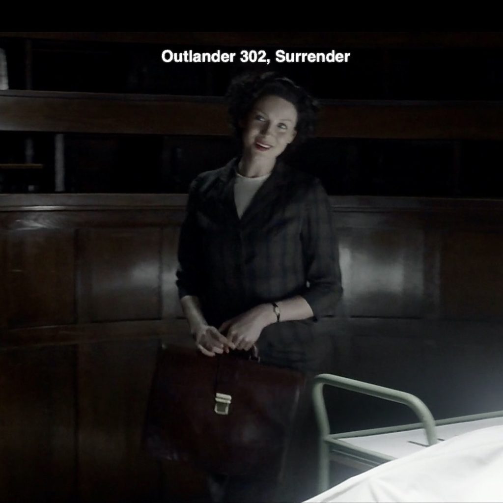
37. Brief Case: A close look at the desk and behold: Claire’s briefcase lays on its side, in front of the typewriter. Is this really an Easter egg? Oh, I think it is! 🤗
Episode 302: Recall Claire first day in medical school? She sussing out the anatomy lecture hall and the prof’s scalpel! He wasn’t amused even though she offers a winning smile. Check out her leather brief case. Yep, that’s the one! 💼
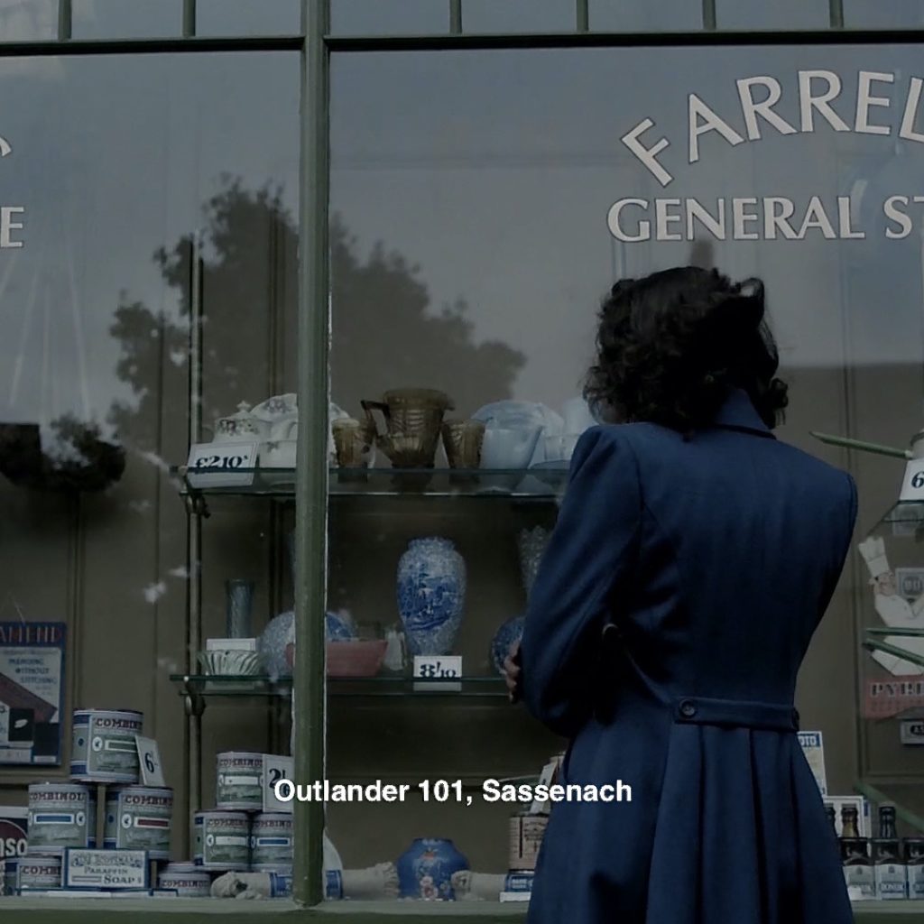
38. Vase: Blame the vase! Claire remembers the blue and white vessel that launched this amazing saga! Would her life have played out differently had she gone into the shop and bought the vase? 🤷♀️
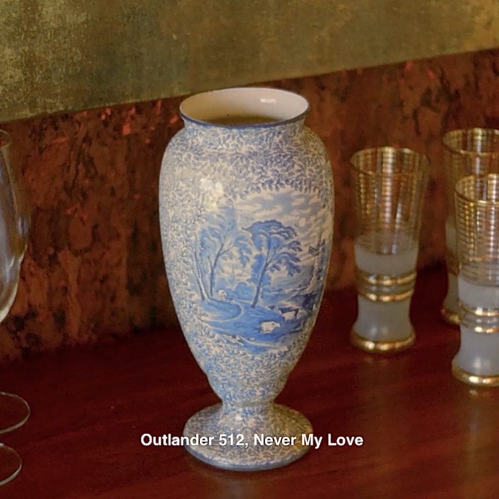
Interesting question, but I dinna think so. In Outlander book, Claire buys a set of three vases but the stones take her, anyway! ⚱️⚱️⚱️
My gaze lingered on a shop window filled with household goods—embroidered tea cloths and cozies, pitchers and glasses, a stack of quite homely pie tins, and a set of three vases.
I had never owned a vase in my life…
Tucking my handbag firmly under my arm, I marched into the shop and bought the vases.
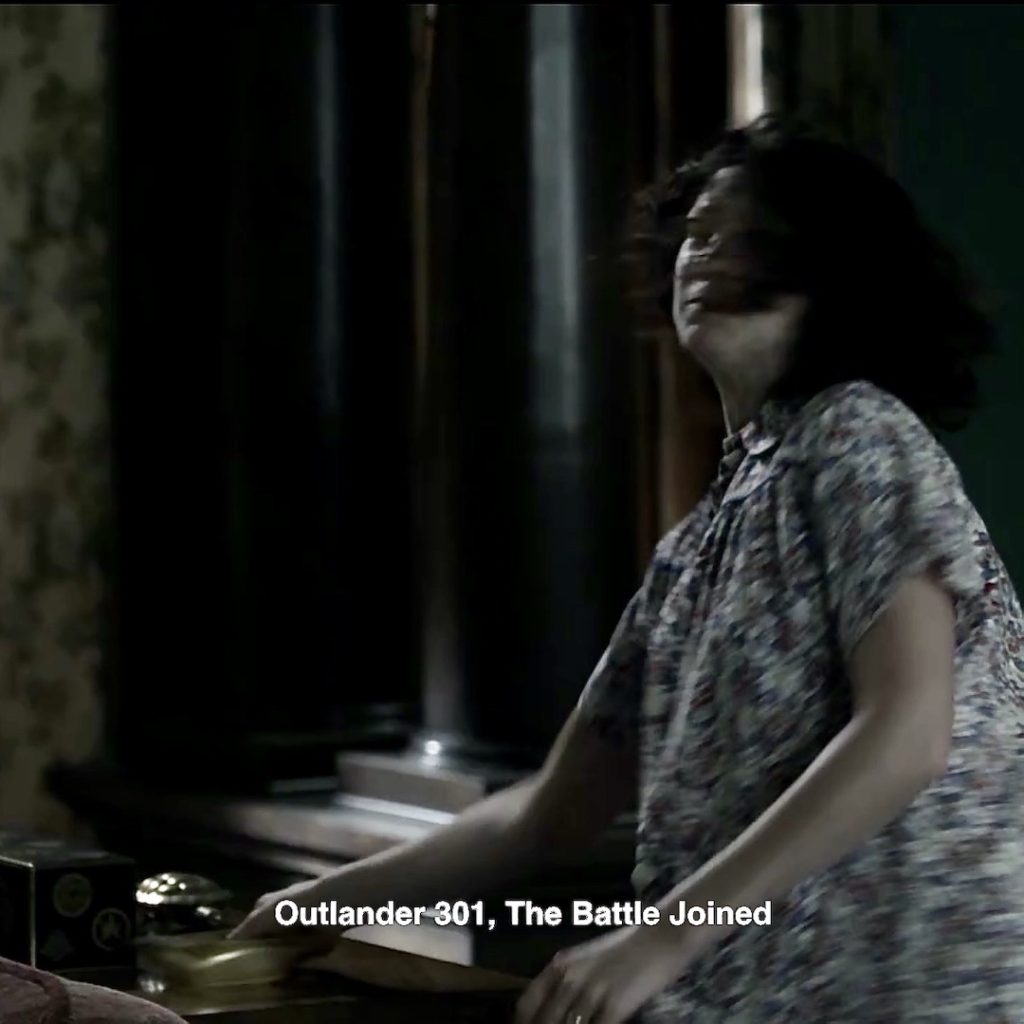
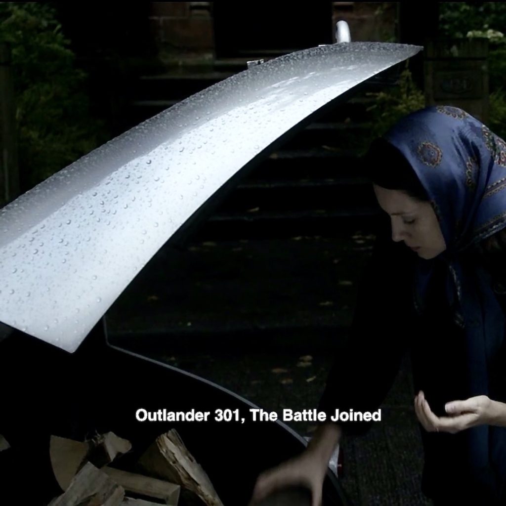
39. Ashtray: Every 60’s house must have an ashtray, although I don’t recall seeing any of these folks smoke.
Episode 301: A callback to Claire hurling an ashtray at Frank’s head? A traumatic event. Yep, the battle joined, indeed! 😮
Episode 101: Oops! I forgot! We did see someone huff-n-puff! 🚬
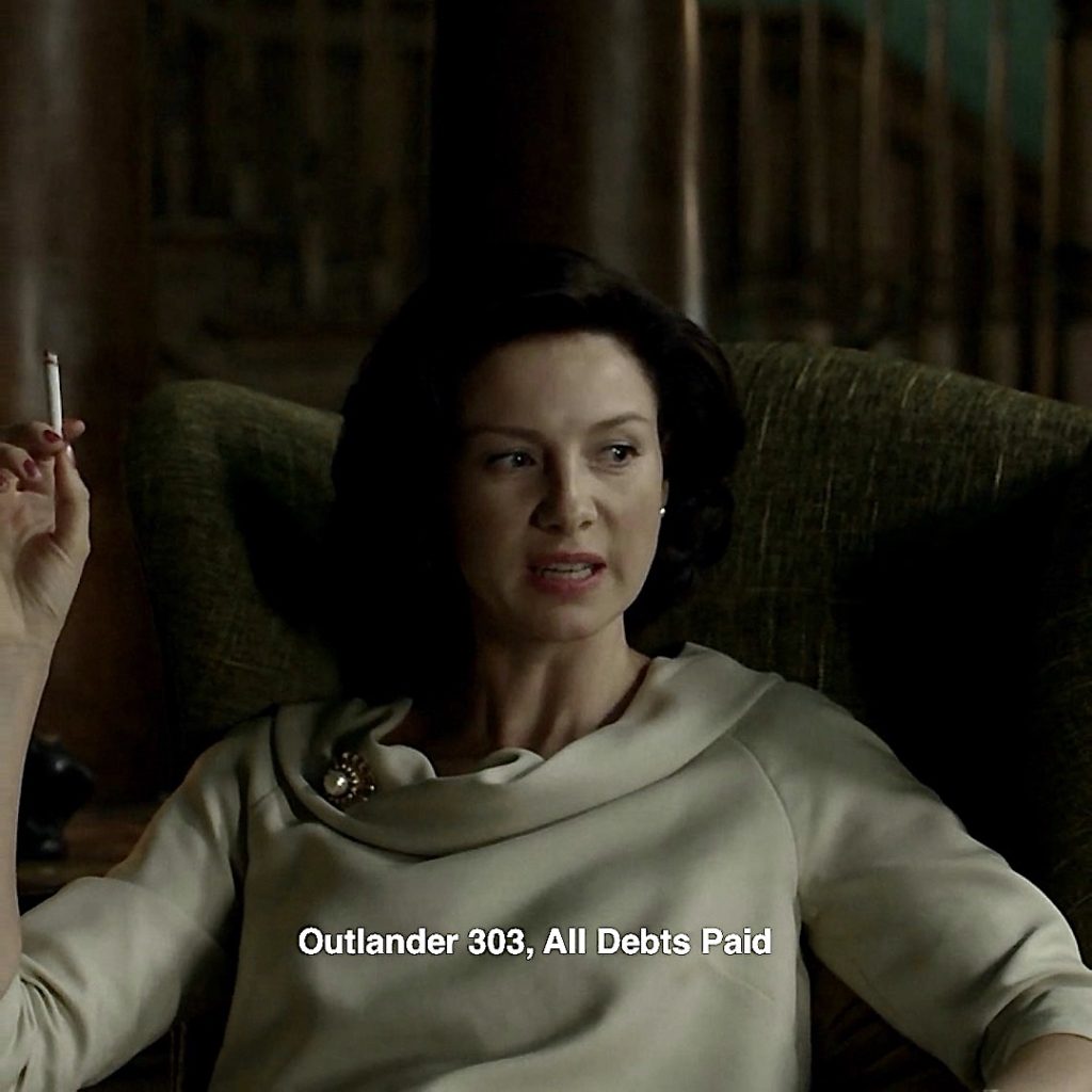
Episode 303: Double Oops! 🙄 Erm… another one! Claire is angry with Frank, embarrassing her on graduation day by the unexpected appearance of Candy-Sandy at her door. So angry, she resorts to a puff or two!
Claire also creates a little animal menagerie for her 60s home! 🐰
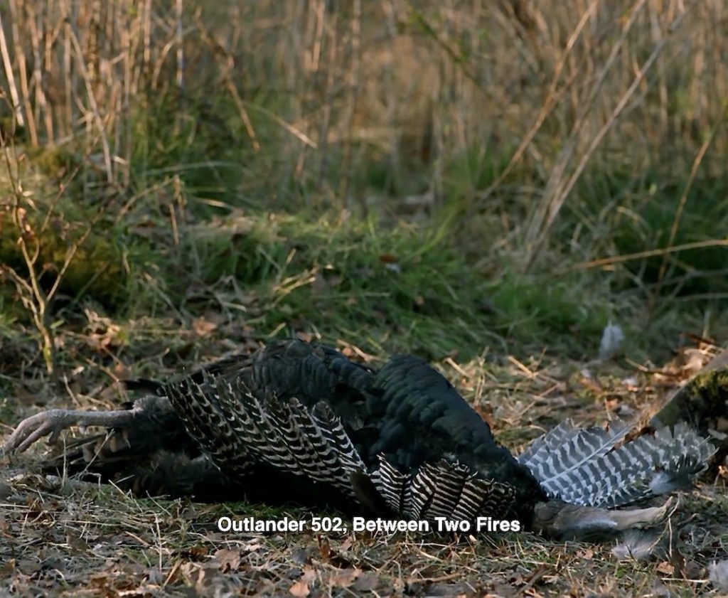
40. Turkey: Back to Jamie’s toast and the turkey roast. That is a delicious-looking bird on the table! 🦃

Episode 502: Gobble, gobble! Who’s a turkey? Dead-eye Bree shoots a turkey after Roger misses. Wonder if it is same one ion the dreamscape table?
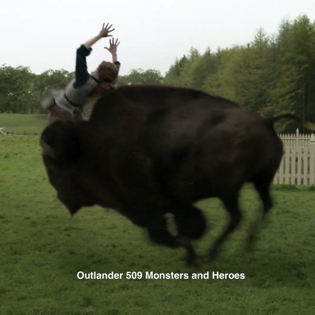
41. Buffalo (American Bison): Claire’s dreamscape includes a Jo 🆚 Germain pillow fight! In the background, a buffalo sculpture adorns a cabinet.

Episode 509: Bree at the Buffalo-Bash!!! 🤸♀️😱
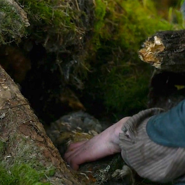
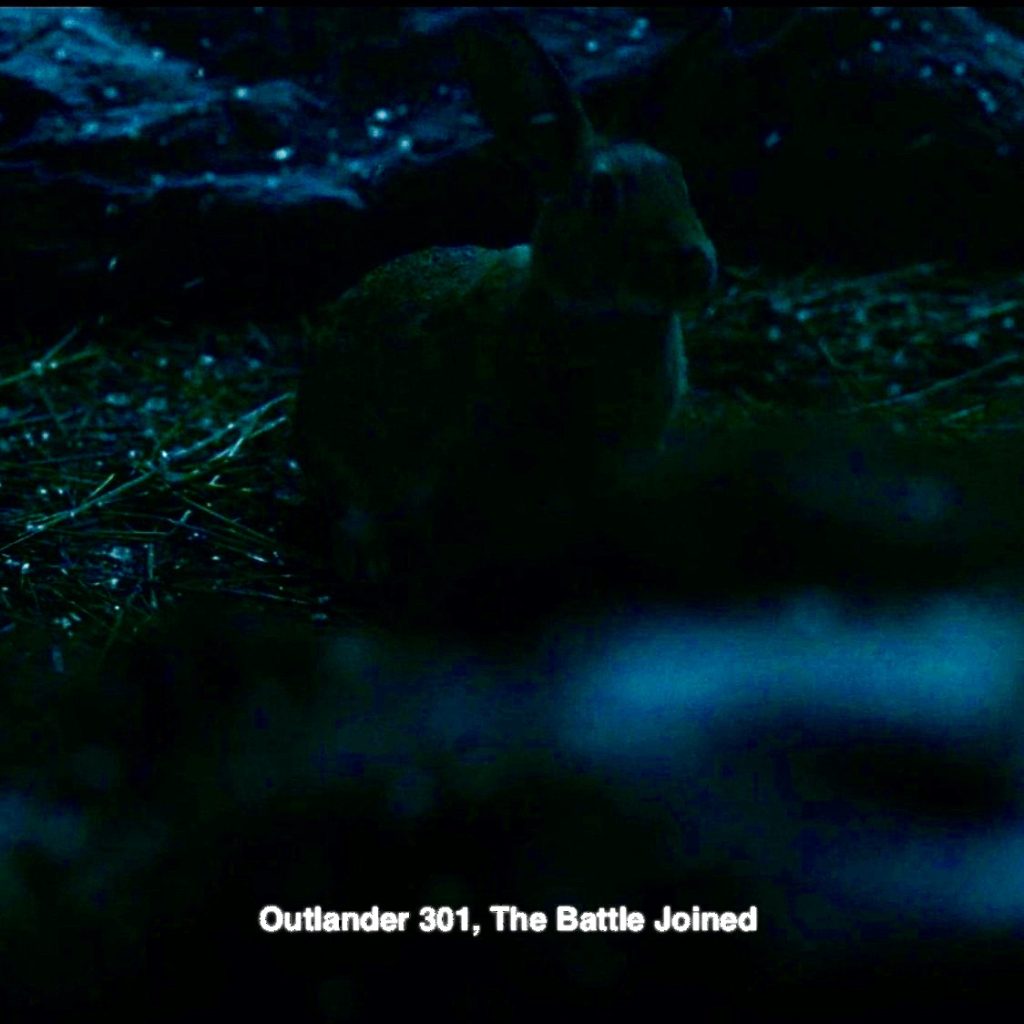
42. Bunny alert! Claire spies a bunny in her imaginary home. So apropos for our Easter Egg hunt! 🐰 🥚
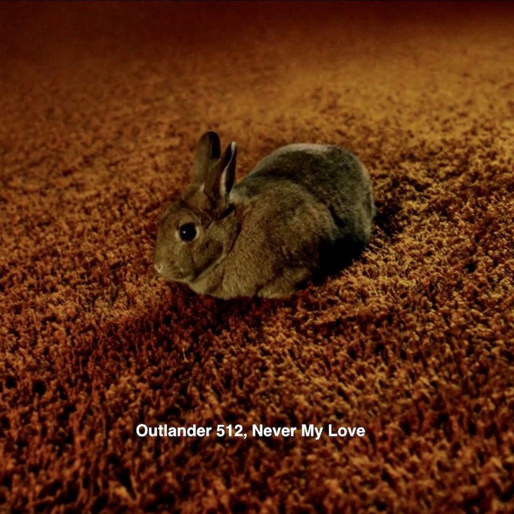
Episode 301: Bunnies are special for Jamie and Claire. This one was chuffing at Culloden Battlefield.
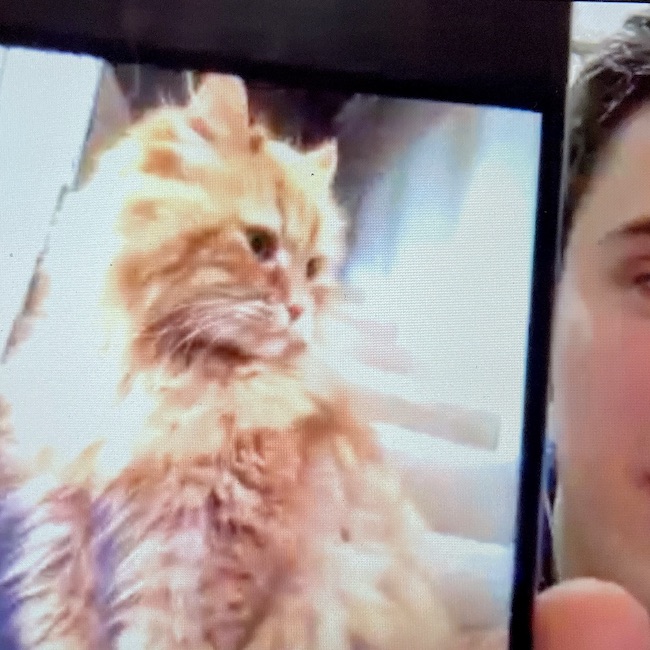
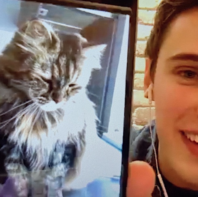
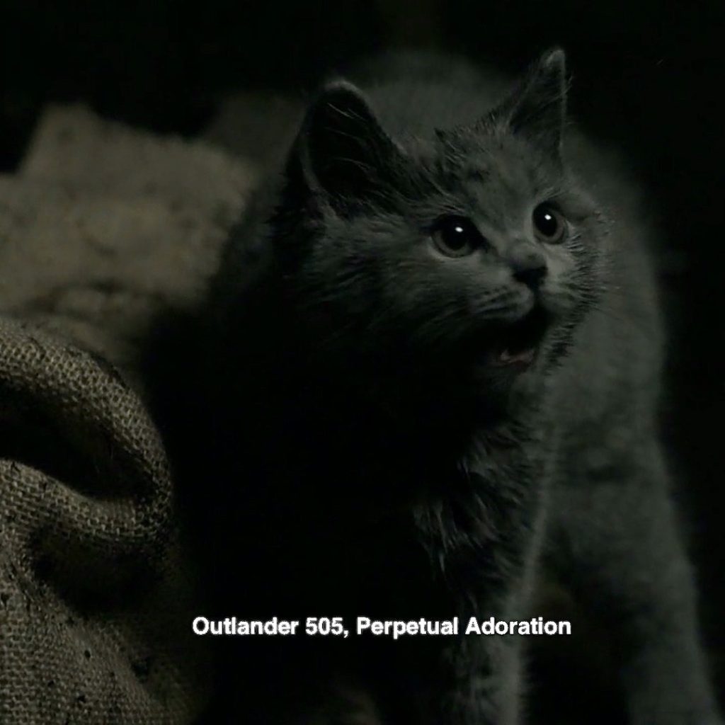
43. Adso! Well, perhaps not Adso perched on the fireplace, but a reminder of the wee cheetie! (This is a Starz promo image)
Episode 505: What a cute, wee little fellow. Jamie finds him in an alleyway after killing Knox and then takes him home to Claire! This quote from The Fiery Cross gives us the background on his name:
“No. I’ve never had a cat before,” I admitted. “Frank was allergic to them—they made him sneeze. And what’s a good Scottish cat name, then—Diarmuid? McGillivray?”
He snorted, then laughed.
“Adso,” he said, positively. “Call him Adso.”
“What sort of name is that?” I demanded, twisting to look back at him in amazement. “I’ve heard a good many peculiar Scottish names, but that’s a new one.”
He rested his chin comfortably on my shoulder, watching the kitten sleep.
“My mother had a wee cat named Adso,” he said, surprisingly. “A gray cheetie, verra much like this one.”
“Did she?” I laid a hand on his leg. He rarely spoke of his mother, who had died when he was eight.
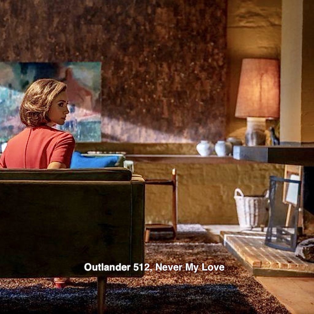
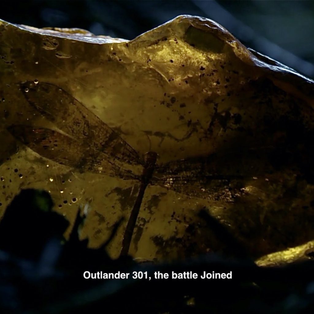
44. Dragonfly: Perched on the TV, near Claire! 😃
Germaine flies off with this stylized dragonfly.
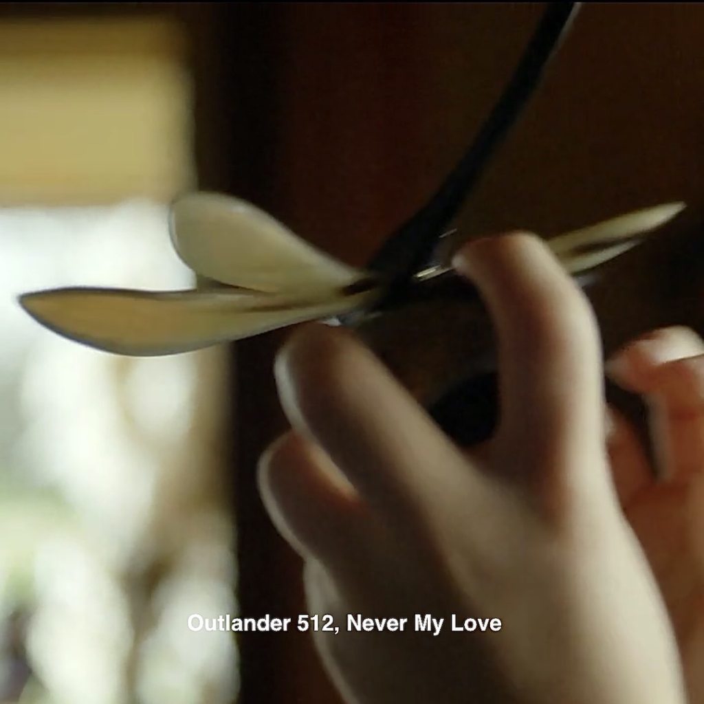
Episode 301: There are many callbacks to the dragonfly that Hugh Munro gives Jamie and Claire as a wedding gift! This one appears in 301, as the piece of amber tumbles from Jamie’s grasp onto Culloden Battlefield.
An excerpt from Dragonfly in Amber book:
“I had not seen Hugh Munro again, but I had wakened in darkness the night before to find Jamie gone from the blanket beside me. I tried to stay awake, waiting for him to return, but fell asleep as the moon began to sink. In the morning, he was sound asleep beside me, and on my blanket rested a small parcel, done up in a sheet of thin paper, fastened with the tail-feather of a woodpecker thrust through the sheet. Unfolding it carefully, I found a large chunk of rough amber. One face of the chunk had been smoothed off and polished, and in this window could be seen the delicate dark form of a tiny dragonfly, suspended in eternal flight.”
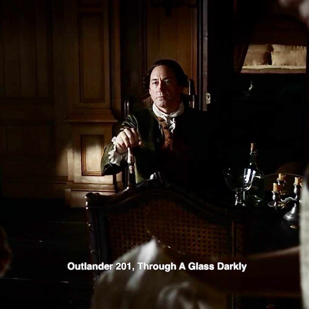
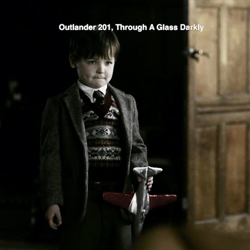
45. Germain’s “Dragonfly Plane:” Germain flies his dragonfly through the air. Look closely at its head, very reminiscent of the nose of an airplane!
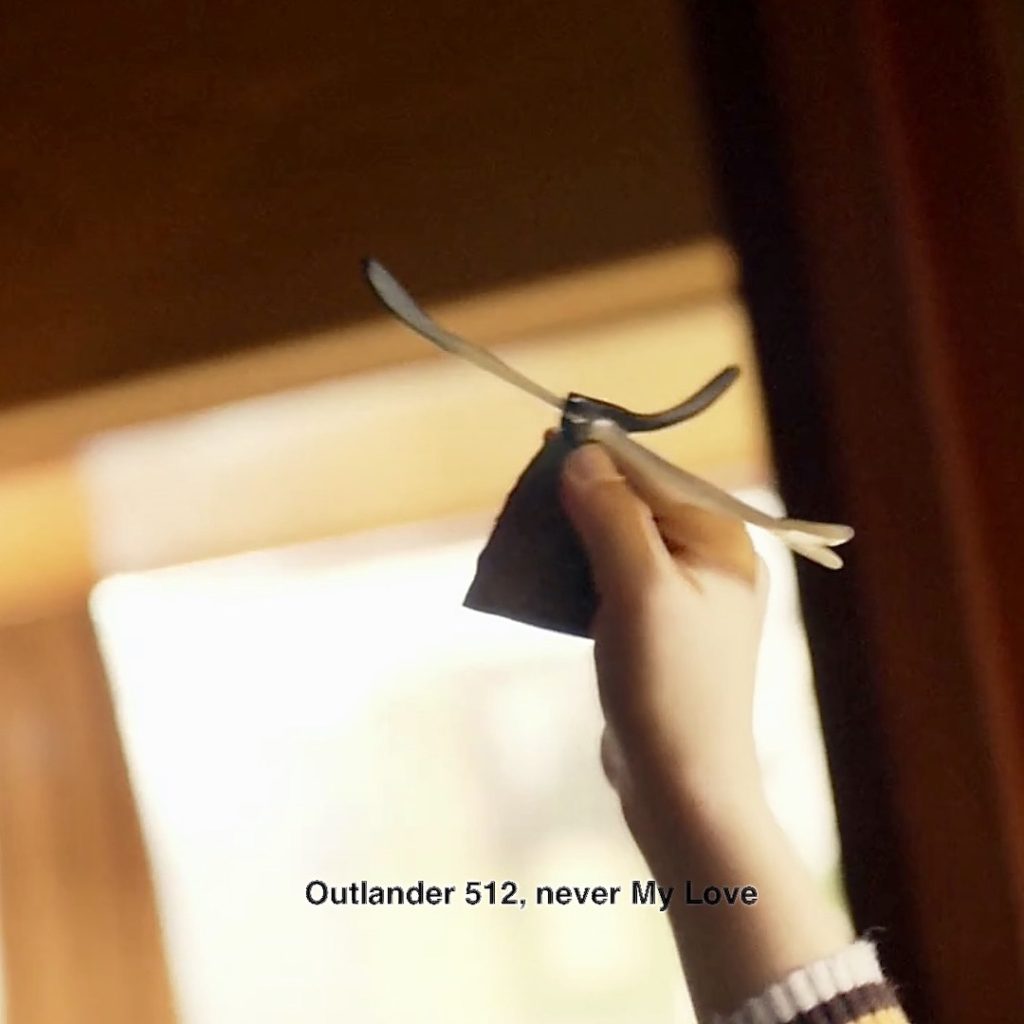
Episode 201, Through A Glass Darkly. Roger holds his beloved plane. He kens Frank just uttered awful words about his own wife!
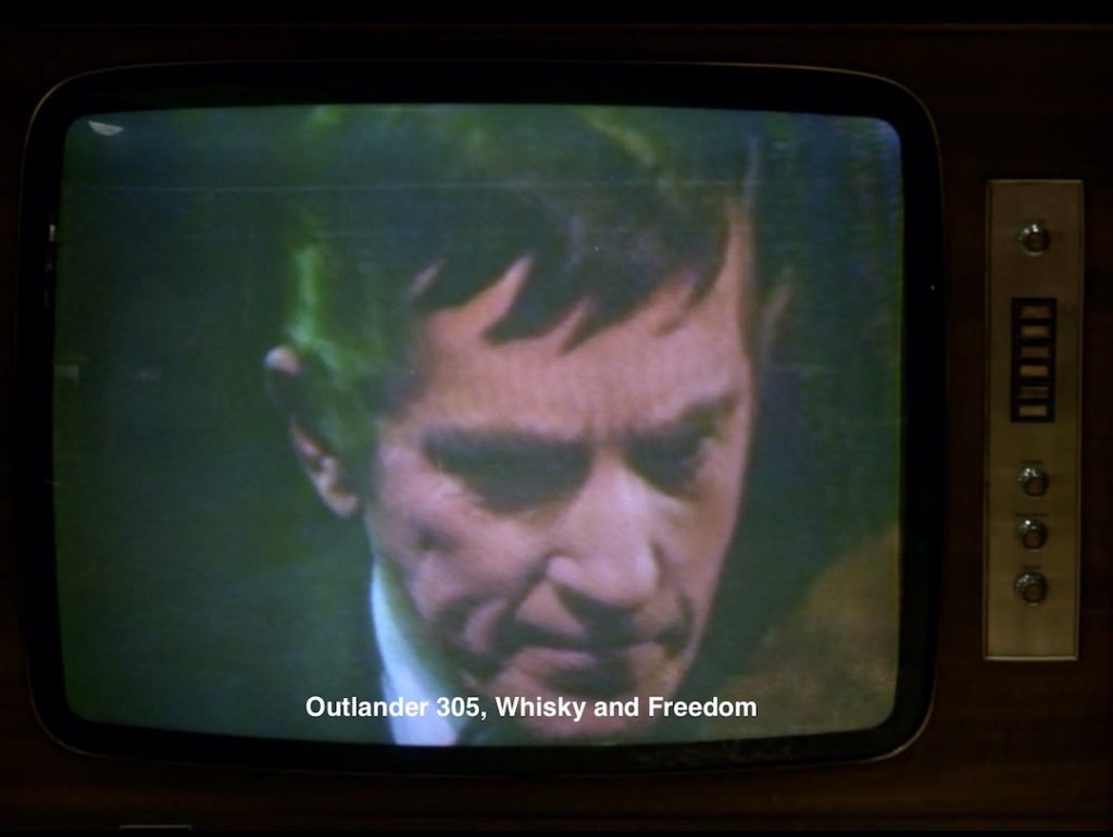
46. TV: Claire kens no 60’s style home would be complete without a TV!
Episode 305, Two different TV shows, show up. 📺
In the doctor’s lounge, Claire and colleagues watch the first orbiting of the moon by the crew of Apollo 8.
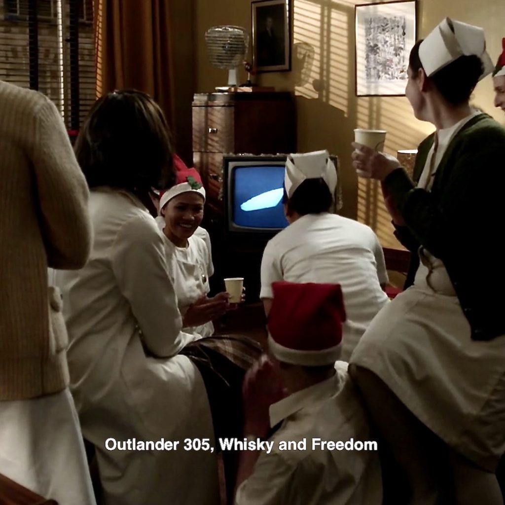
Meanwhile, Roger watches Barnabus Collins of Dark Shadows! 🧛♂️
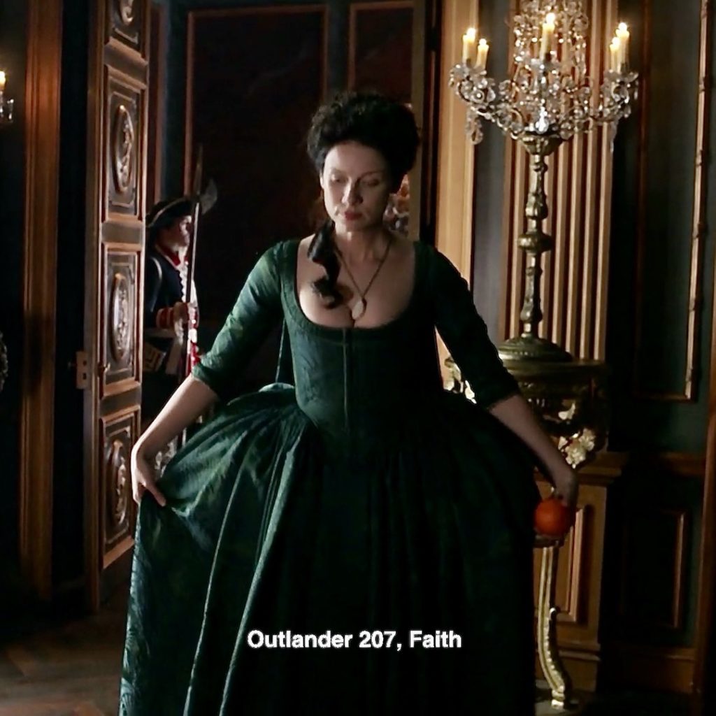
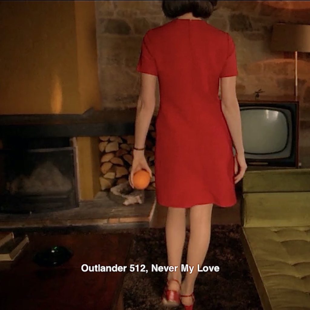
47. The Orange! Mustn’t forget the citrus! Claire picks up a perfect orange and slowly walks away. 🍊
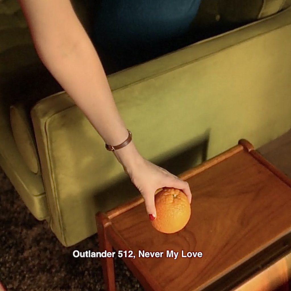
Episode 207: Claire remembers a bowl of fruit in King Louis’ apartments. She leaves with her dignity (in the form of an orange) following her “transaction” with King Louis! It was forgettable! 😜
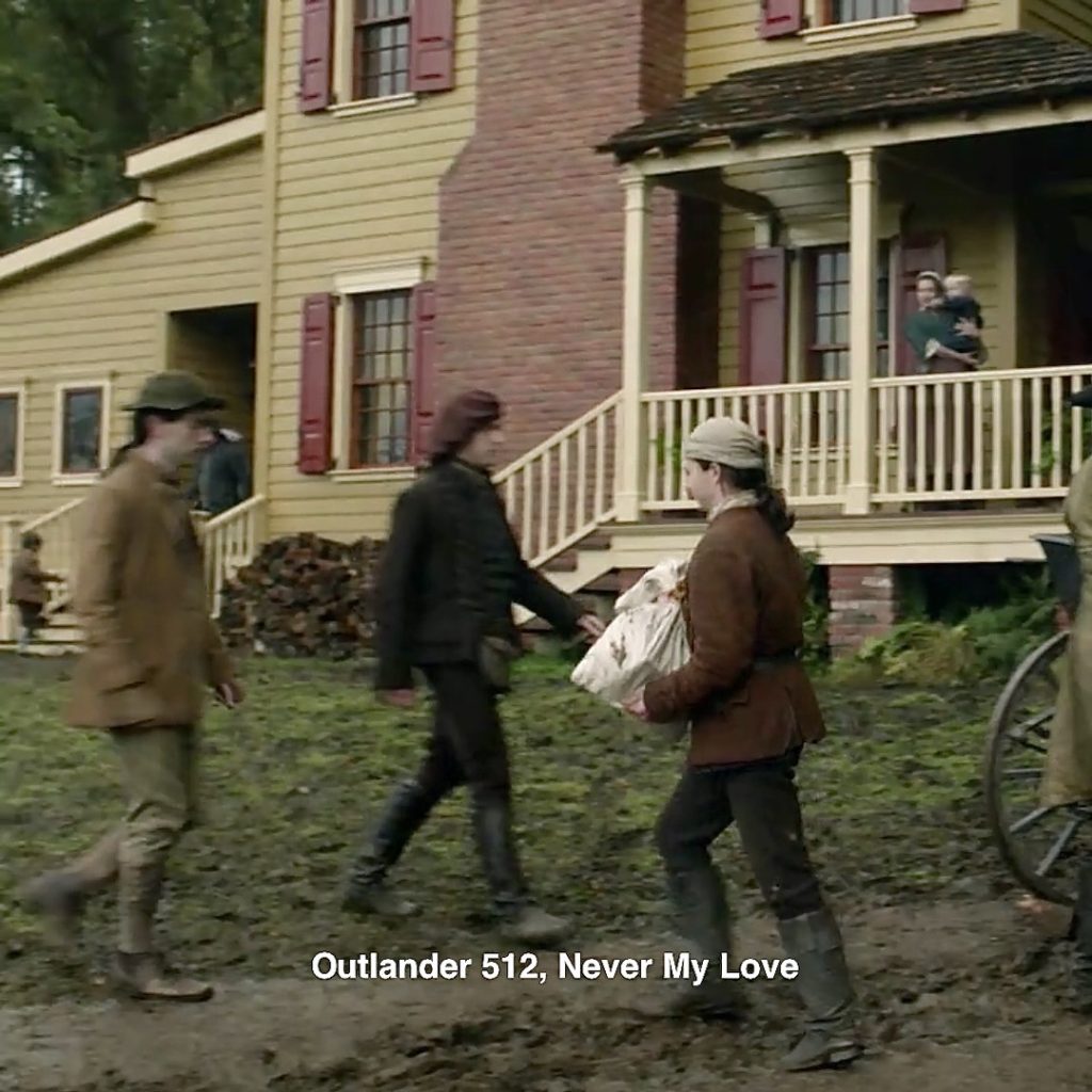
48. Woodpile! Claire grants her imaginary home a woodpile. 🪓
Episode 512, Why not a woodpile? A massive one is maintained at the big house which sports three brick chimneys!
Episode 301, Claire has a small woodpile in the back of her car. Millie offers to help.
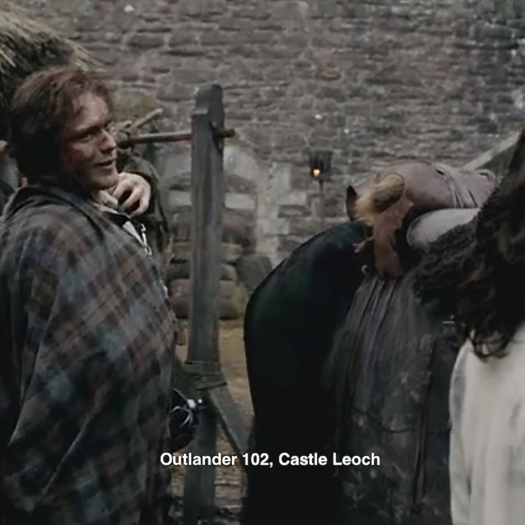
49. Stone Wall: Claire’s imaginary house sports a stone wall feature.
Episode 102: Outlander has lots of stone walls – Lallybroch, Wentworth Prison, Ardsmuir Prison, and Castle Leoch, to name a few. Let’s go with the castle. She will recall those stones! 🏰
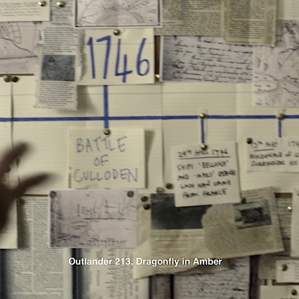
50. Bulletin Board: Everyone needs one in their dreamscape! 😄

Episode 213: Roger’s very, very messy bulletin board waaay back in S 2! 🧹Course, there are a bunch of other bulletin boards, too.
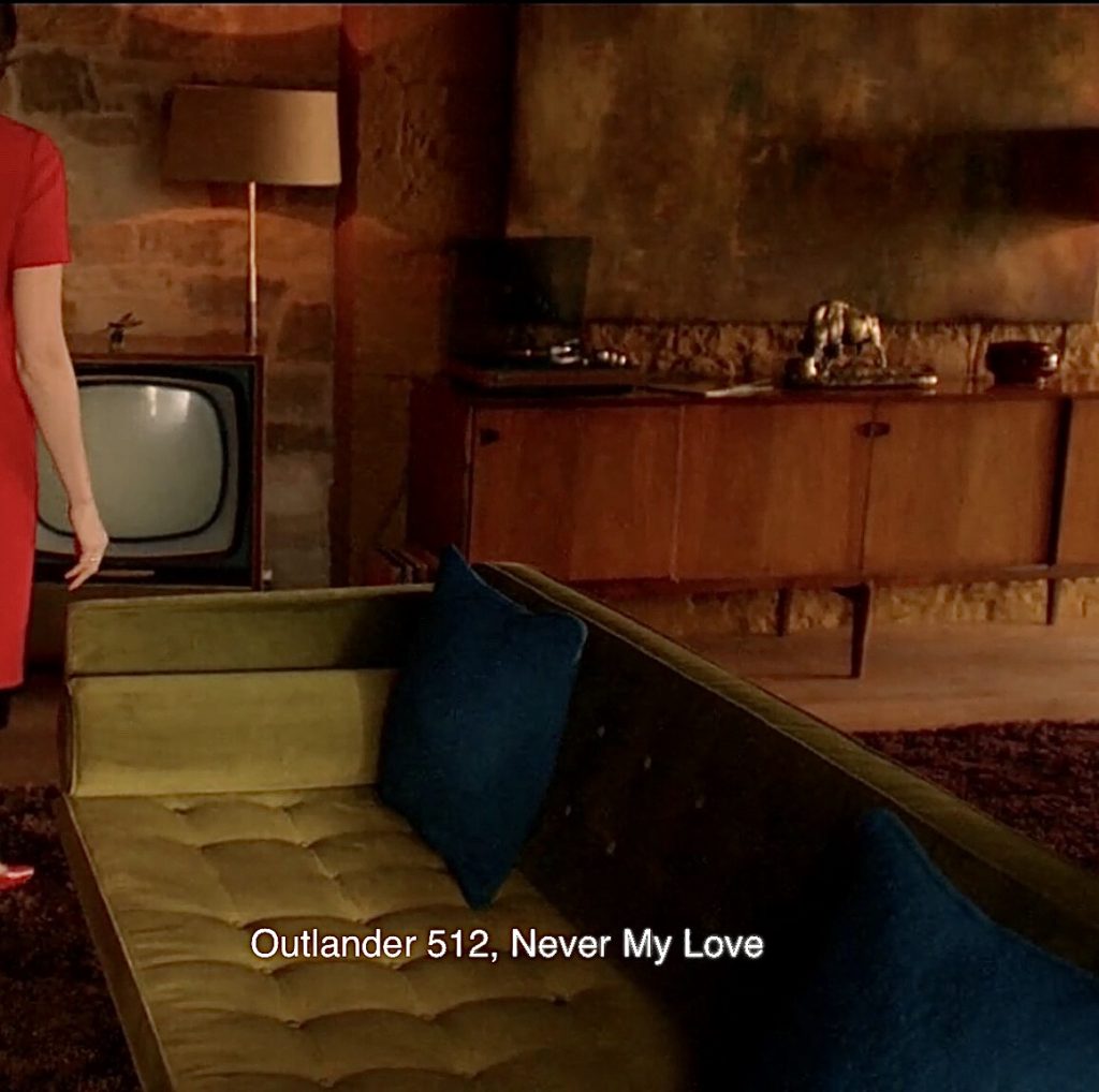
51. Green Couch! Yep. I noticed this beauty. I love the color and so popular in the 60’s. Green washing machines, dish washers, fridges, ovens, etc. The harvest colors were so in!
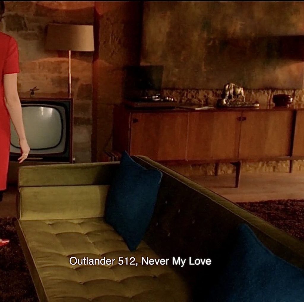 in!
in!
Episode 305: Hearkens back to a green couch in her Boston Home!
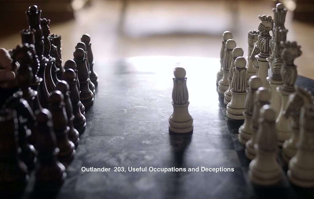
52. Chess: The base of that green lamp behind the battling bolsters looks like a chess piece to some!
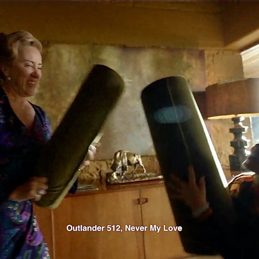
Episode 203: Jamie plays chess with the French finance minister! There is a bit of resemblance. He also played chess with Lord John Grey at Ardsmuir and with Lt. Knox before choking him to death. 😵
53. Cops! Doubtless, everyone recognizes the two cops at Claire’s door delivering tragic news about a car crash. Spoken in flat tones without care or compassion, the lives of beloved Bree, Roger, and Jemmy! Yep, Hodgepodge and the Weasel, a pair of…. 👿👿
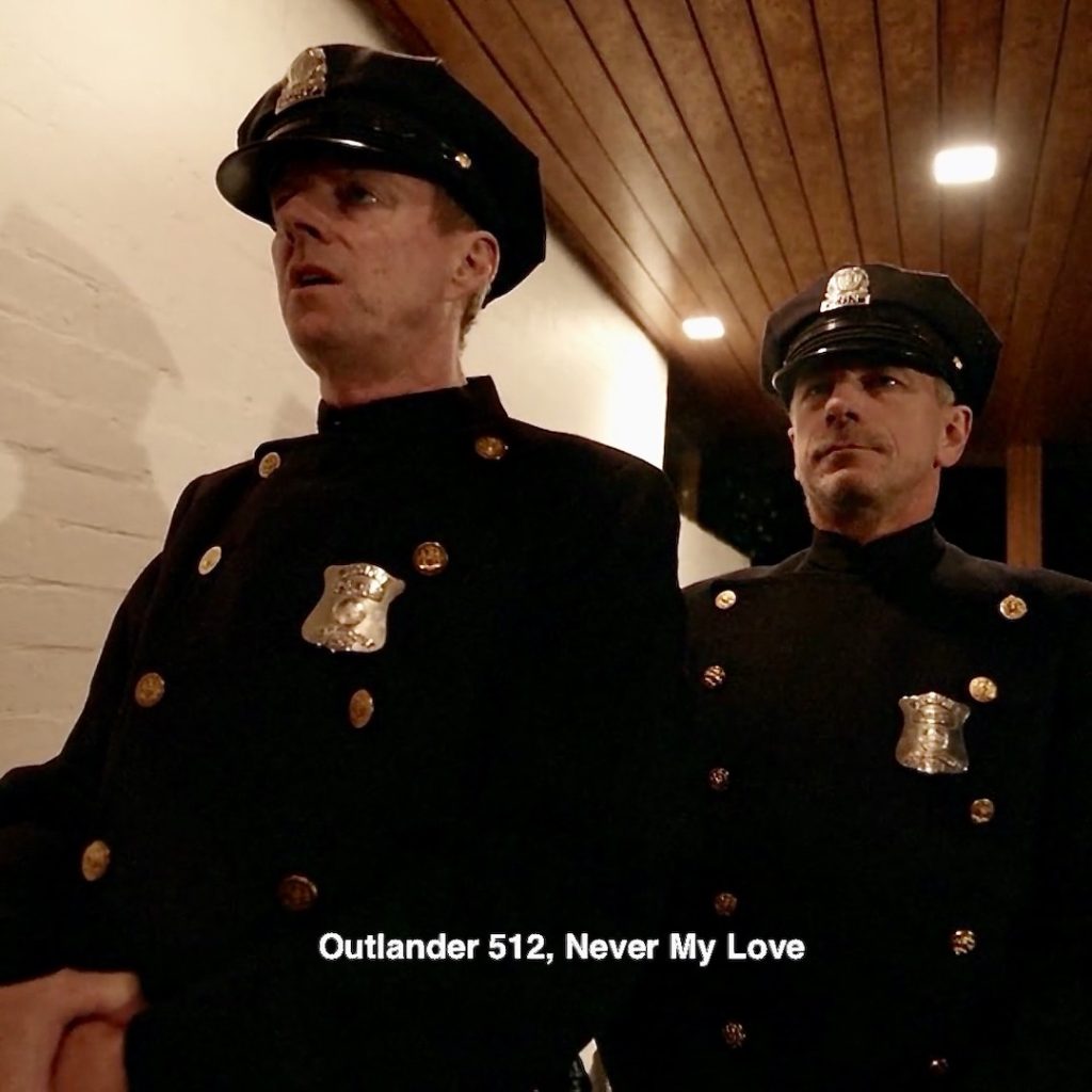
Episode 512: Here are the cowards! Hodgepile listening as dead-eye Lionel delivers his scathing accusation against Dr. Rawlings column, informing women about their reproductive lives! And, depriving him of his god-given rights in the marriage bed. 😡
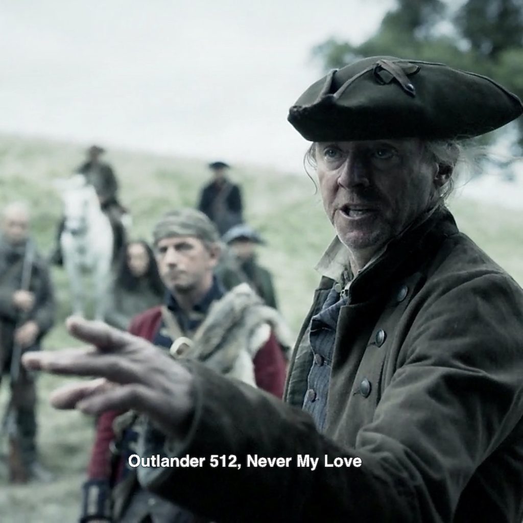
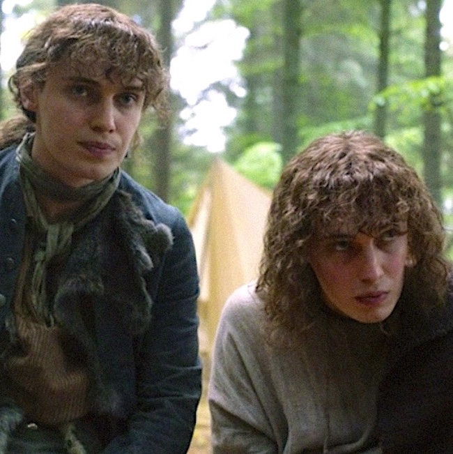
54. Prestonpans: As Murtagh messes with Germain, he mutters:
“Its Prestonpans all over again!”
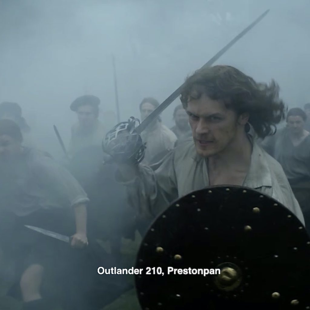
Episode 210: We all remember Highlanders charging through the mists like vengeful ghosts!
55. Painting: I have struggled to find a connection for the stunning painting in the dining area of Claire’s 60s home. But, then, a reader came up with this excellent suggestion!
Episode 213: Here it is! Oh, I can see it! How about you? 🥰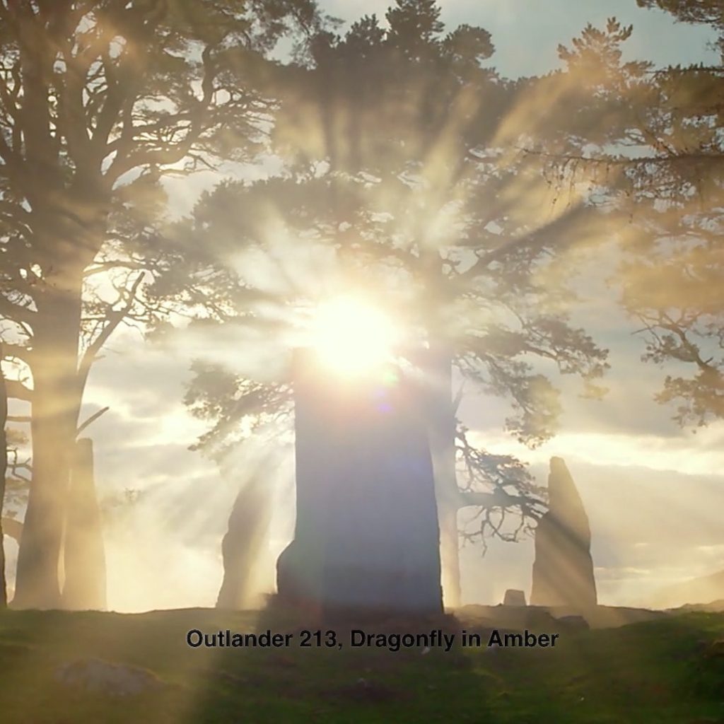
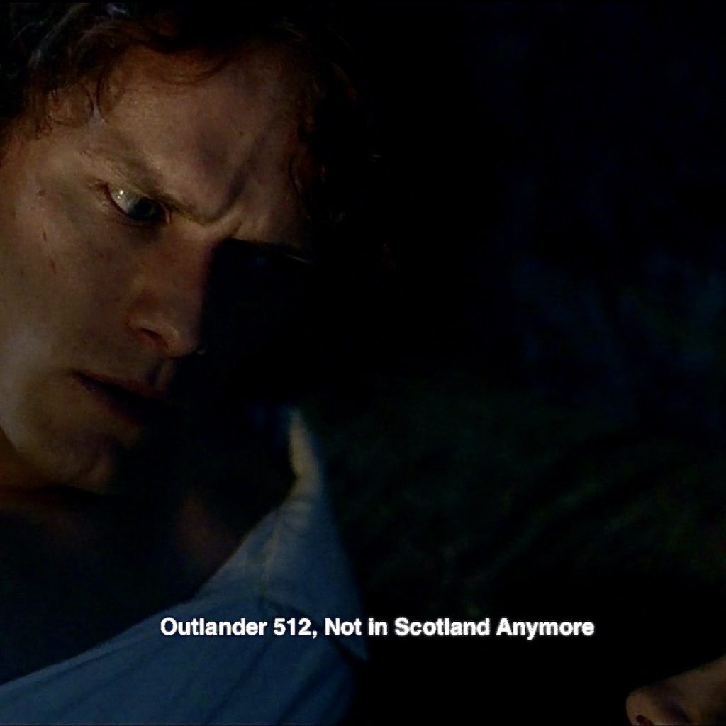
56. Gone: Claire’s tragic face after the news that her beloved child, grandchild, and SIL are gone. killed in a car crash! 🚙
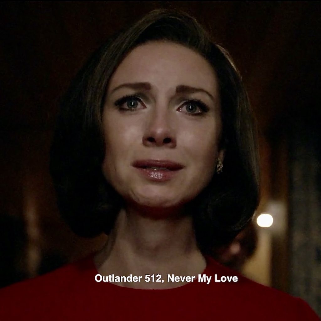
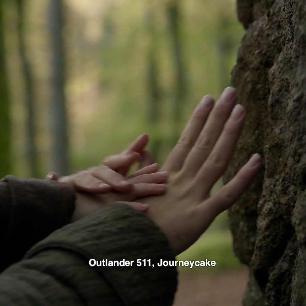
Episode 511: Claire’s mind sifted through the memories and latched onto the tragedy of Bree, Roger and Jemmy having gone back through the stones.
Episode 101: Claire has sad memories of car crashes. In episode 101, she compares traveling through the stones to the weightlessness of a car crash she once experienced.
And, her beloved parents were killed in a car crash causing Uncle Lamb to take her with him on his world travels. This quote from Outlander book:
My father’s only brother, and my only living relative at the time, he had been landed with me, aged five, when my parents were killed in a car crash. Poised for a trip to the Middle East at the time, he had paused in his preparations long enough to make the funeral arrangements, dispose of my parents’ estates, and enroll me in a proper girls’ boarding school. Which I had flatly refused to attend.
Episode 303: More bad news at the door as Claire faces Dandy-Candy-Sandy! Oops! 😳
Episode 303: And , finally, there was Frank. Another fatal car crash. 😥

Let’s finish with three special Easter Eggs just for Jamie and Claire!
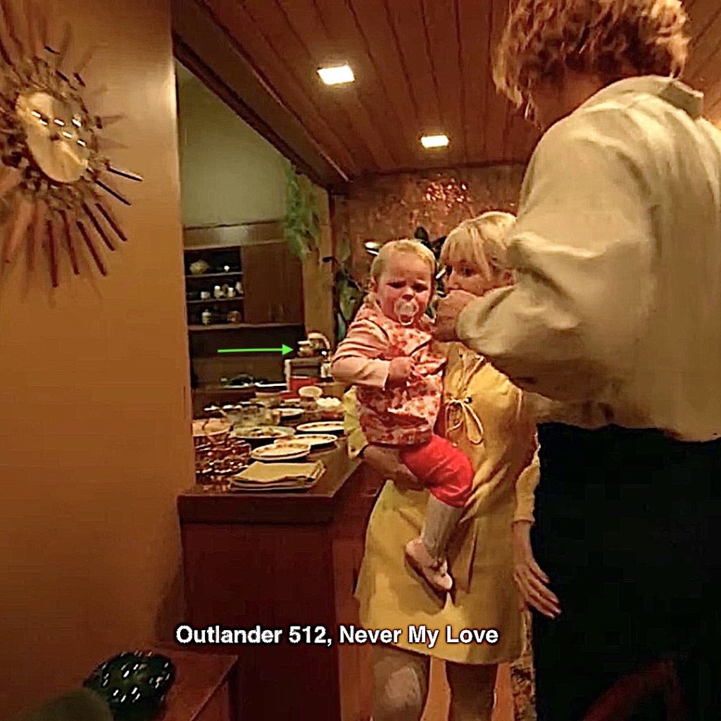
57. Honeypot! Matt B. Roberts declared a honeypot marked with a bee appear in Clair’s 60s home. I may be mistaken but it could be the pot at green arrow. 🍯 Meanwhile, Marsali shoves Jamie out of the way as she goes for more sweet potatoes and marshmallows for wee Jemmy!
Episode 202: Claire visits Louise de La Tour and comes away, to Jamie’s dismay, with a bare honeypot!
“Claire, What have you done to yourself? Your honeypot, It’s bare!” 😃
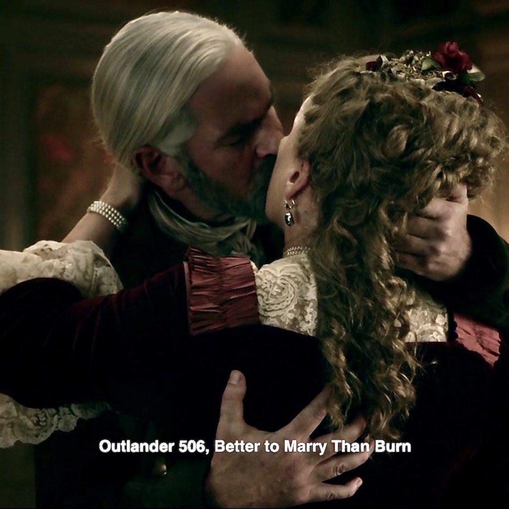
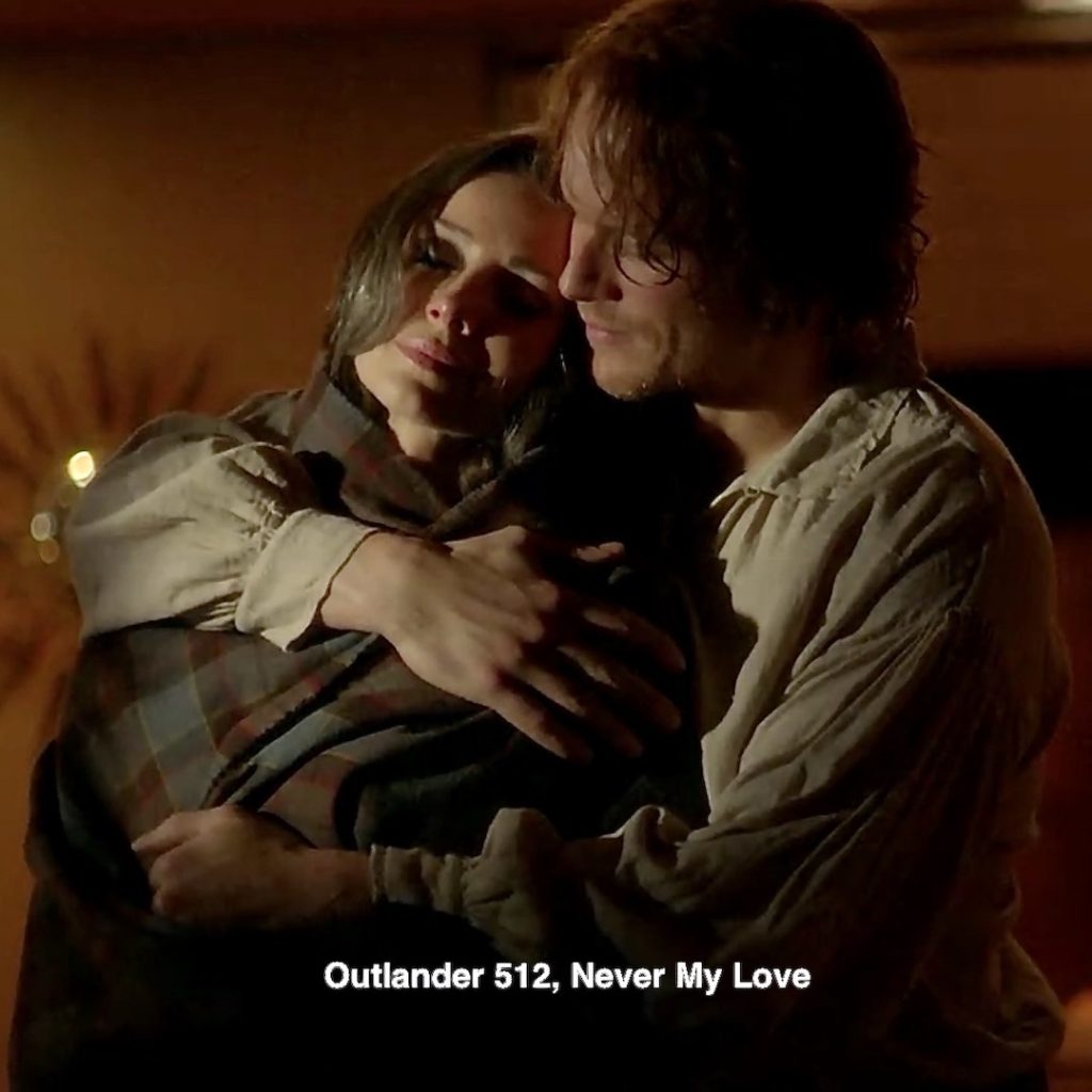
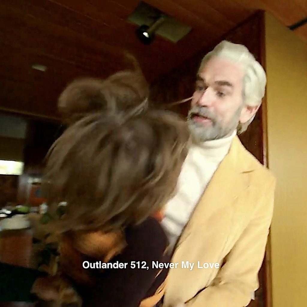
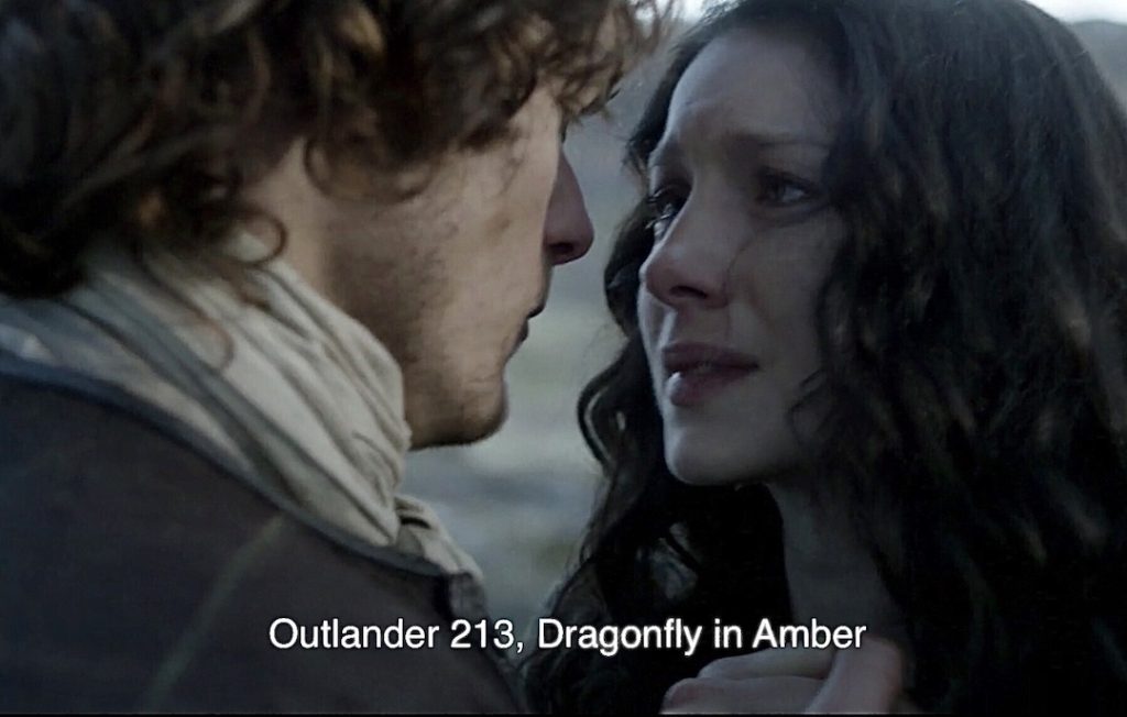
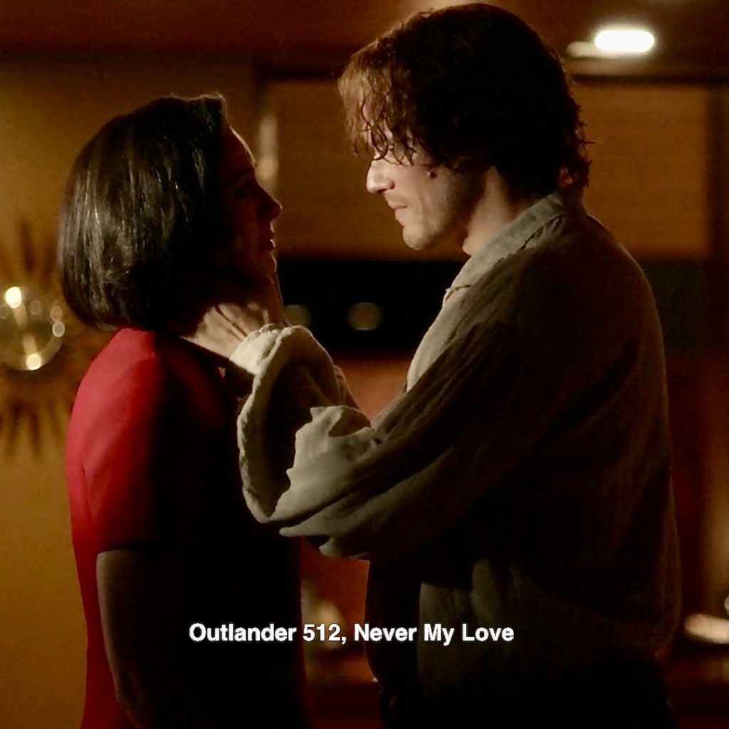
58. Slow Dancing: Claire imagines herself cradled in Jamie arms as they slowly dance together.
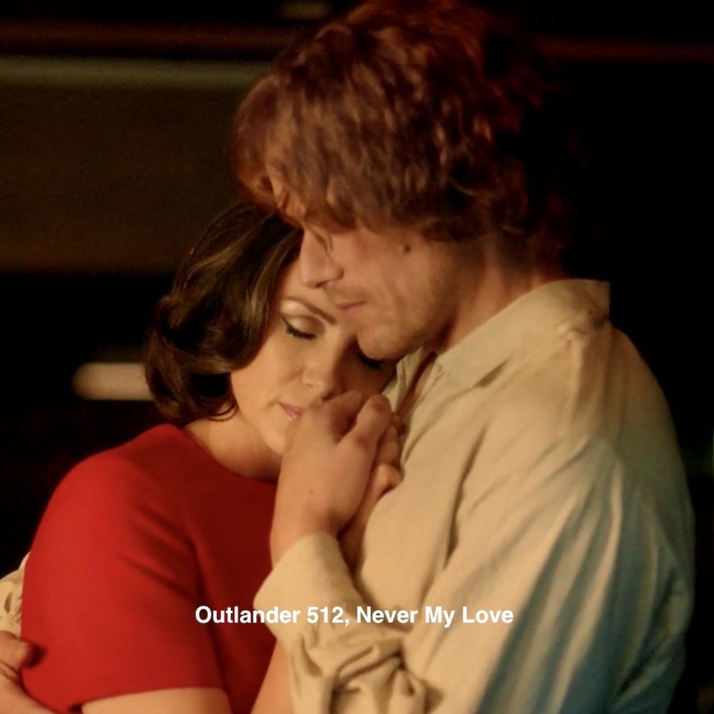
Episode 213: As Jamie leads Claire towards the stones to send her back to Frank, he faces her and grasps her hand as if they are dancing. 🥺
Let’s end with this splendid moment. We all ken it well!
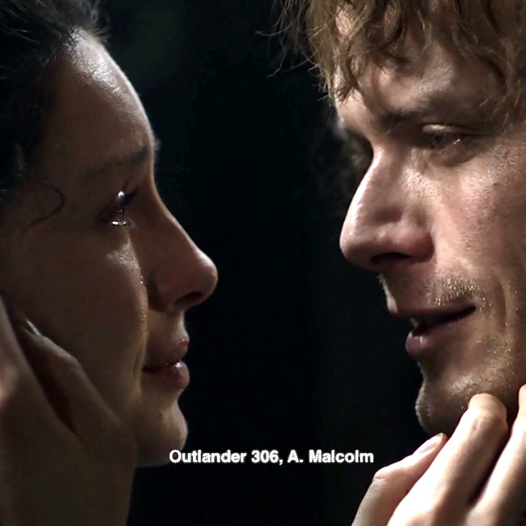
59. Dinna be Afraid: Another call back moment as Jamie cradles Claire’s face between his strong, capable hands. She remembers! 🤔
Episode 306: Whispering together in the print shop after 20 years apart! 😭
From Outlander book: 📖
“Don’t be afraid,” he whispered into my hair. “There’s the two of us now.” I felt warm, soothed, and safe for the first time in many days.
That’s All Folks!
Many thanks to those fans who shared Easter Eggs I originally missed! 🙏🏻
A trip down memory lane for me. How about you?
How many Easter Eggs in your basket?
Mine is full!
The deeply grateful,
Outlander Anatomist
Follow me on:
Photo and Video Credits: Sony/Starz;