Hallo, Outlander fans. Welcome to today’s Fun Fact: Anatomy of Ian’s Uniform!
This Fun Fact is especially appropriate for Americans as May 16th is also our national Armed Forces Day! Pretty timely, no?
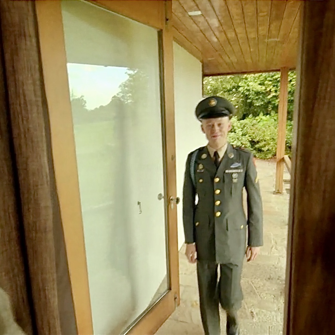
If you are like me, you are fascinated with Ian’s uniform in Outlander episode 512, Never My Love, the splendid finale of Season five! In Claire’s dissociative dreamscape, Ian arrives in full dress uniform for Thanksgiving dinner at the Fraser home! 🦃
I wanted to know more about Ian’s uniform, so I turned to Edward Maloney, Lieutenant Colonel, US Army (retired) for assistance. Know that LTC Maloney is also a faithful, long-time fan of Outlander books and show and was more than willing to share his decades-long expertise in this matter.
Just so you know, Colonel Maloney’s former unit is the 101st Airborne Division of the US Army. Below is its beautiful and dramatic Distinctive Unit Insignia which reads “Rendezvous With Destiny!”
Addendum: I just learned “Hang Tough and Currahee!” is the Battle Cry of the 101st Airborne! Hang tough means “in your parachute harness” and “Currahee” is from the Cherokee word meaning “We stand alone together.” (A good thought for soldiers trained to fight surrounded.)
Thank you, Lieutenant Colonel Maloney, for your service!

So, grab a cuppa (or your fav beverage) and let’s learn about the anatomy of Ian’s uniform, courtesy of LTC Maloney!
Ian’s uniform indicates he belongs with the Infantry, the oldest branch of the US Army. When was this branch formed? Turns out, very close to the date depicted in the Outlander S5 finale!
On 14 June 1775, the Continental Congress authorized ten companies of riflemen, the first infantrymen. Nine years later, the First American Regiment was constituted on 3 June 1784 and it was the 3rd Infantry. Currently, well over two hundred years old, clearly the Infantry is a distinguished branch of the US Army!
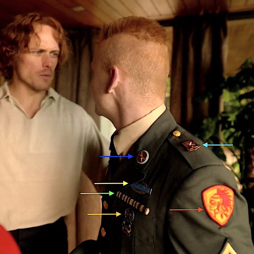
So, follow the colored arrows in the below images to discern the anatomical features of Ian’s uniform. (psst…The following image appears twice so you don’t have to keep moving up and down to follow the arrows and explanations!)
Let’s get on with the dissection! 😉
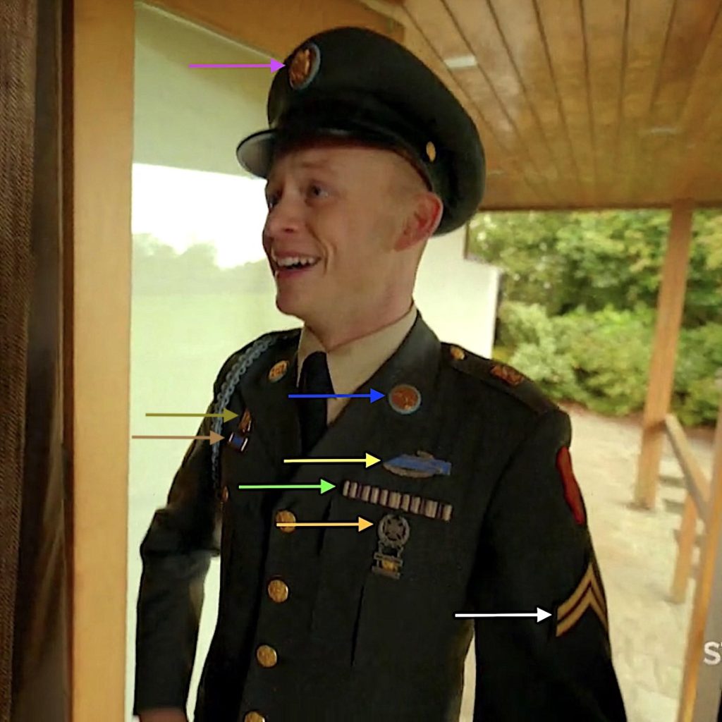
Blue Arrow: A branch insignia on Ian’s L lapel displays two gold crossed muskets, overlying a disk of Saxony Blue. This insignia is unique to infantry and no other branch of the US Army is allowed this distinction.
The crossed muskets are vintage 1795 Springfields, the first official US shoulder arm made in a government arsenal:
-
- caliber .69
- flint lock
- smooth bore
- muzzle loader
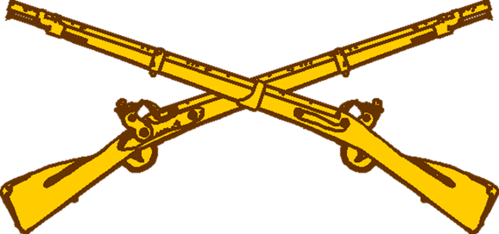
Yellow Arrow: Above his L breast, Ian wears a Combat Infantryman’s Badge, unique to those who have served in combat. It is a single flint lock musket on a blue background.

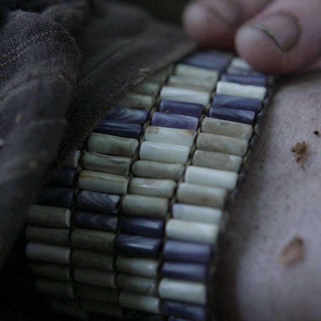
Green Arrow: Just above Ian’s L breast is a line of Decoration & Award Ribbons, also known as “fruit salad” or “Travel Ribbons.” These are worn in lieu of larger full-size or miniature medals which are awarded for service. Here, the Ribbon is a line of Ian’s Mohawk beads!
Gold Arrow: Over Ian’s L breast is The Maltese Cross with a Bull’s-eye surrounded by a wreath, known as the Expert Marksman Badge. This badge is unique to Army and Marine Corps, although the designs differ. The suspended bars underneath the badge are added for each weapon the soldier qualifies as an expert, such as pistol, rifle, etc.
White Arrow: Worn on Ian’s L sleeve, the chevrons signify a soldier’s rank. Two chevrons indicate Corporal, the lowest Noncommissioned Officer Rank (NCO) who leads an infantry fire team.
Violet Arrow: Ian’s service cap bears the US Coat of Arms. It is backed on a brass disk and in the case of infantrymen, backed by a Saxony Blue disk.
Khaki Arrow: Ian’s Regimental Coat of Arms is worn above his R breast. This insignia will indicate his permanent regiment not necessarily the one to which he is currently assigned.
Tan Arrow: Also worn above his R breast, this insignia indicates a Unit award such as Presidential Unit Citation, Distinguished Unit Citation, etc.

Next image, we see Ian’s left shoulder!
Aqua arrow: Indicates the Distinctive Unit Insignia which is usually a variation of the regiment’s coats of arms but unique to each regiment. This one looks like the 2nd Infantry Regiment but the colors are altered.
Red Arrow: L shoulder – Shoulder Sleeve Insignia indicates the current unit of assignment – Ian serves with a Native American unit, the Mohawk. this insignia was created by Outlander.
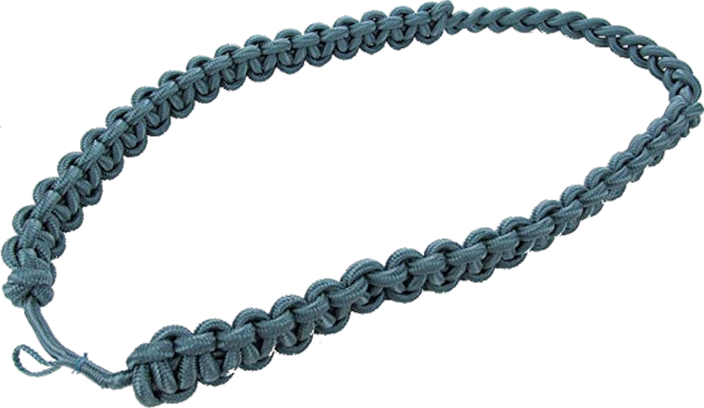
And finally, Ian’s infantry blue cord or fourragere (below) is a military decoration worn over the right shoulder of all infantry-qualified US Army soldiers.
Ian’s fourragere from afar.
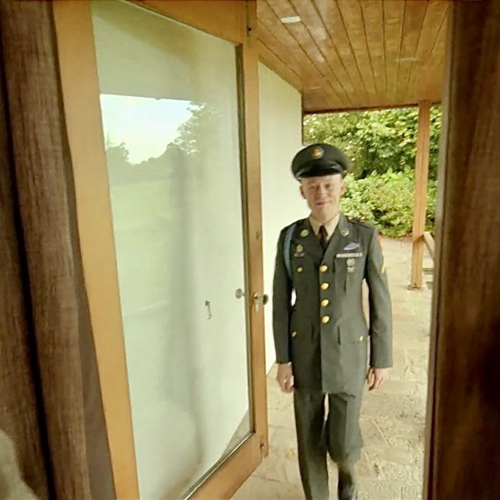
And, a closeup. This is Ian’s fourragere in light blue, (dubbed “Infantry Blue” by the US Army), worn under the right shoulder and under the right epaulette of a US Army infantry soldier’s dress uniform jacket.
The cord is composed of a series of alternating left and right half knots that are tied around a leader cord to form a “Solomon bar”.
Fitting that Ian should wear his dress uniform for Thanksgiving, even if that bird is just an illusion!
In summary, Ian’s uniform tells us he is:
-
- Infantryman
- Decorated soldier
- Served in combat
- Expert marksman
- Corporal of an infantry fire team
- Bears US Coat of Arms for infantrymen on his service cap
- Wears his permanent regiment’s Coat of Arms
- Decorated infantryman
- Member of a distinct infantry unit
- Member of Native American unit
- Qualified infantryman
Whew! I don’t know about you, but I am thoroughly impressed with warrior Ian!
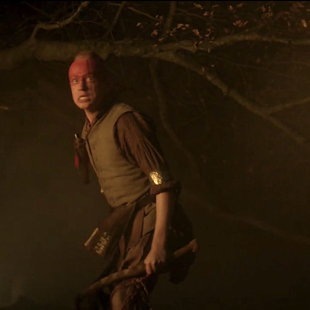
And, I am deeply grateful for the men and women who serve in the US Armed Forces.
I could not have done this fun Fact without the aid of LTC Edward Maloney, US Army. Thanks to his expertise for this brief lesson exploring the anatomy of Ian’s US Army insignia!
I hope all Outlander fans, worldwide, will express their gratitude for the warriors who daily protect them, their families, and their homelands. Please take a quiet moment to honor them!
Disclaimer: If there are any glitches in the insignia descriptions or attributions in this Fun Fact, the fault is entirely my own for not expressing the information with precision.
The deeply grateful,
Outlander Anatomist
Follow me on:
-
- Twitter @OutLandAnatomy
- Join my Facebook Group: OutlandishAnatomyLessons
- Instagram: @outlanderanatomy
- Tumblr: @outlanderanatomy
- Youtube: Outlander Anatomy
Photo and Video Credits: Sony/Starz; Lt. Col. Edward Maloney; www.wikipedia.com; www.amazon.com

