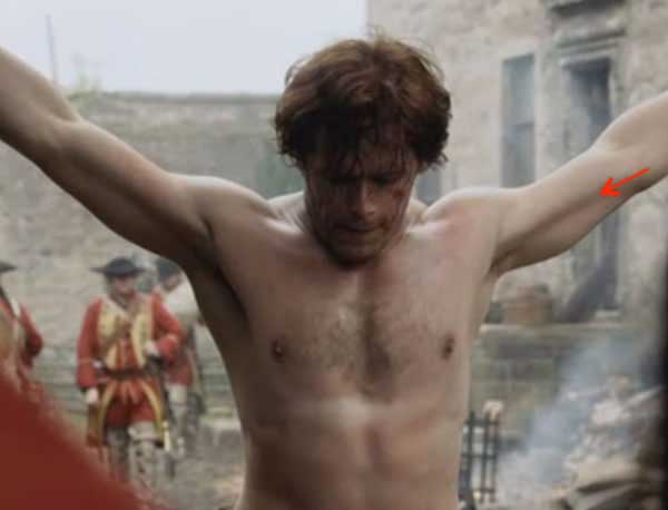
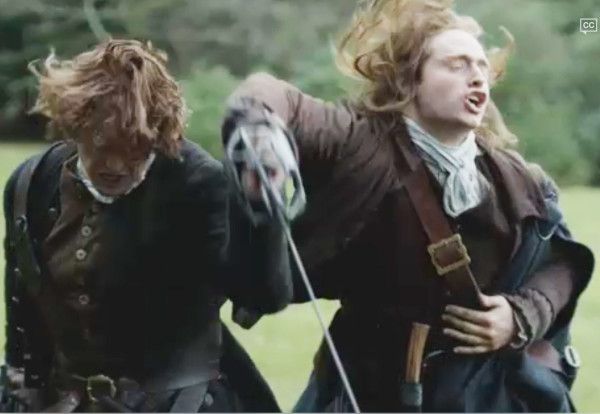
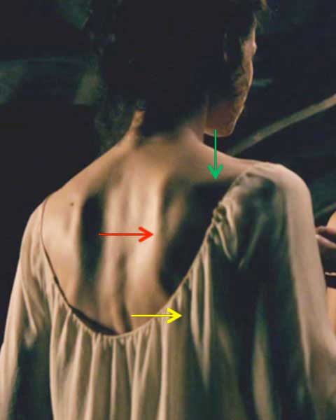
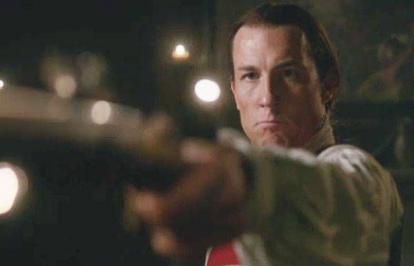
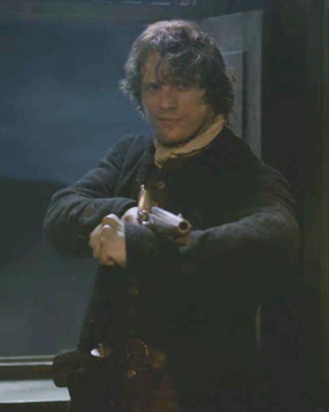
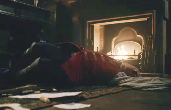
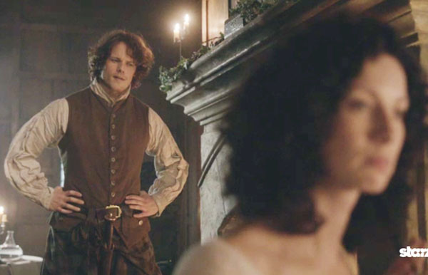
Welcome to today’s Anatomy Lesson #20: Arm and Forearm! Dapper Edward Gowan, romantic soul he, introduces our topic. Bless neat Ned’s heart – he is the only inhabitant of Castle Leoch courageous enough to defend our beloved Claire at the witch’s trial (Starz episode 111, The Devil’s Mark). Not only is he clever and adroit at turning an argument on its head but after Claire and Geillis are sentenced to burn, he brandishes a pistol and threatens the entire assembly: “Wait, you cannot do this. I forbid this!” Who knew he was armed mayhap with the same firearm he used to shoot a Grant from 20 paces (Starz episode 108, Both Sides Now)! Reminds me of an apropos John Wayne quote: “Courage is being scared to death and saddling up anyway.”
Way to go Ned. You are the man!
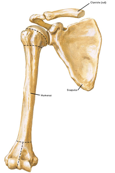
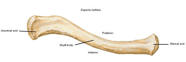
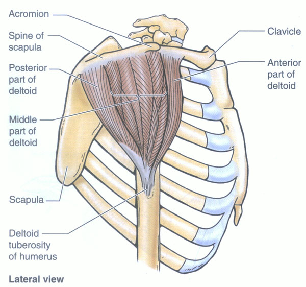
Today’s lesson will build on Anatomy Lesson #19: “To Arms, Too arms, Two Arms” wherein we learned about clavicle, scapula and humerus and the 17 muscles that move these bones. Today’s lesson adds more details of the glenohumeral joint and covers eight additional muscles that move arm and forearm. Do the math and realize that five muscles of arm and pectoral girdle haven’t been covered. We will return to those in a future lesson.
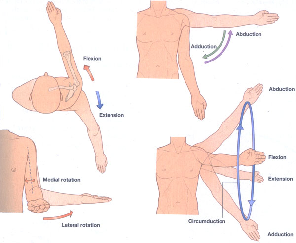
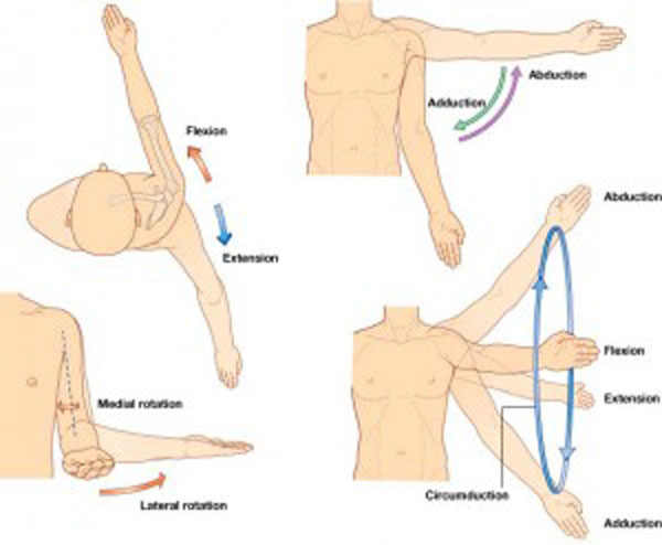
Let’s begin by reviewing arm movements (Photo A). Hopefully you memorized these from our last lesson: flexion, extension, adduction, abduction, medial (internal) rotation, lateral (external) rotation and circumduction. Understand that arm movements can involve combinations of the above.
Photo A
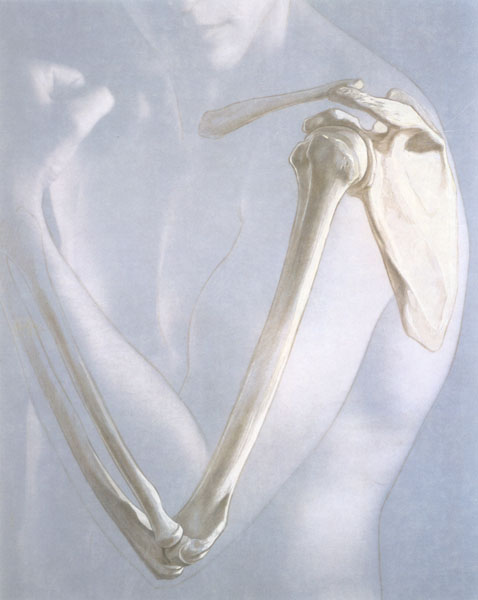
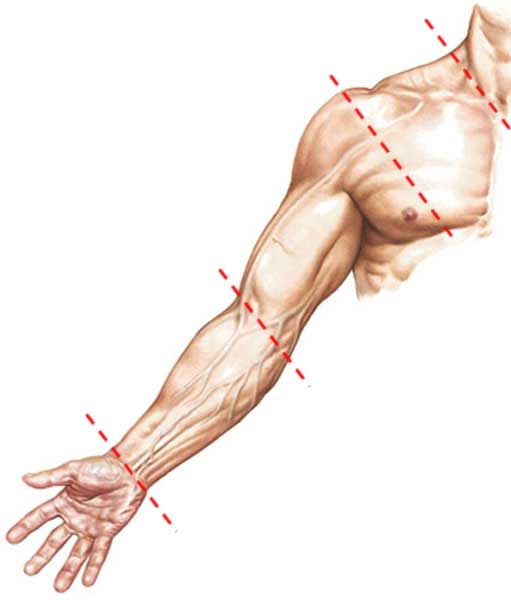
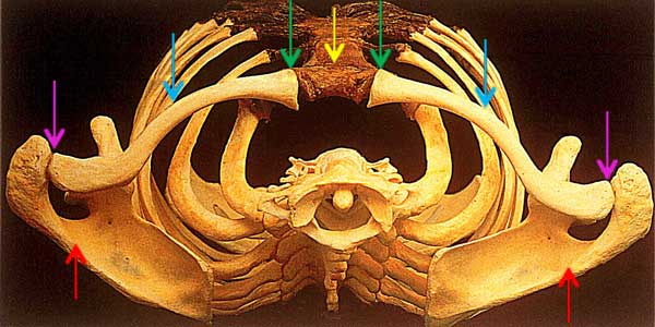
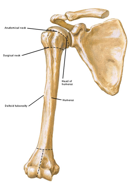
Now, onto the bony foundations of the upper limb; yes, there is more to learn. Movement enjoyed by humans requires a bony endoskeleton (endo from Greek meaning within) for muscular attachments. This haunting artistic rendering reminds us of scapula, clavicle (together the pectoral girdle), humerus and forearm bones all of which provide attachment for upper limb muscles.
Photo B
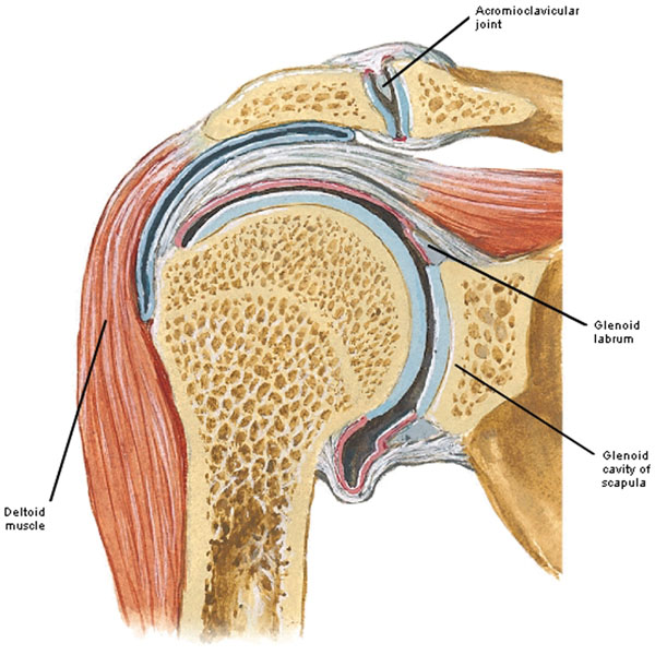
As you may recall the glenohumeral joint is the articulation between humeral head and glenoid cavity of scapula (Photo C). The glenoid cavity is roughly ¼ the size of the much larger humeral head meaning this ball and socket joint is highly mobile but easily dislocated as happened to our bonny Jamie (Anatomy Lesson #2 and Starz, episode 101, Sassenach).
Examine Photo C (slice through shoulder joint) and see that a blue layer covers humeral head and lines glenoid cavity; this is hyaline (articular) cartilage which covers most joint surfaces. The body contains three different types of cartilage but the most common type is hyaline. Cartilage is one of several body tissues that are avascular (lack blood vessels). Thus, in the case of cartilage, oxygen, nutrients and protective/repair cells are delivered and waste products are retrieved more slowly. The significance is this: if damaged, cartilage repair is slower to repair than vascular tissues. Warning: piercing cartilage (e.g. ears, nose) demands extra caution to keep wounds disinfected during the healing process. Once infected, the damage to cartilage can be fulminating because the tissue is avascular. The structure labelled glenoid labrum is important and will be discussed next.
Photo C
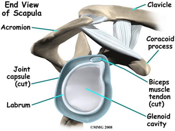
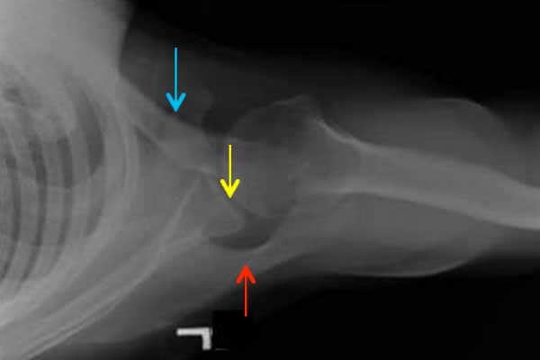
Some readers have inquired about the glenoid labrum, a part of the glenohumeral joint that is made of fibrocartilage (different than hyaline). It enlarges and deepens the glenoid cavity to more effectively surround the humeral head (Photo D – side view of right joint). Labrum injuries are fairly common and include symptoms such as pain, locking, popping, joint instability and decreased range of motion. Because the labrum is made of soft tissue, it cannot be visualized by x-ray and thus requires other imaging modalities.
Photo D
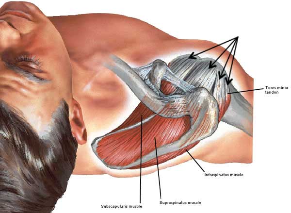
The glenohumeral joint (Anatomy Lesson #2 & Anatomy Lesson #19) is further reinforced by the rotator cuff, an incomplete collar of four muscle tendons (Photo E – superior view, black arrows). Rotator cuff muscles are: supraspinatus, infraspinatus, teres minor and subscapularis. Some anatomists prefer the less common term fibrous cuff because not all cuff muscles rotate the humerus as the name implies. And, in case you are not aware, tendons are connective tissue (collagen) elements that anchor muscle to bone; ligaments are connective tissue elements that attach bone to bone.
Photo E
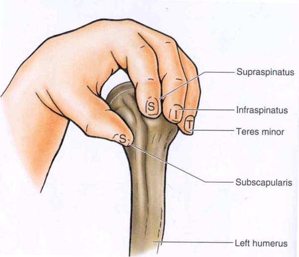
Referred to by the acronym SITS, rotator cuff muscles cover and reinforce top, back and front of each glenohumeral joint. Photo F (left humerus, left hand) employs a simple visual technique for recall: S (index finger) = supraspinatus; I (middle finger) = infraspinatus; T (ring finger) = teres minor and S (thumb) = subscapularis.
Photo F
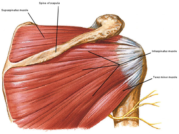
The first SITS muscle is supraspinatus so named because it arises from back of scapula superior to the scapular spine (Photo G). Its tendon passes over the top of glenohumeral joint and inserts into side of humerus. Called the workhorse of abduction, it actively raises the arm from full adduction to full vertical abduction as in hands up, partner!
The second SITS muscle is infraspinatus so named because it arises from back of scapula inferior to the scapular spine and inserts into back of humerus. Upon contraction, it produces lateral (external) rotation of the arm.
The third SITS muscle is teres minor, a small muscle that arises below infraspinatus and inserts into back of humerus. Often fused with infraspinatus, it may be indistinct. Teres minor contributes to lateral rotation of the arm.
Photo G
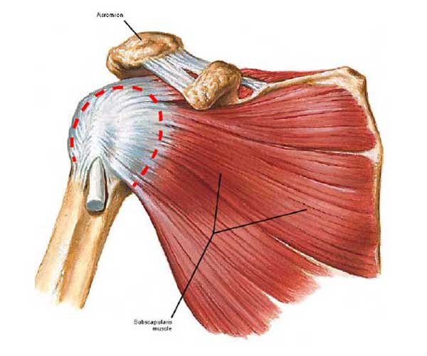
The fourth and last SITS muscle is the fan-shaped subscapularis which takes origin from front of scapula and inserts into front of humerus (Photo H). Its contraction produces medial (internal) rotation of the arm.
In summary, all SITS muscles rotate the humerus except one abductor, supraspinatus.
Again, the tendons of the four SITS muscles reinforce the glenohumeral joint as the rotator cuff (Photo H – dashed red line). One or more of these tendons is a common cause of shoulder pain, tears, impingement and/or inflammation. I cannot demonstrate subscapularis with Starz images because it is situated deeply between scapula and rib cage.
Photo H
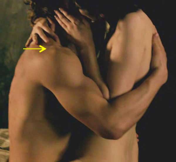
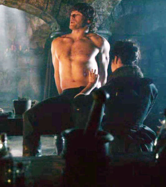
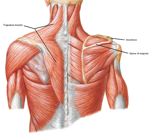
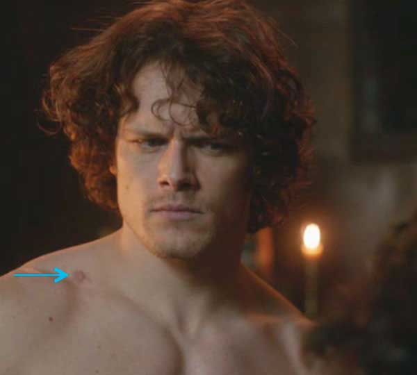
But, I can demonstrate three SITS muscles using our fav model, Jamie! Aye, the obliging laddie hopped up on the dissection table again – he’s so accomodating helping us all learn about human anatomy. Thank ye, Jamie! In the next image, Claire’s graceful right index, middle and ring fingers rest over his awesome trapezius (Starz episode 108, The Wedding). Supraspinatus lies deep to the yellow arrow; it doesn’t create a distinct bulge (hehe) because it is covered and overshadowed by the more powerful trapezius. Nevertheless, we can identify its location.
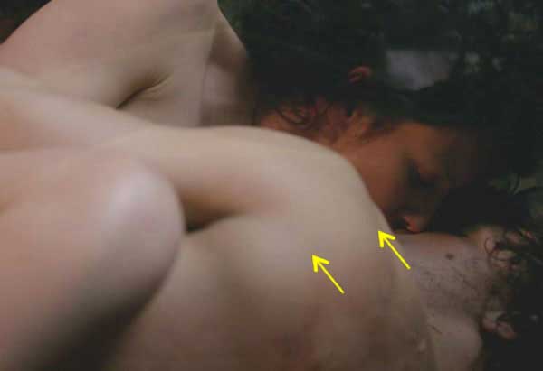
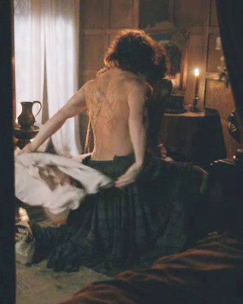
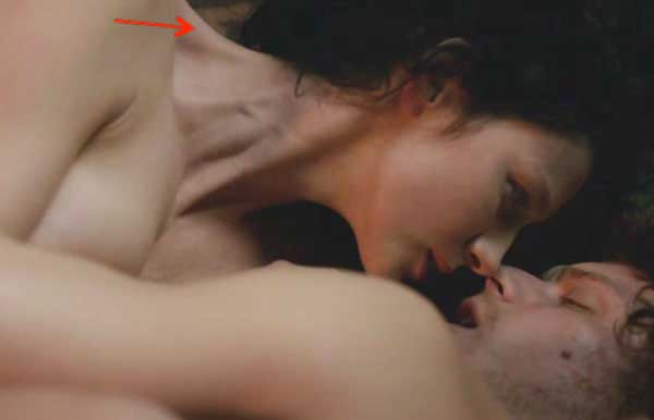
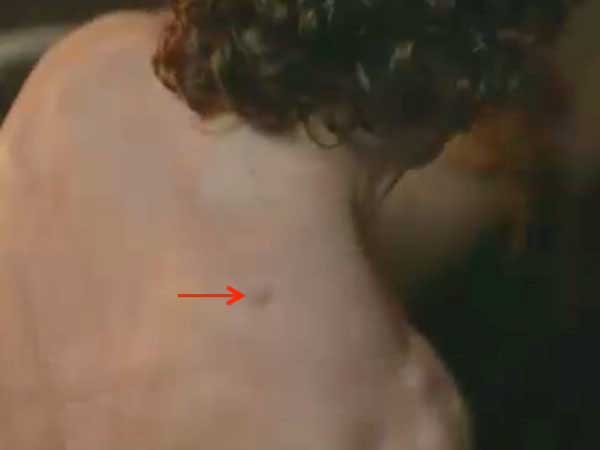
Can we see Jamie’s infraspinatus? Definitely yes! In fact, there are so many fine examples from Starz episodes it is difficult to choose. But here are a couple of favorites. Jamie’s infraspinatus is verra well-developed – doubtless from pitching hay all day at Lallybroch and wielding his, erm, heavy dirk! In the next image, a relaxed infraspinatus underlies the elevated flesh between the yellow arrows. Here Jamie welcomes lovely Claire’s slow and sensuous kiss (Starz episode 109, The Reckoning). Although most of his back is covered with flogging scars, skin overlying infraspinatus remains largely untouched. Herself explains the extent of those scars (Outlander book):
…Even by candlelight and having seen it once before, I was appalled…The scars covered his entire back from shoulders to waist. While many had faded to little more than thin white lines, the worst formed thick silver wedges, cutting across the smooth muscles.
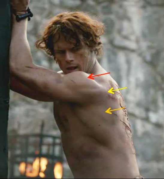
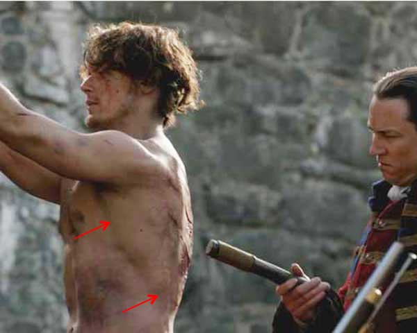
The following image shows a contracted infraspinatus (yellow arrow) as Jamie’s upper limbs strain against his manacles. The red arrow points to deltoid (Anatomy Lesson #19) which is demarcated from infraspinatus by a skin groove. An orange arrow marks teres major, our next muscle. Here Jamie is shackled to the whipping post at Fort William and nearly beaten to death by the unholy vulture (Starz episode 6, The Garrison Commander). Herself describes it (Outlander book):
….“Well, I was tied to that post, tied like an animal, and whipped ’til my blood ran! I’ll carry the scars from it ‘til I die.”
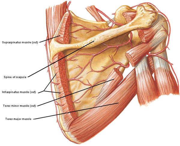
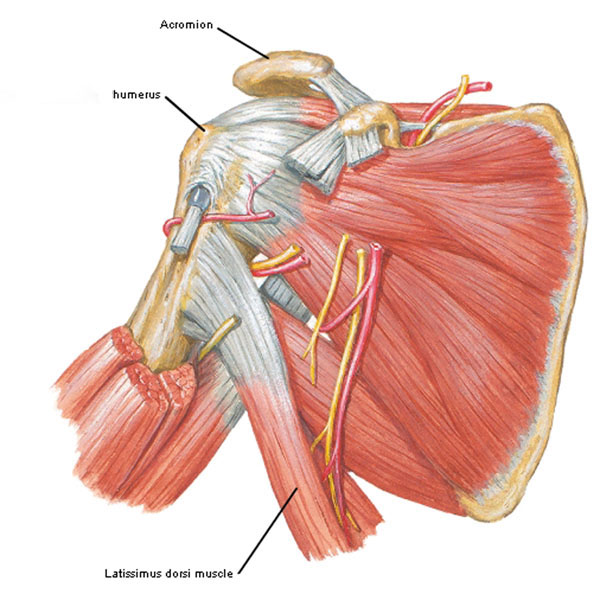
The next muscle moving the humerus, teres major, is not a rotator cuff muscle (Photo I). This strong and substantial muscle arises from the back of scapula just below teres minor and inserts into the front of humerus. Teres major has three functions: it adducts, extends and medially rotates the humerus.
Photo I
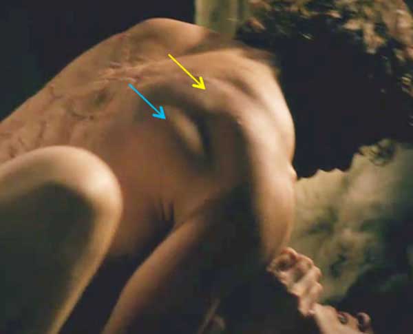
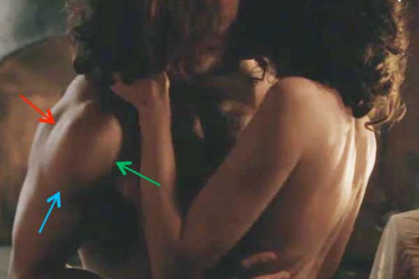
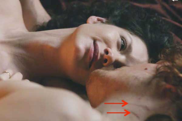
Oops! Just lost me train of thought gawking at the next image (Starz episode 107, The Wedding)! What was the topic? Ah, I remember….. Here on their wedding night, Jamie gets the idea – aye, the lad is a quick study! He no longer crushes Claire as he contracts teres major (blue arrow) and infraspinatus (yellow arrow) to help support his weight.
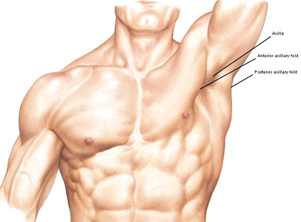
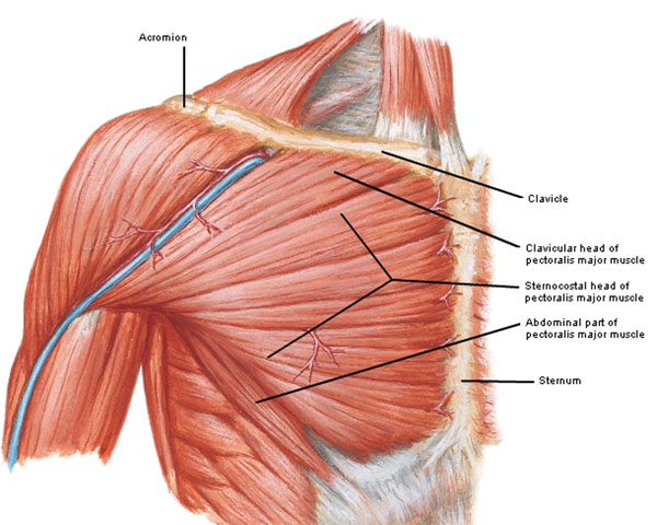
Teres major also helps form an important topographical landmark. In Anatomy Lesson #10, The Chest, we learned that pectoralis major forms the anterior axillary fold as it passes from chest to humerus (Photo J). Now, in anatomy, if there is an anterior there is a posterior so the posterior axillary fold contains both latissimus dorsi (Anatomy Lesson #19) and teres major muscles. The armpit or axilla (anatomical term) lies between these two folds.
Try this: If you can, abduct one arm to the vertical position. Place the opposing fist into your axilla. Now open the hand and grip the front fleshy fold; this is the anterior axillary fold formed by pectoralis major. Move your fingers backward and grip the back fold of tissue: this is the posterior axillary fold containing teres major and latissimus dorsi muscles and scapula. Good job, students!
Photo J
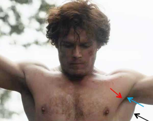
Let’s find the above features using a Starz image. Here, Jamie’s wrists are bound and his arms elevated to about 95° of abduction (slightly above horizontal). In this position, the hollow of the axilla (turquoise arrow), anterior axillary fold (red arrow) and posterior axillary fold (black arrow) are clearly visible. Here, at Lallybroch, he receives his first beating from that ghastly and gruesome garrison commander (Starz episode 2, Castle Leoch). Herself writes (Outlander book):
They stripped off my shirt, bound me to the wagon tongue, and Randall beat me across the back with the flat of his saber. He was in a black fury, but a wee bit the worse for wear, ye might say. It stung me a bit, but he couldna keep it up for long.”
Try this: Grab a friend right inbetween the red and blue arrows and see what happens… You might want to jump back quickly once you make the grab. Just joshing! You probably dinna want to do that after all…..although I bet Rupert and Angus would be game.
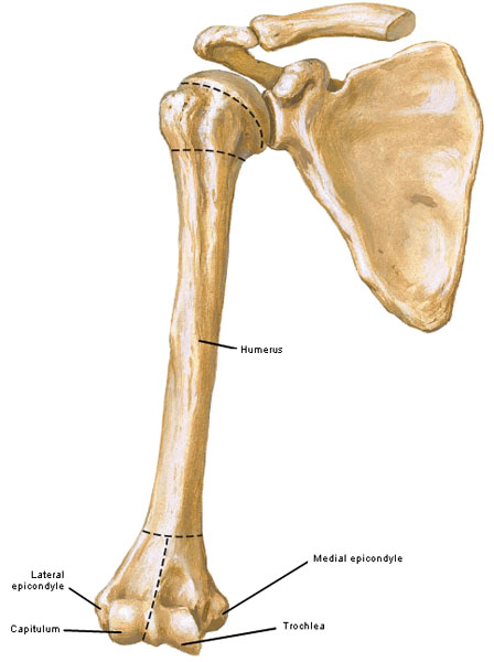
Lets return to the humerus and study its distal (far) end which helps form the elbow joint (Photo K – anterior view). The shaft of the humerus flares near the elbow ending in two bony side knobs, medial epicondyle and lateral epicondyle. The tip of humerus bears two oddly shaped parts: trochlea (Latin meaning pully) and capitulum (Latin meaning small head), structures that articulate with two forearm bones.
Try this: Grip one arm about midway down with the opposite fingers and feel the hard bony shaft of humerus. Now, with elbow bent and palm turned to the sky, grip the elbow joint between thumb and index finger. Feel the bony knobs? The medial epicondyle lies nearest the body and the lateral epicondyle is away from the body. You cannot palpate trochlea or capitulum as they lie too deeply.
Photo K
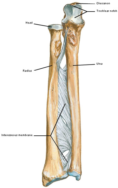
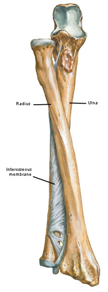
In Anatomy Lesson #19, we learned that the upper limb between elbow and wrist joints is the forearm and it contains two bones, radius and ulna (Photo L – anterior view of right forearm). With palm up or facing forward, the longer ulna lies medially (little finger side) and the shorter radius lies laterally (thumb side). The bluish layers represent hyaline cartilage that covers the joint surfaces. The bones are united by a tough fibrous interosseous membrane. The top of ulna bears a bony projection, the olecranon with a large trochlear notch. With the bones parallel, the forearm is in a position known as supination: palms face forward or upward.
Try this: With a bent elbow and palm facing up palpate the bone along the back of forearm; this is the ulna. Most people can only palpate the distal (far) half of the radius as it nears the wrist on the thumb side; please feel yours. Find the bony olecranon or point of the elbow.
Photo L
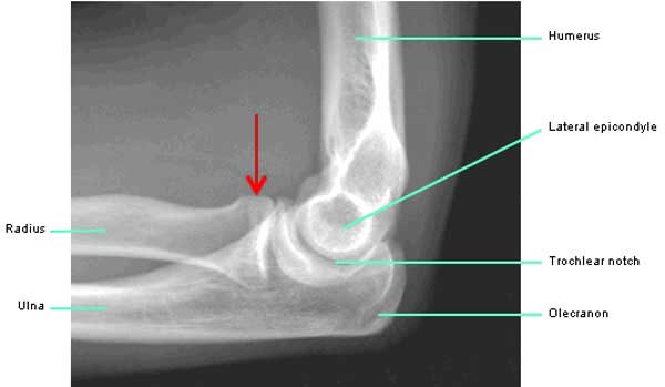
Both ulna and radius participate in the elegant elbow joint. Here, the trochlear notch of ulna grips the trochlea of humerus like a pipe wrench; together they form a hinge joint that opens with extension and closes with flexion (Photo M – flexion). The disk-like head of radius (red arrow) cups the humeral capitulum and pivots on it during supination and pronation (described below). Thus, radius, ulna and humerus together form the two-part elbow joint.
Photo M
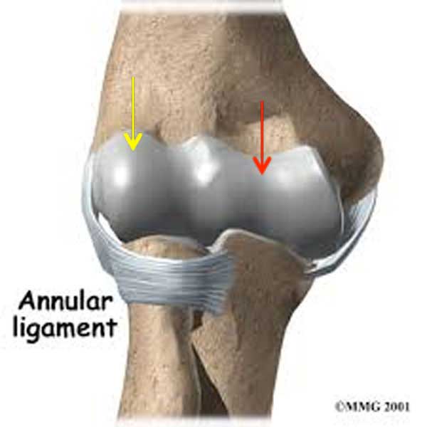
Although the head of radius and capitulum are loosely associated, a strong annular ligament holds the radial head against the ulna (Photo N – anterior view right elbow joint): red arrow marks the trochlea and yellow arrow indicates the capitulum of humerus.
Photo N
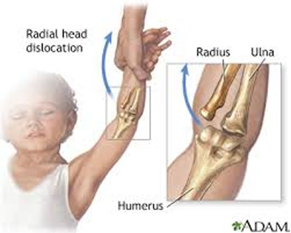
Now for a wee but important Clinical Correlation: In young children, the radial head is small and loosely seated in the annular ligament (Photo O). A quick jerk of a child’s upper limb can subluxate (partially dislocate) the radial head meaning it is pulled from the grip of the annular ligament. Also known as pulled elbow or nursemaid’s elbow, radial head subluxation is the most common upper-extremity injury in infants and young children who present at an emergency room (ER). It most often occurs as an adult holds a child’s hand and jerks the arm or pulls on it to prevent a fall; understand that the jerk can be minor or even trivial. Happily, reduction of the subluxated radial head is easily performed in the ER.
Photo O
Now let’s discuss forearm movement. Returning to this earlier image (Photo P), note radius and humerus lie roughly parallel in a forearm position known as supination. Here, palms may face either upward (flexed elbow joint) or forward (extended elbow joint).
Photo P
With palm turned downward (flexed elbow) or backward (extended elbow) the distal (far) end of radius crosses in front of distal ulna, a forearm position known as pronation (Photo Q). Due to ligament and muscle tension, pronation is the typical resting position of forearm and hand as it is more comfortable than supination. Lastly, with elbow joint extended and palms facing the body, the forearm lies between pronation and supination.
Try this: With bent elbow, turn palm up and palpate the forearm bones: they are parallel, the thumb points away from the body and the forearm is supinated. Keeping the bent elbow slowly turn forearm so the palm faces down. Watch the distal radius move from the side to the front of ulna and come to rest so the thumb points toward the body; the forearm is now pronated. Feel the radius move as you alternately supinate and pronate the forearm. Notice that the hand is passively carried with the forearm movements. Pronation and supination are possible because the radial head spins freely on the capitulum as distal radius swings back and forth. This ingenious design adds significant mobility to the upper limb enabling us to bring our hands in front of the face for fine manipulation of the environment!
Photo Q
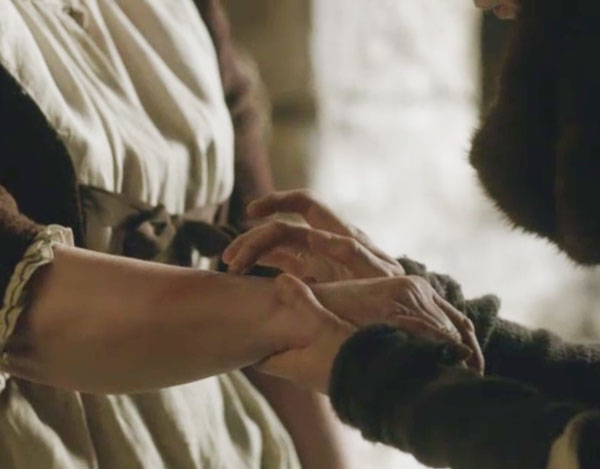
Okey dokey, let’s find pronation and supination using dazzling images from Starz episodes. The first image is of Herself! Yes! And, this is the first time I have used Herself to teach anatomy: here in the form of fine-fabulous-fictitious Iona MacKenzie (Starz episode 104, The Gathering). Diana can now add actress to her prodigious resume!
Question: Is Iona’s left forearm pronated or supinated? Is her elbow joint flexed or extended?
Answer: Elbow joint is flexed. Forearm is pronated (pronation) meaning distal radius is crossed over distal ulna. You can determine this immediately even though her forearm is covered by lace and wool because the palms face down and thumb points toward the body. Ummm, Mrs. Fitz are ye are being a wee bit meow-meow towards Iona?
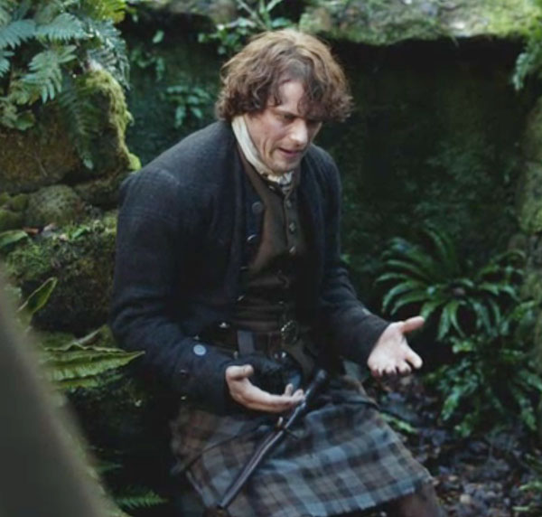
In the next image, Jamie is dismayed, defeated and deflated after his passionate battle of words with Claire (Starz episode 109, The Reckoning). His palms face up in an attitude of supplication. Herself describes it best (Outlander book):
…His voice cracked. “And when ye screamed, I went to you, armed wi’ nothing but an empty gun and my two hands.” … “You’re tearin’ my guts out, Claire.”
Question: Name the position of Jamie’s forearms.
Answer: Supination (supinated) meaning ulna and radius are parallel, palms face up, and thumbs point away from the body.
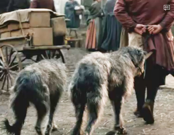
Let’s challenge ourselves with more complexity in arm and forearm movements. Here a gentleman walks his hounds at the farmer’s market (Starz episode 110, By the Pricking of My Thumbs). Forearms are behind his back. Palms face backwards.
Question: Name the position of his arms. Name the position of his elbow joints. Are forearms pronated or supinated?
Answer: Arms are extended. Elbow joints are flexed. Forearms are pronated. Remember: if palms face downward or backward, the forearm is in pronation. Do you get the idea? You can assess the overall position of the upper limb by considering its component parts and determining their individual positions.
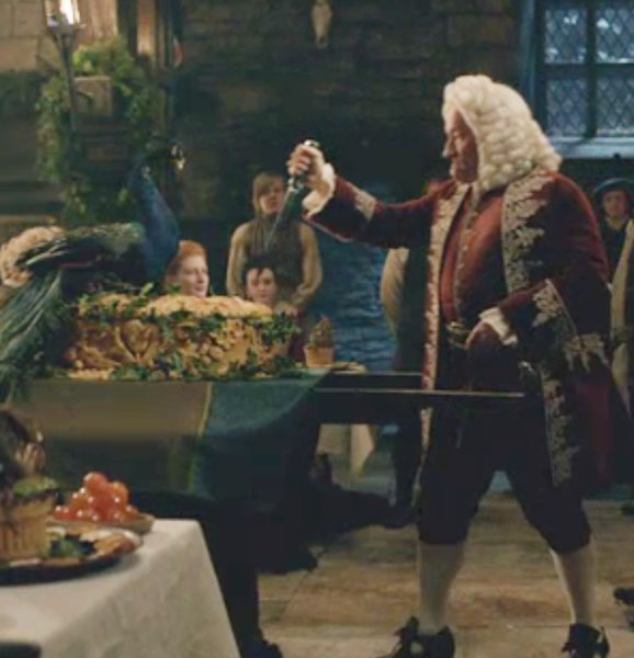
Simple Simon met a pieman. Ha! Well, this Simon is anything but simple; he is the dirty Duke of Sandringham! Have ye ever witnessed a more dramatic pie cutting? Ye would think he was preparing to stab a boar! Hmmm….wish Black Jack Randall was hiding under that crust.
Question: Name the position of the Duke’s right arm and forearm. Hint: palms face toward the body.
Answer: His right arm is flexed. Now the forearm position is tricky. Place your own forearm in the same duke-position and judge the relationship of ulna and radius. Weel, it turns out that with the palm facing the body, the radius is between supination and pronation. Whaaat? No fair ye say? Dinna kill the messenger! The body does its own thing and we do our best to explain. Snort!
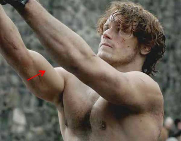
Enough of bones, let’s now examine three more muscles of the humerus and forearm: biceps brachii, brachialis and triceps brachii. Muscles of the arm are divided between two compartments: biceps and brachialis occupy an anterior compartment (in front of humerus) and triceps brachii occupies a posterior compartment (behind the humerus). Another image of Jamie at the Lallybroch beating (Starz episode 102, Castle Leoch) clearly shows the pronounced groove between the arm compartments: biceps and brachialis muscles lay above the red arrow and triceps lies below. Appreciate how beautifully muscles of chest, shoulder, arm and forearm interweave to produce the bonny ebb and flow of body contour; each muscle plays its part in this anatomical ballet.
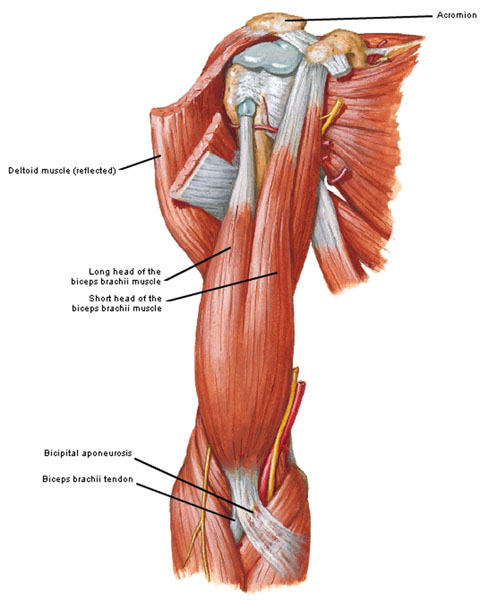
Now for the muscles: biceps brachii (Latin meaning two-headed muscle of the arm) has a long head and a short head both of which take origin from the scapula and cross the glenohumeral joint. The two heads fuse into a single muscle belly that splits into two tendons that cross the elbow joint: a flat tendon joins the superficial forearm fascia (bicipital aponeurosis) and a sturdy tendon inserts near the head of radius (Photo R – anterior view). Because biceps brachii crosses two joints, it acts on both: it aids arm flexion but its major action is on the forearm where it supinates and then flexes the supinated forearm. Think about opening a bottle with a corkscrew: first the biceps unscrews the cork (supination) and then it pulls out the cork (flexion).
Try this: With bent elbow, grip your biceps and supinate the forearm (palm up). Now, alternately flex and extend the elbow joint. Feel the biceps contract and relax with this movement? Now pronate the forearm (palm down) and repeat; the biceps is mostly flaccid because it activates with the forearm in supination.
Photo R
Jamie provides a splendid example of his right biceps brachii as he is manacled to the flogging post at Fort William (Starz episode 106, The Garrison Commander)! BRJ circles like the depraved jackal he is. Most of the bulge seen at the red arrow is created by biceps brachii although some is due to the deeper lying brachialis muscle. Read on, please.
A short cautionary tale: A couple of years ago, a lady at my gym avulsed (detached) the short head of biceps from its scapular origin giving the muscle a “Popeye” appearance. The cause: she was lifting weights that exceeded her biceps strength. Unfortunately, she delayed seeking medical care for a few months by which time the biceps had scarred and the tendon was no longer eligible for surgical reattachment. If you suspect a tendon tear, prompt assessment and treatment promotes optimal outcome! A better weight-lifting strategy is to reduce weight load but increase number of repetitions.
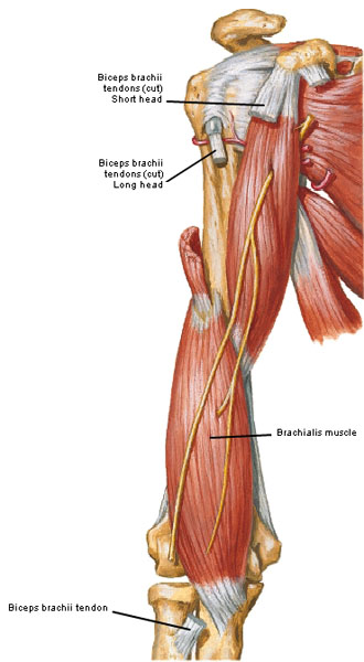
The next muscle is brachialis; it lies deep to biceps brachii (Photo S – anterior view). Brachialis arises from humerus and inserts on ulna. Although overlaid by biceps, brachialis is actually the workhorse of the elbow joint because it flexes the joint regardless of forearm position.
Try this: With a bent elbow, return forearm to pronation, grasp the biceps region, now flex the elbow joint. The deep contraction you feel is brachialis.
Photo S
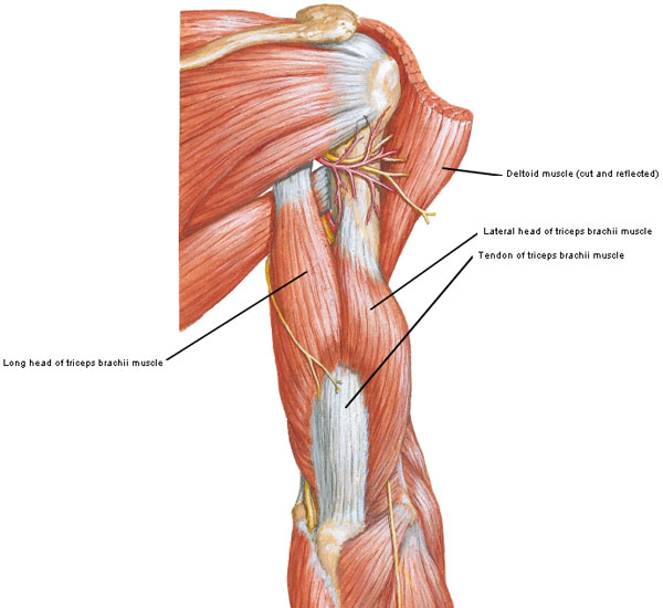
I can’t demonstrate brachialis with Starz images because it lies too deeply so let’s move on to the last muscle of this lesson, triceps brachii. As the name implies, triceps has three heads: a long head arises from scapula, a lateral head arises from upper humerus and a medial head arises from lower humerus (Photo T – posterior view). The heads unite as one muscle mass that turns into a stout tendon above the elbow joint and then inserts into the olecranon of ulna. The long head helps adduct and extend the shoulder joint but together all three heads extend the elbow joint especially against resistance such as with push-ups.
Try this: Grip your triceps in back of the humerus and alternately flex and strongly extend the elbow joint. Feel the triceps contract with extension and relax with flexion?
Photo T
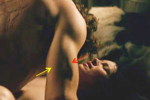
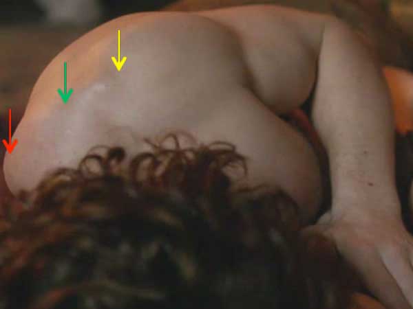
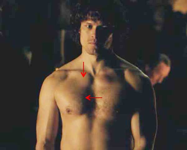
The triceps brachii are clearly visible in well-muscled individuals such as JAMMF. Here the long head of triceps is marked by the yellow arrow and the red arrow points to the lateral head of triceps (Starz episode 107, The Wedding). The medial head cannot be identified because it lies deeply. Claire, the expression on your face … wowzers!
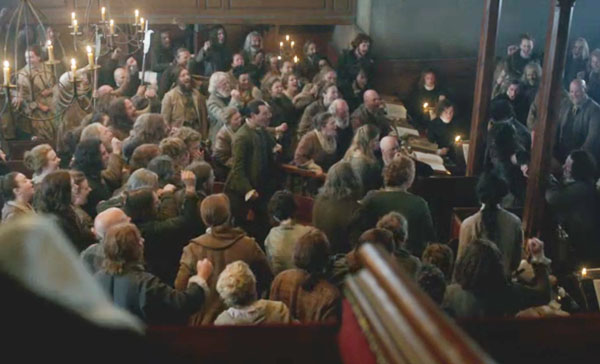
How about we entertain a short pop quiz using images from the latest Starz episodes? To set the mood, let’s consider the beautiful murmuration of starlings at the outset of Starz episode 111, The Devil’s Mark. If you have yet to see a significant murmuration, take a few and watch this one set to Johann Pachelbel’s Canon. Here, the murmuration dips, sways and undulates like a single living organism.
In the thieves’ hole, Claire recalls seeing starling murmurations as a child visiting Brighton. She explains that starlings perform their aerial ballet because there is safety in numbers from predators. Intentional or not, I swear I saw a murmuration of the mob at the trial and it is bloody brilliant! Watch again and see the crowd shout and surge forward each time Claire is skelped with the whip! Between blows, they fall back moving as a single organism. Safety in numbers to be sure, but this time the mob is predator and two accused are victims. Riveting and fascinating. Props to the extras and Claire!!!
Now for the pop quiz:
The first image is of that lousy, long-haired lass Laoghaire looking verra guilty, as she should (Starz episode 110, By the Pricking of my Thumbs)! Weel, she tried her best to seduce Jamie and left a nasty ill wish under Claire’s bed. If she had a firearm right now, she would likely fire it at Claire! Puir Mrs. Fitz, she hasna a clue about her cunning, manipulative “darling granddaughter.”
Question: What is the position of Laoghaire’s arms? What is the position of her elbow joints?
Answer: Both arms are abducted to 45°. If you answered just abduction, that is fine. But, feel free to modify an answer for further accuracy. Both elbow joints are flexed.
Here’s kindly Mrs. F. anointing motherly Mrs. F. with salve. Och, those deadly ovens at Castle Leoch have burned her yet again (Starz episode 110, By the Pricking of my Thumbs)! But, as per Letitia, she bakes yummy bannocks!
Question: Name the position of Mrs. Fitz’s right forearm.
Answer: Right arm is pronated (palm faces downward).
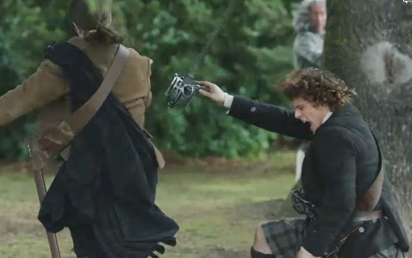
Our fabulous hero barely has time to draw his dirk after a sneak attack by Alexander MacDonald (Starz, episode 110, By the Pricking of my Thumbs)! Fear not, his retaliatory move is breath-taking! Like Joshua or David of the Old Testament, Jamie drops to his knee and fells his opponent with a single blow to the right hamstring. Take my word for it, Alex willna be walking well after that. Hummmm, cowardly Duke cowers behind the tree.
Question: Name the positions of Jamie’s right arm, elbow joint and forearm.
Answer: His right arm is flexed, elbow joint is extended and forearm is pronated (palm down). By golly, this is fun!
Jamie didn’t emerge Scot free (haha) from that “common brawl” – his own blood was spilled (Starz, episode 110, By the Pricking of my Thumbs)… “You’re not normally a closed-mouth woman, Claire. I expected noisier displeasure!” Claire’s lips are sealed as she stitches Jamie’s wound. He attempts to sooth her with “’Tis but one more scar, Sassenach. Nothing to brood over.” You wanna bet? Ouch! Her stitching of that gaping wound sans anesthesia isna tender. She is royally pissed! He kens he’s getting the silent treatment; that needle speaks more eloquently than a thousand words! Stalwart Jamie sits with shoulders back and weight supported by his hands.
Question: Name the position of Jamie’s elbow joints and arms (this question harkens back to the arm movement review at the start of this lesson).
Answer: Lateral rotation of arm. Elbow joints are extended.
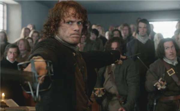
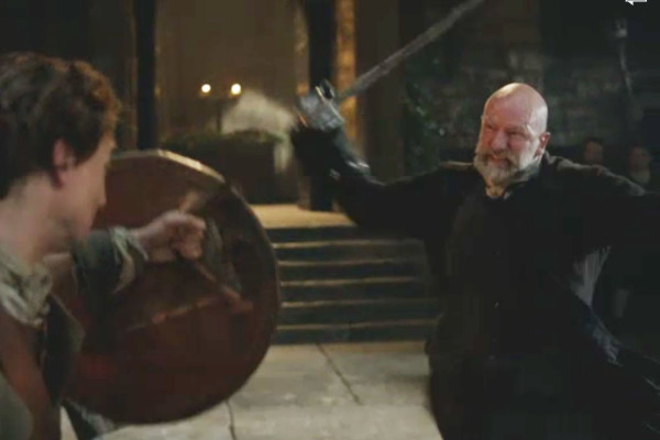
Here’s our Highland hero with both sword and dirk drawn (Starz episode 111, The Devil’s Mark). He barreled down the steps scattering men like pins in a bowling alley! His intent is clear “first man forward will be the first man down.” Lads, odds may be five or six against one but Jamie means it! Back off! Outlander book sets the stage verra well:
“I draw it in defense of this woman, and the truth,” he said … Jamie looked the judges over coolly…”I swore an oath before the altar of God to protect this woman. And if you’re tellin’ me that ye consider your own authority to be greater than that of the Almighty, then I must inform ye that I’m no of that opinion, myself.”
Question: Name the position of Jamie’s arms, elbow joints and forearms:
Answer: Arms are abducted to 90 ° (a.k.a. horizontal abduction), elbows are extended, forearms are between pronation and supination (palms face toward the body if arms are lowered).
Students, you did very well on the pop quiz. Thanks for playing along!
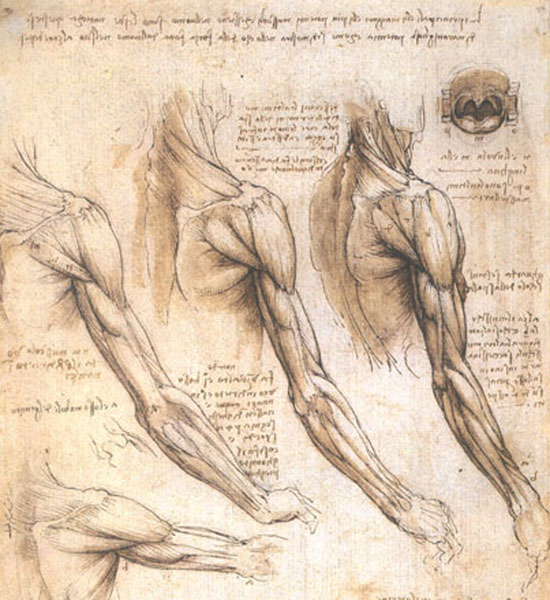
Now, let’s end this lesson with a bit of history. Human anatomy (Greek meaning to cut away) is considered the oldest medical science. Millennia passed before western culture permitted and embraced dissection of the human body thus keeping in place centuries of inaccurate concepts and teachings. More pertinent to today’s topic, accurate anatomical drawings of the upper limb have been available for not quite 500 years. Some of the earliest and most elegant anatomical drawings were done by Renaissance genius, Leonardo Da Vinci (1452-1519). Although not the first to draw the upper limb, he broke ground because his images included comparative anatomy between species as well as drawings of the living and the dead. Although his paintings were widely known during his lifetime, only a few associates were aware of his anatomical research. He never worked as a professional anatomist, he never taught the subject and unfortunately, he never published his anatomical observations which would have greatly advanced the science of anatomy. Photo U shows his wonderful progressive drawings of shoulder, arm and forearm musculature. Although not quite anatomically correct, they are very close. This image is a replica of one of 600 Da Vinci drawings housed in The Royal Library at Windsor Castle, one of the finest private collections in the world. BTW, upper right image is a rendering of hard and soft palates and tongue. Not sure why it appears on the same page as the upper limb; mayhap to save paper?
Photo U
Hope you enjoyed the lesson on the arm and forearm. Knowledge is power: forearmed is forewarned!
A deeply grateful,
Outlander Anatomist
Photo creds: Starz, Netter’s Atlas of Human Anatomy, 4th ed., Clinically Oriented Anatomy, 5th ed., Human Anatomy, Martini and Tiimmons, 1st ed., Gray’s Anatomy for Students, 1st edition, www.baycarehealth.adam.com, www.eorthopod.com, www.methodistorthopedics.com, www.stephanierose156.blogspot.com, www.Wikipedia.com

