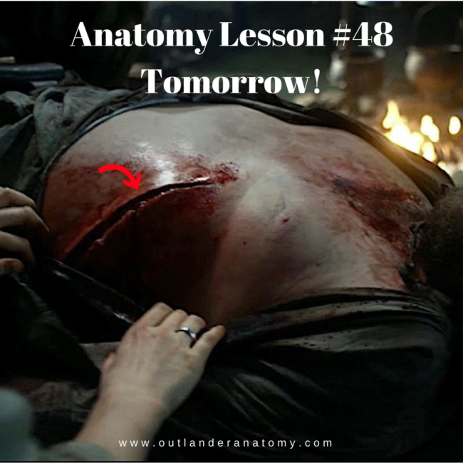
Anatomy Lesson #61: Jamie’s Mars and Scars

Hallo, anatomy students. First lesson posted in a while – I have been waiting for new S4 material!

Meantime, “Me keep do.”  So, here’s one last lesson from S3.  Welcome to Anatomy Lesson #61, Jamie’s Mars and Scars!

Getting right to it…. Many events transpired in ep 306, A. Malcolm, wherein Jamie meets Claire after two hundred and twenty-two years apart. Woot!

Like you, the intimate scenes of this episode immediately drew my eyes to Jamie’s body –  Oops, I mean his mars and scars. Come on, I’m a scientist! 

My anatomist’s eye immediately began calculating… Weil, actually not until the second viewing, but you get my drift…. Are Jamie’s scars all present? Are any scars missing? Are they accurately depicted? Did they change?

What say you, Outlander fans, shall we investigate?

This is a chronological list of injuries over three seasons of Outlander TV and most produce scars. All but one appear in the books. Do you ken the outlier?

  1. dislocated shoulder joint
  2. gunshot wound, front
  3. gunshot wound, back
  4. BJR backlash
  5. McDonald’s sword work
  6. BJR hand smashing
  7. BJR burn
  8. BJR Culloden scar

Truly, this red-heided lad has suffered his share of owies! 

We are all too aware of horrific wounds from modern warfare, but battlefield wounds have always been appalling. In the 16th century, French barber surgeon Ambrose Paré became famous for his ingenious repairs of battlefield injuries. One of his famous illustrations is a composite drawing that depictes the range of wounds suffered by warriors of the past (Image A). Seems as if our Jamie may have posed for this image. Gasp!
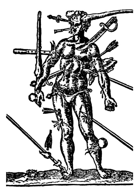
Image A

Just for fun and because you have nothing else to do <g>, let’s explore the laundry list of Jamie’s Wounds.  We’ll jump backward and forward 20 years as we analyze his mars and scars in chronological order.

Strap yourselves in. Here we go!

1. Dislocated Shoulder Joint:

Claire first spies Jamie perched atop a stool in a crofter’s cottage (Starz ep 101, Outlander). His hunched form cradles right forearm. Her keen nurse’s eye is horrified by his injury. Here from Outlander book:

I gasped, as did several of the men. The shoulder had been wounded; there was a deep ragged furrow across the top, and blood was running freely down the young man’s breast. But more shocking was the shoulder joint itself. A dreadful hump rose on that side, and the arm hung at an impossible angle.

Q: What happened to Jamie?

A: He suffered an anterior dislocation of the right shoulder joint.

His fellow Highlanders move to force the joint into place but Claire-Declares, NO!  No qualified chirurgeon in this grubby group of lads!

The dreadful “hump” is the acromion, a feature of the scapula (shoulder blade). Read about the shoulder joint and Claire’s treatment of its dislocation in Anatomy Lesson #2, When Claire Meets Jamie or How to Fall in Love While Reducing a Dislocated Shoulder Joint!

Footnote #1: In Outlander book, Jamie suffered both a dislocated shoulder and a gunshot wound. Diana wrote that the musket ball knocked him from the saddle and he fell on his hand. Crunch went the shoulder joint! 
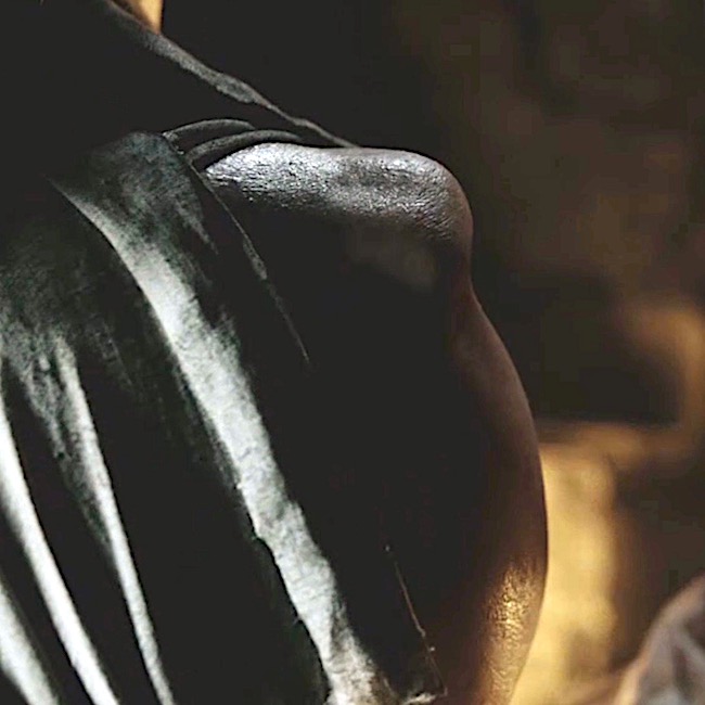
Outlander ep 101, Sassenach

Although bruising and swelling often accompany such dislocations, both may resolve without sequelae. Jumping forward 20 years, witness Jamie’s fine form at the brothel reunion in Starz ep 306, A. Malcolm. Evidence of a shoulder dislocation? Nada! Fortunately for Claire, this shoulder works perfectly fine. In fact, everything works perfectly fine. Wink, wink!
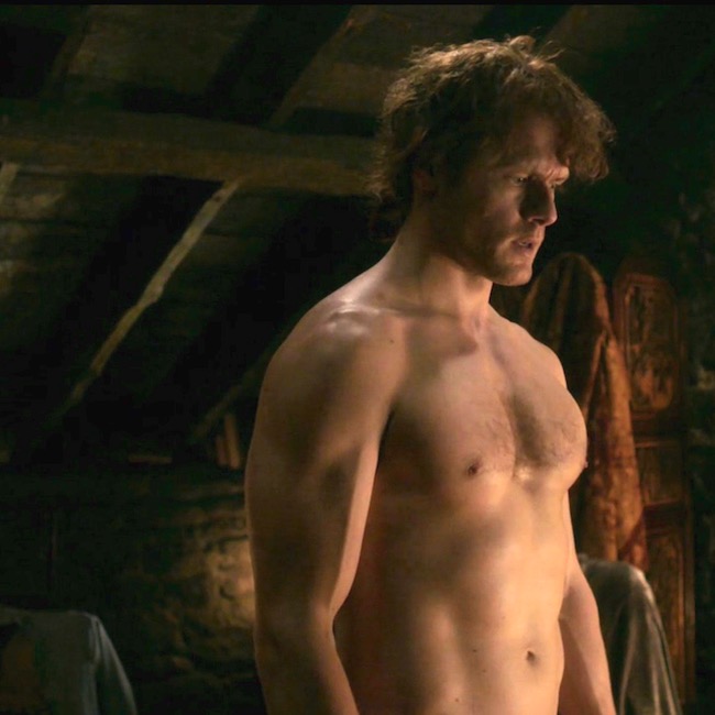
Outlander ep 306, A. Malcolm

Before we leave the shoulder issue, several fans have asked if the TV depiction of Jamie’s dislocation is realistic. Well, mostly. An actual anterior dislocation of the shoulder joint looks like Image B (left shoulder). Here, the rounded contour of the shoulder is lost because head of the humerus has been displaced forward into the armpit area (axilla).  The bony knob (red arrow) is the acromion but, clearly, it is not as prominent as depicted in the TV version.

The difference can be explained because in the TV episode, the “dreadful hump” was a prosthetic applied to the top of Jamie’s shoulder. Thus, the hump sits higher and appears more prominent than in life. But, otherwise, kudos to the special effects team!
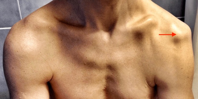
Image B

Moving on!

2. Gunshot Wound, Front:

Back to the crofter’s cottage! Hightailing it towards home, the Highlanders are ambushed by Redcoats at Cocknammon Rock. But, the English are swiftly dispatched. Hours later, Claire shouts:

“Stop! Help! He’s going over!”

Timber!!! – Jamie topples from his steed.

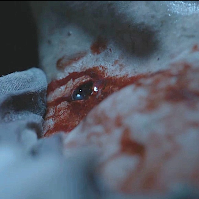
Claire swiftly discovers a gunshot wound. Jamie, why didn’t you fess up? Now, Claire must deal with a nasty hole left by a musket ball plowing a path through Jamie’s trapezius muscle! Claire explains to Dougal’s merry band of men (Outlander book): 

“The gunshot wound has been bleeding again, and the idiot’s been knifed as well. I think it’s not serious, but he’s lost quite a lot of blood. His shirt is soaked through, but I don’t know how much of it is his. He needs rest and quiet; we should camp here at least until morning.” 

Fat chance Dougal will stop for his nephew to rest; not with Captain Randall prancing through the heather.

“On your feet, soldier!” 

Footnote #2: The TV version shifted the gunshot wound later in the episode and completely ignored the knifing. Probably figured Claire had enough on her plate. Hah!
Outlander ep 101, Sassenach

Fast forward 20 years: Delightful! Starz FX team remembered to include a faint scar as evidence of that entrance crater left by the musket ball. 

Can’t you just see a Starz employee with a wound list?

  • Dislocated shoulder joint, check!
  • Front gunshot wound, check! (
  • etc.

Outlander ep 306, A. Malcolm

3. Gunshot Wound, Back:

Back to the past. Next morning, in Castle Leoch’s keep, wary Claire meets-and-greets wary Mrs. Fitz – immortalized  in this masterful excerpt from Outlander book:

“But he’s hurt. He was shot yesterday and stabbed last night. I bandaged the wound for riding, but I didn’t have time to clean or dress it properly. I must care for it now, before it gets infected.” 

“Infected?”

“Yes, that is, I mean, inflamed, you know, with pus and swelling and fever.”

“Oh, aye, I know what ye mean. But do ye mean to say as ye know what to do for that? Are ye a charmer then? A Beaton?”

“Something like that.”

This terse little tête-à-tête, wherein WWII combat nurse and 17th century castle housekeeper face-off, reveals Claire’s nimble brain! The term infected, meaning invasion by micro-organism, won’t come into common use for another century. Oops, she quickly course-corrects! 

Then, off she goes to properly tend Jamie. (One might think the lad is accident prone!)

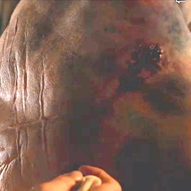
The wound at back of shoulder marks the exit site of the musket ball. Serious bruising from gunshot wound and dislocated shoulder and horrific criss-crossing scars (see below) definitely catch her eye. Eek!
Outlander ep 102, Castle Leoch

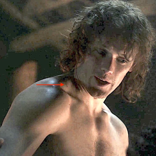
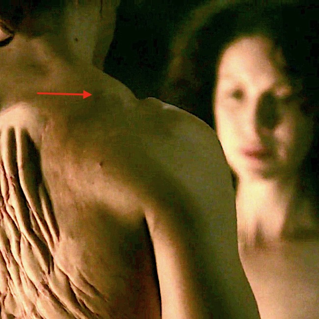
Spring forward 20 years. Was the exit wound depicted at the brothel reunion? Aye, it was. Take a really close keek at Jamie’s back….there it be (red arrow).

Psst…let’s just ignore the unfortunate pleating of Jamie’s back prosthesis – best save those pleats for his kilt.
Outlander ep 306, A. Malcolm

Next!

Back to Castle Leoch. We can consider Rupert’s hearty beating of Jamie who gallantly choses fists over strap (enough straps) to protect loose-behaving Missy Laoghaire. Once again, mars are treated by Outlander Nurse. But, none of these blows caused permanent damage, so let’s move on!
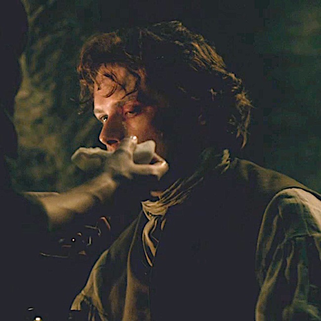
Outlander ep 102, Castle leoch

4.  BJR Backlash:

Just right for Hallowe’en – foul work by a foul one! Fast forward to ep 106, The Garrison Commander, wherein Captain Blackguard informs Claire of his beautiful masterpiece. Produced four years earlier, BJ floged Jamie’s back into a bloody mess using a cat-‘o-nine tails with lead tips. Yep, these do leave scars!  And, dinna forget, Jamie already had been flogged days earlier! 😳

If you can stomach more flogging details, read Anatomy Lesson #10, Jamie’s Back – Aye, Jamie’s Back!

Described by Dougal in Outlander book: 

Dougal grimaced. “A pitiful sight, it was, too—still raw, no more than half-healed, wi’ the weals turned black and the rest yellow wi’ bruises. The thought of a whip comin’ down on that soreness was enough to make me blench, along wi’ most of those watching.”
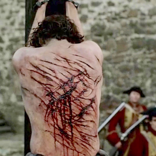
Outlander ep 106, The Garrison Commander

So naturally, the back scars persist two decades later.

A splendid quote from Voyager book; Jamie’s scars have healed and contracted as scars normally do. 

I curled up behind him, knees fitting neatly behind his own. The firelight shone dully from behind me now, gleaming over the smooth round of his shoulder and dimly illuminating his back. I could see the faint lines of the scars that webbed his shoulders, thin streaks of silver on his flesh. At one time, I had known those scars so intimately, I could have traced them with my fingers, blindfolded. 

Footnote #3: To my eye, the prosthesis at Madam Jeanne’s establishment appears identical to the original. Over a 20-year span, such scars should contract and thickness diminish.  I suggest a new prosthesis to document these changes, an expected progression of wound healing and scar formation.
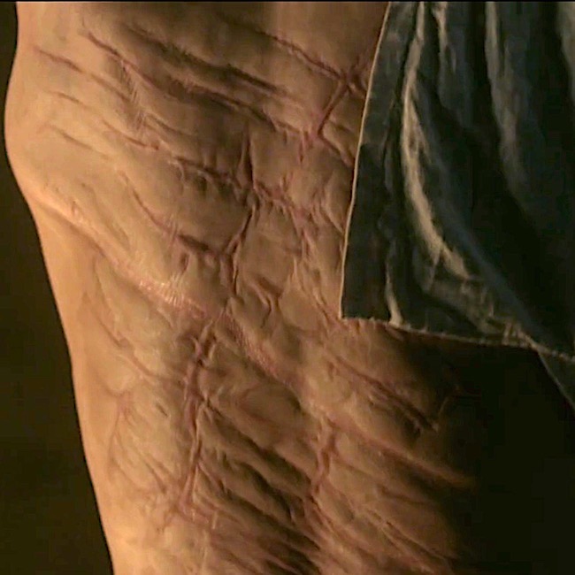
Outlander ep 306, A. Malcolm

5. MacDonald’s Sword Work:

Back to the early years. Ye ken the MacDonald clan with its three nasty-mouthed bros? Best ignore them, Jamie.

Nope. He gives them sass and in return, suffers a slice and dice Big Mac Attack! Three against one? No fair!
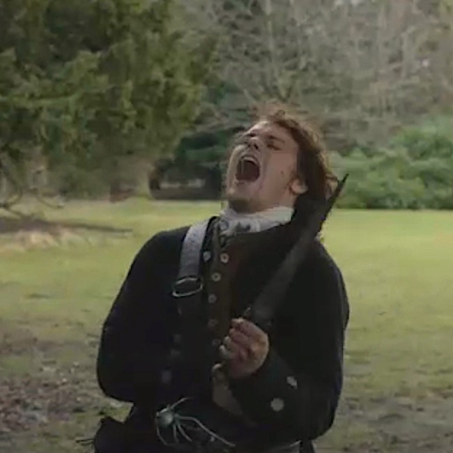
Outlander ep 110, By The Pricking of My Thumbs

The moral to this event is arguable, but consider something along the line of: dinna get mouthy with the MacDonald clan, especially if three well-armed laddies are against one!

But, Jamie exacts his revenge as he slices the hamstrings of one attacker, who from henceforth will hobble. And, who is cowering behind the tree? The cowardly Duke, who soon has a fate-date with The Godfather! Yes!!!
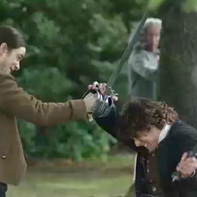
Outlander ep 110, By The Pricking of My Thumbs

Later, Claire stitches Jamie’s sword wound using the biggest needle she can find – royally POed!  Most of you ken that she accidentally stuck that needle into “Jamie’s” skin during filming. Och! 😱

Footnote #4: If you chose this as the only injury not in the books, score! Although Diana didn’t write the MacDonald fight, Jamie does suffer a saber-slash at Prestonpans. Here from Dragonfly in Amber:

It was a saber-slash, slanting across the ribs. A lucky angle; straight in and it would have gone deep into the intercostal muscles between the ribs. As it was, an eight-inch flap of skin gaped loose, red beginning to ooze beneath it again with the release of pressure. It would take a goodly number of stitches to repair, but aside from the constant danger of infection, the wound was in no way serious.
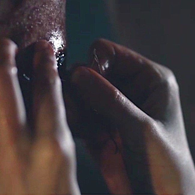
Outlander ep 110, By The Pricking of My Thumbs 

Did the Outlander FX team include the scar from the Sandringham kebby-lebby? Yep! There it is basking in Claire afterglow!
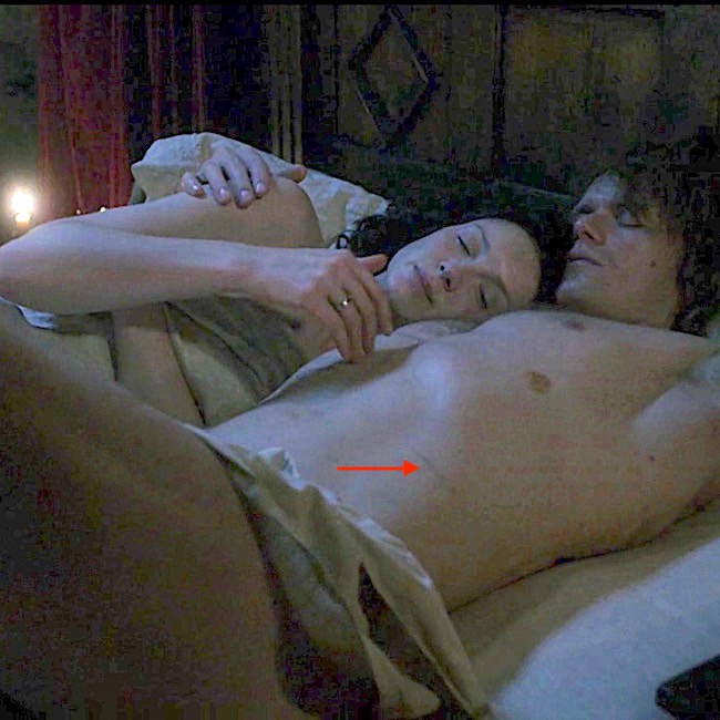
Outlander ep 306, A. Malcolm

6. BJR Smashing:

You ken about smashing pumpkins? BJR has his version, whacking Jamie’s hand with a mallet, smashing metacarpals and phalanges.  

Read more about BJR atrocious pastimes in Anatomy Lesson #22, Jamie’s Hand – Symbol of Sacrifice!
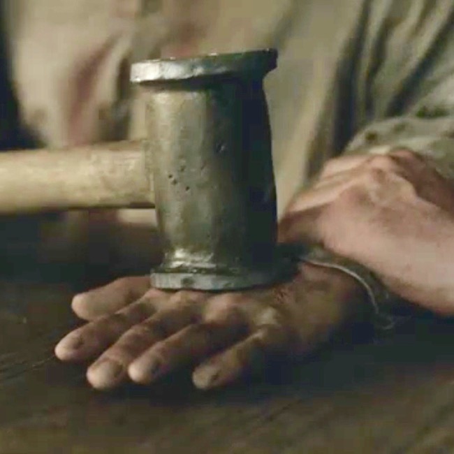
Outlander ep 115, Wentworth Prison

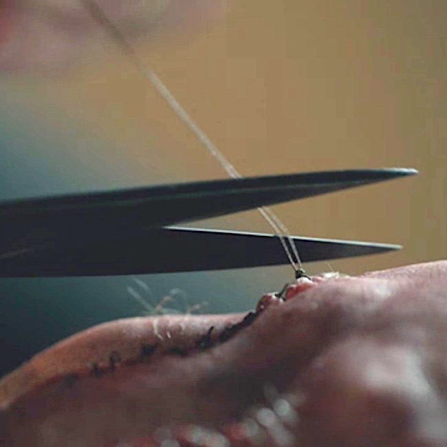
Once again, Claire to the rescue! She stitches Jamie’s wounds and splints broken metacarpals and phalanges. Herself clearly explains the fate of Jamie’s paw after BJR’s maniacal ministrations (Outlander book):

I began to lose myself in the concentration of the job, directing all my awareness to my fingertips, assessing each point of damage and deciding how best to draw the smashed bones back into alignment. Luckily the thumb had suffered least; only a simple fracture of the first joint. That would heal clean. The second knuckle on the fourth finger was completely gone; I felt only a pulpy grating of bone chips when I rolled it gently between my own thumb and forefinger, making Jamie groan. Nothing could be done about that, save splint the joint and hope for the best.

The compound fracture of the middle finger was the worst to contemplate. The finger would have to be pulled straight, drawing the protruding bone back through the torn flesh. I had seen this done before—under general anesthesia, with the guidance of X rays.

Outlander ep 116, To Ransom A Man’s soul

And, heroically repairs his mangled hand. Not a surgeon yet, but she did well!
Outlander ep 116, To Ransom A Man’s soul

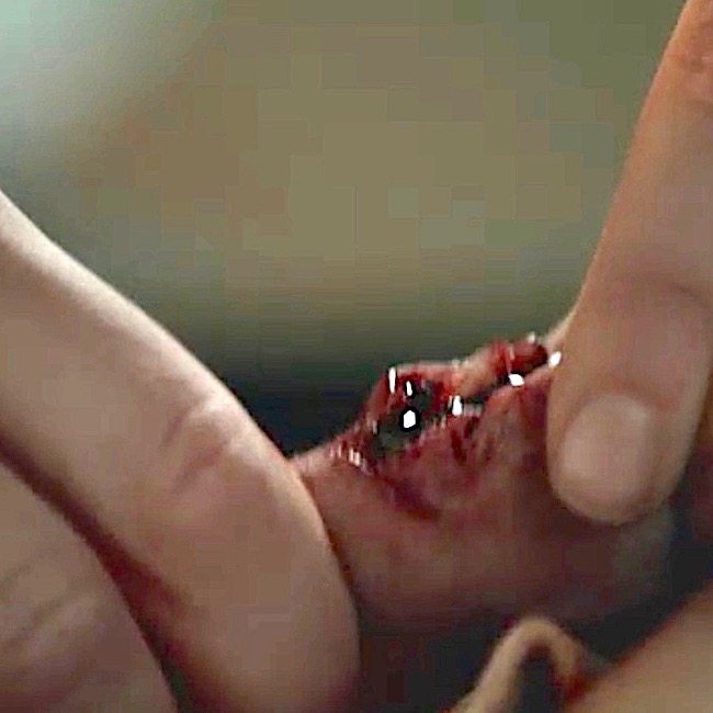
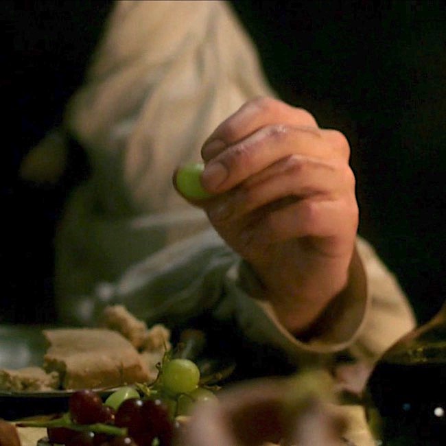
Are Jamie’s finger scars visible after 20 years? Oh, indeed they are! Look closely; nothing shows them better than eating grapes.

FX peeps were on their game. Plain as the back of your hand. Not that Claire was paying much heed to those scars, mind ye. Snort!
Outlander ep 306, A. Malcolm

7. BJR Burn:

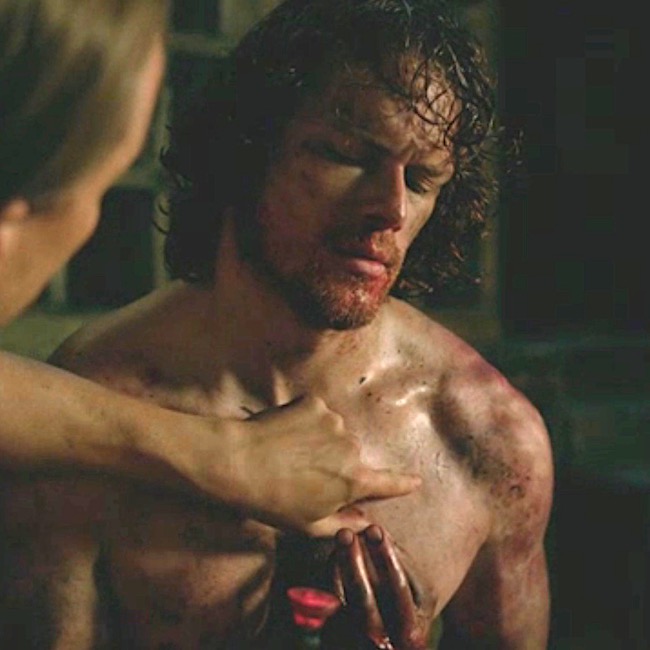
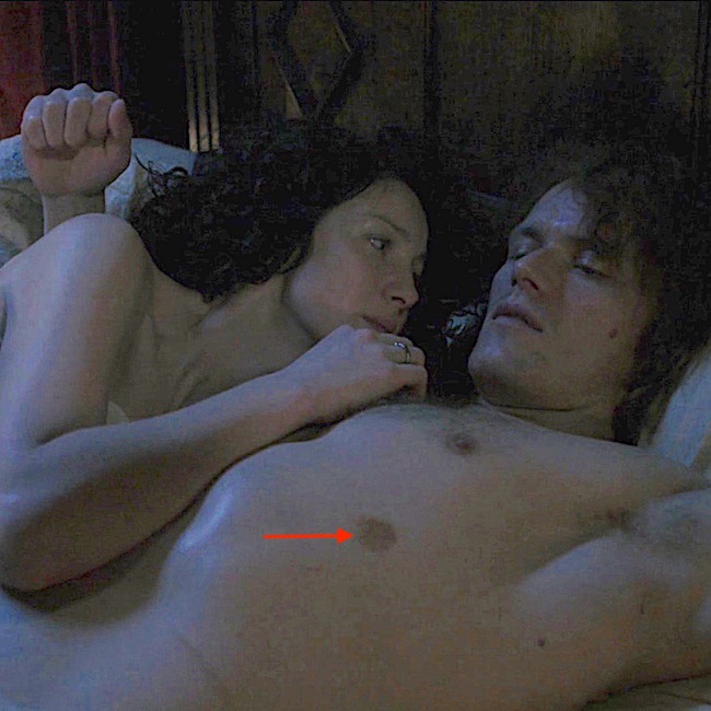
Oh, and among those grim hours in the hands of a mad man, let’s not forget the ghastly image of BJR directing Jamie to burn initials into the flesh overlying his heart. Randall offers a red hot wax stamp to “seal” the job! Gah!
Outlander ep 116, To Ransom A Man’s Soul

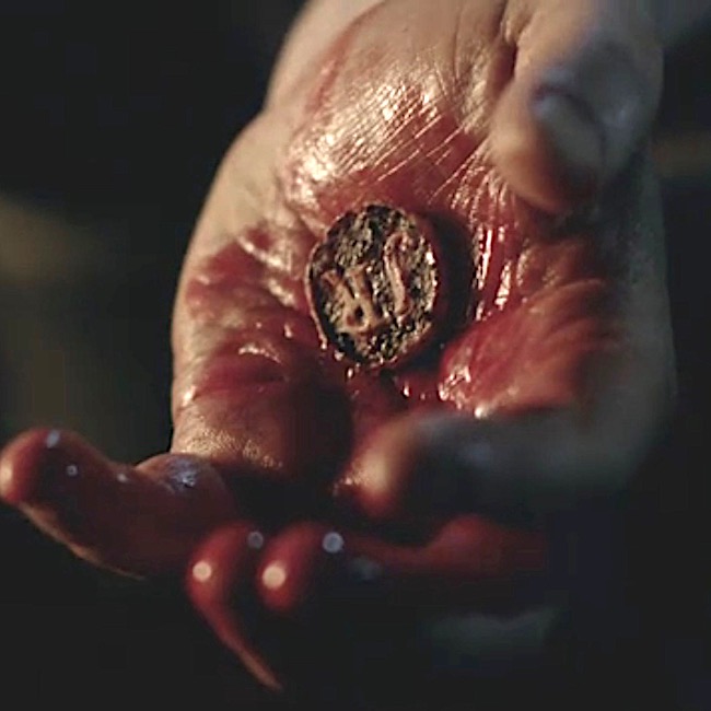
Those initials do not survive because TV Godfather excises them and casts  into the fiery pit. Go Murtagh! Rah! 

Footnote #5: In Outlander book, Sir Marcus removes the offending button of skin: 

“Best let me help ye, man. Ye’ll fall on it in a moment.” After a moment’s pause, Jamie reluctantly surrendered the knife and lay back against the wadded blanket. He touched his chest an inch or two below the nipple. “There.” 

Sir Marcus reached to the sideboard and snagged a lamp, setting it on the stool he had vacated. At this distance, I couldn’t see what he was peering at; it looked like a small red burn, roughly circular in shape. He took another deliberate pull at his whisky glass, then set it down next to the lamp and pressed the tip of the knife against Jamie’s chest. I must have made an involuntary movement, because the Lady Annabelle clutched my sleeve with a murmured caution. The knife point pressed in and twisted suddenly, flicking away in the motion one uses to cut a bad spot out of a ripe peach. Jamie grunted, once, and a thin stream of red ran down the slope of his belly to stain the blanket.

Talk about branding! Deeply grateful, today’s entrepreneurs never met Johnathan Wolverton!  
Outlander ep 116, To Ransom A Man’s Soul

Back at the brothel, the burn scar is on full display (red arrow), looking a bit like a supernumerary nipple! He, he. The Starz crew didna forget!
Outlander ep 306, A. Malcolm

And, finally, the last wound!

8. BJR Culloden Slash: 

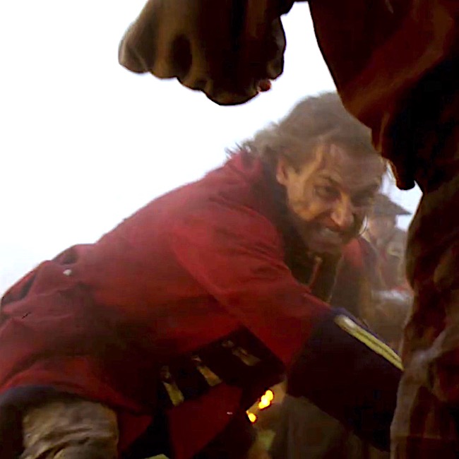
Black Jack is at it again – fiendish fiend! At Culloden, Jamie glances away from a fallen Randall and swish, the blackguard’s blade strikes and bites verra close to Jamie’s femoral artery! 
Outlander ep 301, The Battle Joined

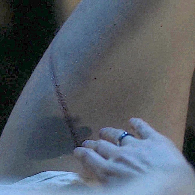
Twenty years later after the reunion-union, Claire espies the long thigh scar. Not quite as described in Voyager, but close. Outlander team snagged this one, too!

The scar ran from midthigh nearly to his groin, an eight-inch length of twisted, whitish tissue. I couldn’t repress a gasp at its appearance, and dropped to my knees beside him. I laid my cheek on his thigh, holding tight to his leg, as though I would keep him now—as I had not been able to keep him then. I could feel the slow, deep pulse of the blood through his femoral artery under my fingers—a bare inch away from the ugly gully of that twisting scar. 

….Soft as a moth flying in the dark, my hand skimmed his leg, and found the thin deep runnel of the scar. My fingers traced its invisible length and paused, with the barest of touches at its end, wordlessly asking, “How?”

His breathing changed with a sigh, and his hand lay over mine.

“Culloden,” he said, the whispered word an evocation of tragedy. Death. Futility. And the terrible parting that had taken me from him. 

Foot Note #6: In Voyager book, the scar extends to Jamie’s groin. The TV scar is closer to the knee – offering a modicum of modesty as millions take notice??? 😉
Outlander ep 306, A. Malcolm

So, eight wounds and FX folks nailed them all. Go team!

Think about it: BJ delivered almost 40% of Jamie’s wounds. However, if one counts each back scar, he owns well over 90% of Jamie’s mars and scars!

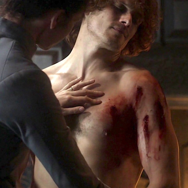
Oh, and here’s a wee bonus! Dinna forget loving Laoghaire’s contribution to Jamie’s landscape when the mad matron peppers him with bird shot. Of course, this event occurs after the reunion so those scars must await Season 4! Whew!  
Starz ep 308, First Wife

Let’s close with this lesson with a snippet from big book seven, An Echo in the Bone, which aptly describes Claire as Jamie’s healer and helpmeet. Not really a spoiler as it reveals no plot points.  So, safe to read.

A shadow fell across the floor in front of me and I looked up. Jamie was standing there with a most peculiar look on his face. 

“What?” I said, startled. “Has something happened?”

“No,” he said, and advancing into the study, leaned down and put his hands on the desk, bringing his face within a foot of mine.

“Have ye ever been in the slightest doubt that I need ye?” he demanded. It took roughly half a second of thought to answer this. “No,” I replied promptly. “To the best of my knowledge, you needed me urgently the moment I saw you. And I haven’t had reason to think you’ve got any more self-sufficient since. 
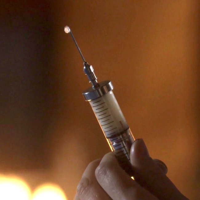
Outlander ep 308, First Wife

Final Points: Generally, society considers scars to be unwanted and unsightly. However, from anatomy and pathology POVs, scar formation is a normal process. Yes, indeed!  This is the way mammalian creatures typically repair tissue disruption –  like a biological bandaid. Although a scar may not perform as well as the original tissue, it keeps the body functional and responsive. To learn more about injury and repair, visit these anatomy lessons:

Recall the start of this lesson?

  • Were all scars accounted for? Yes.
  • Are any scars missing? No.
  • Were they accurately depicted? Mostly.
  • Did they change? Mostly.

Jamie’s mars and scars paint a poignant history. Claire was there for him from the get-go, treating broken bones, gunshot wounds, contusions, lacerations, incisions, dislocations, burns, and emotional abuse. Cheers to the gallant lass and her braw lad (and their creator)!

The deeply grateful,

Outlander Anatomist

Follow me on:

Photo Credits: Sony/Starz , adc.bmj.com (Image A); sghs.org (Image B)  

“A Real Eye Opener – The Eye, Part 4”

Welcome all students to today’s Anatomy Lesson #32, The eye – Part 4! Remarkable sensory organ that it is, this will be our fourth lesson of the eye series. Although I thought this would be the last eye lesson, a 5th and last lesson on eye anatomy is required. But, you’ll see why this fascinating and wonderful organ requires yet another lesson!

We know that the eye is designed to provide us with vision. Understand that the air around us is filled with a sea of electromagnetic energy from the shortest gamma waves to the longest radio waves. Humans are blind to almost all of this energy because our eyes detect only that part of the electromagnetic spectrum known as the visible spectrum or, in more common terms, visible light.

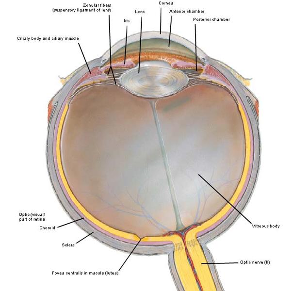
Let’s begin the lesson with a quick review. In Anatomy Lesson #31, we learned parts of the eyeball. Photo A is a horizontal section through a left eyeball (as viewed from above). The sclera is the white outer coat. The choroid is a coat of blood vessels internal to the sclera. The retina with its specialized sites is inside the choroid. The transparent cornea projects from the front surface. Iris and pupil (Anatomy Lesson #31) are behind the cornea. The biconvex lens is behind the iris. The ciliary body with its ciliary muscle forms a ring around the lens. The anterior chamber is a space between cornea and iris that is filled with the watery aqueous humor. A space between iris and lens is the poorly named posterior chamber which is also filled with aqueous humor. Behind the lens is the largest space of the eyeball, the vitreous chamber containing vitreous humor or vitreous body.

Figure0083-a-KLS-editede

Photo A

NOTE: this lesson shows images of the eyeball in horizontal section oriented with the cornea directed upward (Photo A) or rotated 90° so the cornea faces left or right (Photo C). Don’t be distressed when you see this flip flop; it just reflects preference by the creator of the image.

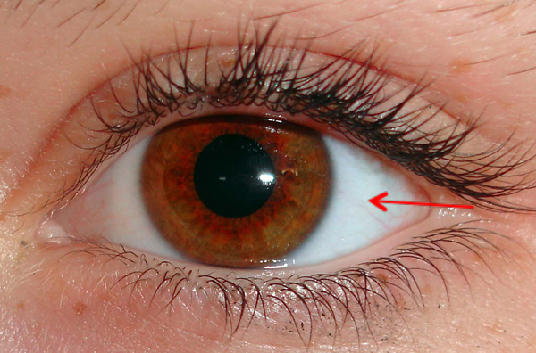
This lesson will cover sclera, cornea and lens. The sclera is the tough, white outer fibrous coat of the eyeball (Photo B – red arrow); it provides shape and resistance as well as attachment for extraocular muscles (Anatomy Lesson #30) and some intraocular muscles (Anatomy Lesson #31). The tiny red blood vessels seen in Photo B are actually located in the bulbar conjunctiva (Anatomy Lesson #30) which adheres to the anterior sclera.

Hazel-Eye-Morguefile2-KLS-edited

Photo B

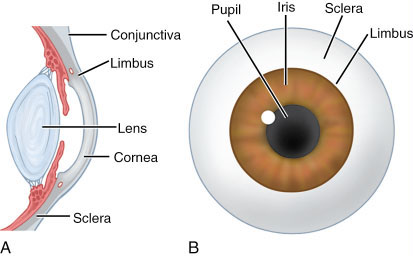
The cornea (Latin meaning horn) is a transparent dome covering the anterior 1/6 of the globe. It bulges because its radius is smaller than that of the remaining eyeball. This creates a small sulcus (groove) at the corneoscleral junction known as the limbus (Latin meaning edge or border; Photo C).

Try this: Ask a partner to sit while you look at one of their eyes from a side view. Identify the white sclera. Find the transparent cornea bulging from the front of the eyeball. Find the limbus, the groove where cornea and sclera meet.

862

Photo C

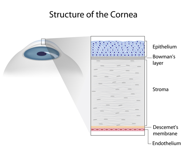
The cornea is a truly fascinating part of the eye being composed of cells and extracellular molecules arranged in five microscopic layers (Photo D). With the exception of the stroma or middle layer, the names of the other layers don’t concern us today. A normal cornea is transparent because the stroma is made of regularly arranged layers of collagen molecules and because it lacks blood vessels.

Question: if it has no blood supply, how does it get oxygen and nutrients?

Answer: these are delivered to the superficial cornea by the tear film and to the deep cornea by aqueous humor (or humour). The cornea is well-equipped with pain fibers as anyone knows who has injured it. And, as you realize, if the cornea is touched an automatic reflex is evoked that closes the eyelid for protection.

shutterstock_127500686

Photo D

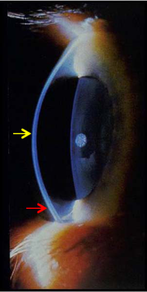
The cornea is not of the same thickness throughout: it is thinnest at its middle (~0.5 mm) and thickest near its rim (~0.8 mm). This change in thickness is clearly evident in this magnificent image from Lesson #31 (Photo E – yellow arrow at middle of lens; red arrow at rim).

Picture3-KLS-edited

Photo E

The normal cornea is smooth and clear like glass, but it is also strong and durable so it helps the eye in three ways:

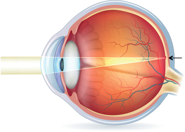
  • Refracts (bends) light rays to help focus them on the retina (Photo F – black arrow).
  • Shields the eyeball from germs, dust, and other harmful matter.
  • Filters out some of the damaging ultraviolet (UV) wavelengths in sunlight.

Light rays entering the eyeball must be bent to focus them on the retina. The cornea is powerful providing roughly 2/3 of the overall refractive power of the globe. But, understand that its curvature is fixed and normally does not change.

normal%20vision%20diagram

Photo F

Corneas are subject to a variety of diseases and conditions including allergies, conjunctivitis (pink eye), infection, dryness and dystrophies (cloudiness) to name a few. A seriously damaged cornea may be injured such that light cannot penetrate the eyeball to reach the light-sensitive retina. Sometimes a corneal transplant is required wherein a surgeon removes the central portion of the cloudy cornea and replaces it with a clear cornea, usually donated through an eye bank.

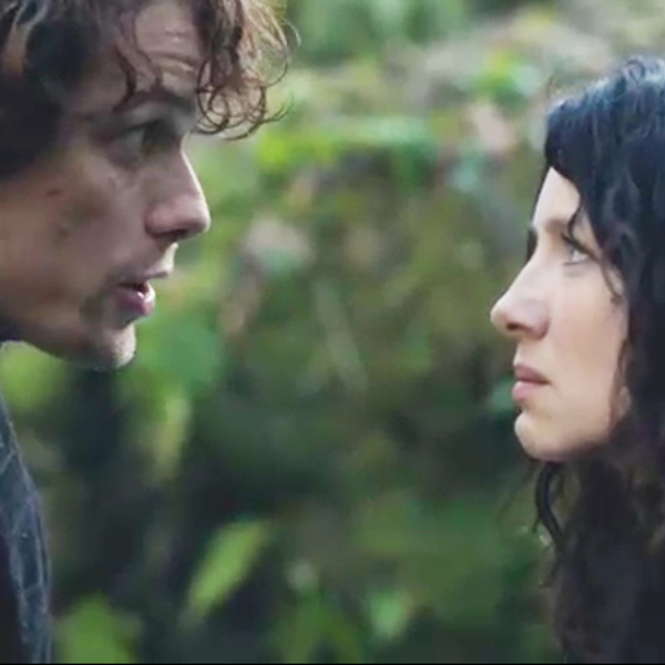
Here are two pairs of clear corneas going at it eye-to-eye (Starz episode 109, The Reckoning). Look me in the corneas lass, when I’m talking to ye! I am your 18th century hubby and ye will obey me! This  Sassynach looks Jamie in the eye and while she’s at it, she’ll spit in it too! She doesn’t have to do what Jamie says. Aye ye do; If ye dinna do what I say then there’s a price ye’ll have to pay! Ouch!

ep-109-Jamie-and-Claire-cornea-KLS-edited

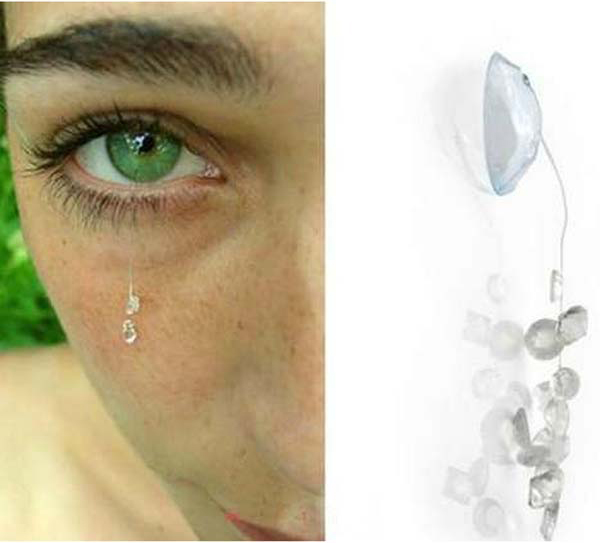
And, just when we think that we have seen it all, well, we haven’t! Have you ever heard of corneal jewelry? Yep, small crystals and other gewgaws hang via a thread from a contact-type lens! Not sure that swinging counterweight would feel very good dropping from the cornea but can’t say as I’ve never tried one.

fashion-eyeful-contact-lens-jewelry-KLS-edited

Photo G

Moving along, the lens (Latin meaning lentil) is next. The lens is a transparent, bi-convex structure suspended between iris and vitreous humor that continues to grow throughout life. The lens is shaped like a flying saucer although its front surface is flatter than its back surface (Photo H). Near the pupillary rim, the iris is in contact with the front surface of the lens but the two diverge at the sides. The back surface of the lens sits in a divot of the vitreous humor. Like the cornea, its purpose is to refract light rays to focus on the retina and is responsible for about 1/3 of the overall refractive power of the eyeball.

eye KLS edited

Photo H

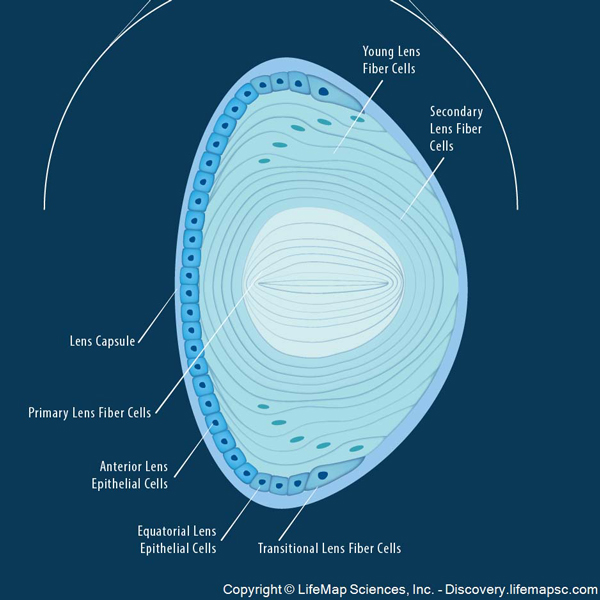
Lens structure is amazing and complex but simplified for this lesson. Like the cornea, the lens is avascular to assist with transparency. It is made of very long (12 mm) and very thin cells known as lens fibers (Photo I). These cells are arranged in layers around a central core much like the rings of an onion. The lens has also a capsule, some cube-shaped cells in front and a few other features but these do not concern us for this lesson.

The_Anatomy_and_Structure_of_the_Adult_Human_Lens-copy-KLS-edited

Photo I

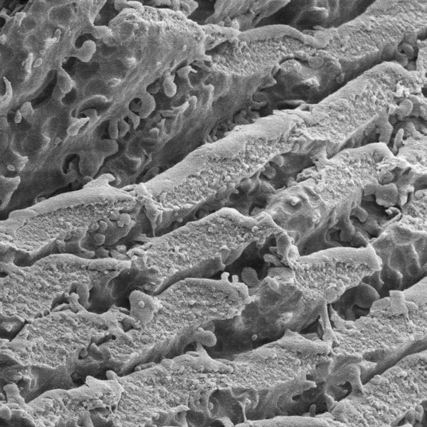
Photo J is an image of lens fibers taken with a scanning electron microscope (Anatomy Lesson #6), an instrument that takes 3-D images of microscopic structures. Very cool! With this type of microscopy, lens fibers look like tightly-packed lasagna noodles. Nuclei and other cell structures (organelles) are absent from mature lens fibers to boost transparency. Lastly, the fibers are filled with crystalin protein and their water content is very low.

fig_1_1_big-KLS-edited

Photo J

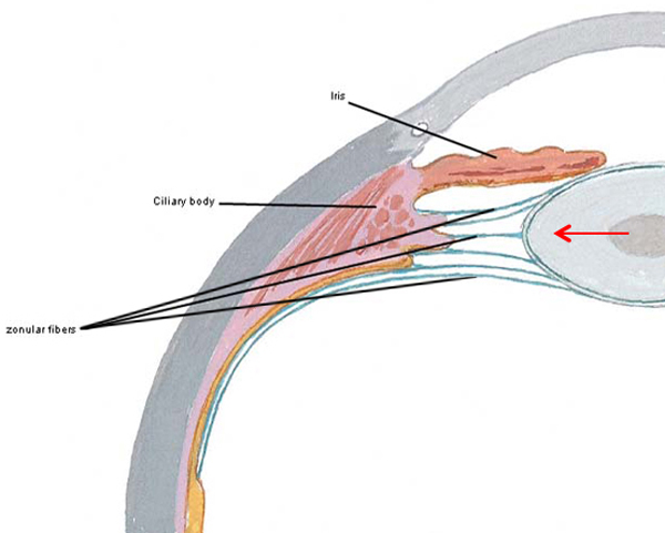
The lens is suspended from the ciliary body by a ring of fibrous strands known as zonular fibers. Although difficult to appreciate in a two dimensional image, zonular fibers attach all the way around the equator of the lens (Photo K – red arrow) much like a round trampoline pad that is suspended from a frame by its coils.

Figure0085B-KLS-edited

Photo K

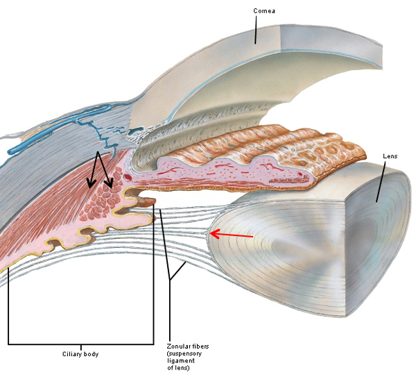
A closer look at the suspensory mechanism reveals that several layers of zonular fibers attach to the lens at various points at or near its equator (Photo L – red arrow). Collectively zonular fibers form the suspensory ligament of the lens. Another important feature, the ciliary body contains bundles of (involuntary) smooth muscle cells (Photo L – black arrows).

Figure0084-KLS-edited

Photo L

Like the cornea, the lens refracts (bends) light rays to help focus them on the retina. Unlike the cornea with its fixed curvature, the lens is flexible and its curvature can be adjusted to fine tune focus, an ability which decreases with age. Based on the distance from a viewed object, the lens flattens or fattens.

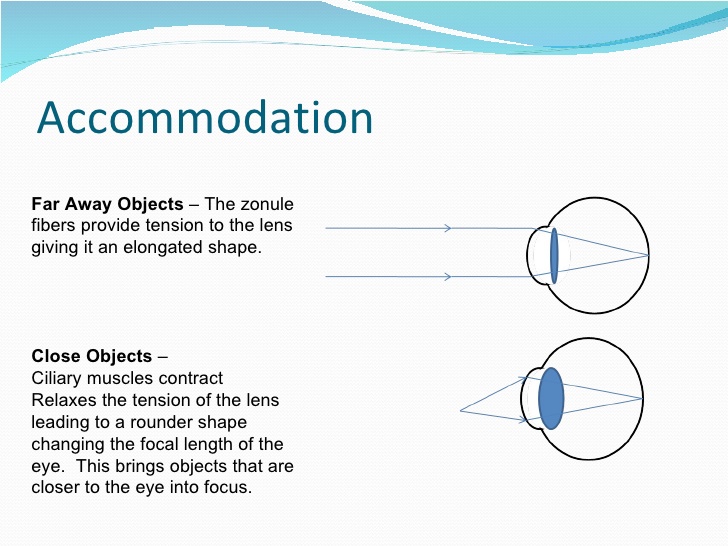
Simply put #1: light rays from far objects enter the eyeball roughly parallel (Photo M – top image) and not much lens refraction is required to focus them on the retina. With this scenario, the ciliary muscle is relaxed and the lens is flat because it is under tension by zonular fibers of the suspensory ligament.

Simply put #2: light rays from near objects (Photo M – bottom image) diverge before reaching the eye so the lens thickens to refract these rays and bring them into focus at the retina (if the lens doesn’t thicken, near light rays would come to focus behind the retina and the image would be blurry). With this scenario, ciliary muscles contract releasing tension on zonular fibers (i.e. the suspensory ligament) so the lens fattens, an action known as accommodation of the lens. Accommodation is accomplished by, yes, the parasympathetic division (PSNS) of the autonomic nervous system causing ciliary muscles to contract!

eye-anatomy-15-728

Photo M

A clarification is important here – accommodation for near vision actually involves three important changes, all of which occur automatically:

  • Eyeballs converge because both medial rectus muscles contract (Anatomy Lesson #30)
  • Pupils shrink in size because the sphincter pupillae muscles contract (Anatomy Lesson #31)
  • Lens thickens because ciliary muscles contract, releasing tension on zonular fibers and the lens assumes its natural fat state (This description may sound counterintuitive but it is correct)

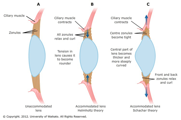
Although scientists know that lens curvature increases with near vision to ensure that the light entering the eye focuses on the retina, the way this happens is still unresolved. Photo N, left panel shows the flattened shape of the lens with far vision. The middle and right panels show two theories about the shape change of the lens during near vision/accommodation. The Helmholtz theory (1855) proposes that the ciliary muscle contracts, all zonular fibers relax and the lens thickens. The Scharchar theory (1992) suggests that the ciliary muscle contracts, selective zonular fibers relax and only the central part of the lens thickens. There is yet a third theory but too technical for today’s lesson. It has been claimed that most modern eye specialists hold with the Helmholtz theory.

Try This: Have a pal sit at one end of a room and focus on something at the far end. Now, step in front of the pal, bend down and ask him/her to look at your nose. If you do this carefully, you can see the eyeballs draw together and the pupils shrink with accommodation. You will not see the lens change shape because it lies too deeply. Try it a couple of more times with the pal looking at something over your shoulder and then at your nose to be sure you got it. This is accommodation.

Theories-of-eye-accommodation

Photo N

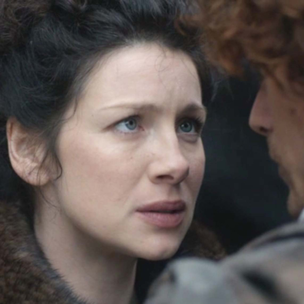
Claire presents us with a wonderful example of accommodation as she looks into the bonny orbs of her handsome Highland Hero (Starz episode 110, By the Pricking of My Thumbs)! Her pupils are constricted and her eyes converge to focus on the object of her desire. Thickening of her lens is not visible. Come back to her, James Fraser!

ep-110-Claire-eyes-KLS-edited

Not to be outdone, Jamie’s eyes can accommodate too (Starz episode 111, The Devil’s Mark). Ahhhh, where did ye say ye were from, Sassylass??? Pupils constricted and eyes convergent…great job, laddie!

ep-111-Jamie-eyes-KLS-edited

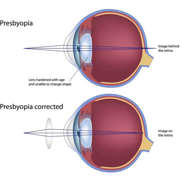
With age, the lens and ciliary muscle undergo changes. Known as presbyopia (Greek meaning old eye), the lens gradually loses its flexibility and the ciliary muscle weakens so focusing on near objects is impaired (Photo O – top image) and the image comes to focus behind the retina. Thankfully, distance vision usually remains unaffected. This condition is easily treated with eyeglasses or contact lenses (Photo O – bottom image) which refract the light rays to bring them to focus at the retina. Please note that at present, presbyopia cannot be treated effectively with surgery. Presbyopia usually becomes apparent in our early- to mid-40s, but this is actually a gradual process that often begins earlier.

presbyopia-treatment-copy-KLS-edited

Photo O

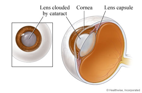
Next, let’s consider cataracts: not a torrent of water but opacities of the lens that can result in impaired vision or blindness (Photo P). Cataracts have been around for a very long time with the earliest documented case being depicted in a small wooden Egyptian statue from about 2400 B.C.E.

A cataract can occur in either or both eyes but does not spread from one eye to the other. Eye cataracts are common in older people as the aging lens and its fibers become more and more opaque, but cataracts can also form congenitally, after injury, following treatment with some medications, or from disorders such as glaucoma. A startling statistic: half of the US population 80 years old and older have cataracts or have had cataract surgery. Now days, there are several different surgical approaches to cataract treatment but they typically involve removal of the natural lens followed by implanting an artificial intraocular lens.

cataracts%203

Photo P

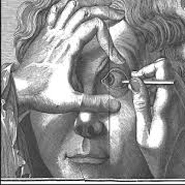
Let’s take a look at some interesting history about the cataract. It may be difficult to believe, but the earliest recorded surgical treatment of cataracts was 800 B.C.E., in India. Using a procedure known as couching, a curved needle was inserted through the coats of the eyeball. The lens was pushed downwards and backwards into the vitreous humor and out of the field of vision. Later, couching spread from India to China, Babylonia and Egypt. Centuries later, couching was practiced in Europe (Photo Q) by traveling barber-surgeons with little or no training and the procedure often left patients blind or at best with only partially restored vision. Other strange and startling treatments were also popular. Nicholas Culpeper records a cataract treatment in his School of Physick (1659):

“The head of a cat that is all black burned in a new pot or crucible, and made into fine ashes, and a little of it blown with a quill into an eye that hath a web or pearl growing in it, three times a day is a sovereign remedy”

couching cataracts copy KLS edited

Photo Q

During Outlander’s time, John Taylor (1703-1772) was the first in a long line of British oculists. Dubbing himself “Chevalier” and “Ophthalmiater Royal,” Taylor became the personal eye surgeon to King George II (Yep; same sovereign that Claire mentions to Colum in Starz episode 102, Castle Leoch), the pope, and a number of European royal families (Photo R). A colorful character who many consider a charlatan, he traveled throughout Europe in a coach painted with images of eyes to advertise his expertise. Taylor removed cataracts either by couching or breaking them up and extracting the bits and pieces. Prior to performing each surgery, he delivered to the audience (yes, groups of people often observed such surgeries) a long, grandiose self-promoting speech.

Purportedly, Taylor once confessed that he had blinded hundreds of patients. Two of Taylor’s botched cataract surgeries included famous composers George Frederic Handel (1685–1759) and Johann Sebastian Bach (1685-1750). Taylor performed couching on Handel but it was unsuccessful and the composer suffered blindness throughout his remaining years. Taylor also used couching to treat Bach’s bilateral cataracts. A week later, Bach was re-operated on but he remained blind and, weakened by the surgeries, he died four months later.

Joannes_Taylor_Medicus KLS edited

Photo R

Many of us are indeed fortunate to be able to choose good eye surgeons with superior training and expertise than those of Jamie’s time. But, I would be amiss not to note that today couching is still practiced in remote areas of third world countries. Let’s give a shout out (and a donation if possible) to those surgeons who travel to dangerous regions of the globe and often at their own expense to bring current therapies to those who are less fortunate.

Let’s close the lesson with a parting keek at Jamie as he keeks at Claire (Starz episode 102, Castle Leoch)! Herself reveals in her 8th book, Written in My Own Heart’s Blood, that Jamie often surreptitiously appraised Claire with his bonny orbs before they wed. He declares that anyone would have thought them well-suited for each other had they noticed his thorough perusal of her:

“If they’d seen the way I looked at ye, Sassenach, when ye didna see me lookin’—then, aye, they would.”

OR, to quote Rick’s famous line from Casablanca:

“Here’s looking at you kid!”

looking 5

Yum! Yum! Gah!

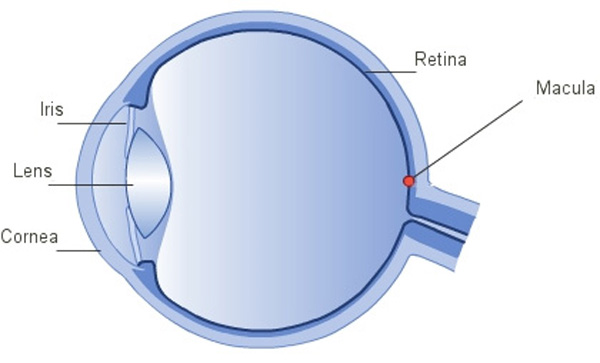
Next lesson will be the last one on the eye: we’ll cover the retina and its parts, nearsightedness, farsightedness and some grand experiments with our own vision. For now, let’s end with a fun poem about eyes by Mr. R! The last Anatomy Lesson to feature one of his delightful science poems was in Anatomy Lesson #24.

Eyes Poem: sense of sight

I need my eyes,
So I can see,
I don’t have 5,

poem 1

Or 1,

poem 2

Or 3…

poem 3

Just 2 eyes,
On my face,

poem 4

It seems to be,
A common place,
For eyes to sit,
On animals,
From frogs, to fish,
From bears, to gulls…

poem 5

2 eyes,
1 face,
That’s a match,
But really here’s,
A little catch…
Spiders see,
With 8 bug-eyes,
And here’s another,
Eye surprise,
Scallops have,
Most of all,

poem 6

More than 50,
Must see all!
I’ll stick with 2,
They let me see,
My 2 eyes,
Are good for me…


A deeply grateful

Outlander Anatomist

photo creds: Starz, Netter’s Atlas of Human Anatomy, 4th ed. (Photos A, K, L), Wheater’s Functional Histology, 4th edition (images of structure of eye and of retina), Outlander Anatomy collection (Photo E), www.archopht.jamanetwork.com (Photo Q – couching lenses), www.discovery.lifemapsc.com (Photo I – lens anatomy), www.en.wikipedia.org (Photos R – John Taylor), www.lasik-center.com (Photo H – lens), www.medicine.academic.ru (Photo C – limbus), www.optics.rochester.edu (Photo J – SEM of lens fibers), www.rosineyecare.com (Photo O – presbyopia), www.sciencelearn.comm.nz (Photo N – theories of accommodation), www.sciencepoems.net (-Eyes poem, Sense of Sight; Mr. R’s World of Math and Science), www.slideshare.net (Photo M – accommodation), www.timelessnaturalhealth.com (Photo B – sclera), www.toovia.com (Photo G – corneal jewelry), www.visioncarespecialists.com (Photo D – corneal structure), www.webmd.com (Photo P – cataracts), www.wynneeeyeassociates.com (Photo F – refraction of light rays by cornea)