Anatomy Lesson 17: “The Wedding” or “Hallelujah Chorus”

Greetings, dear readers! Today, we will enjoy a short Anatomy Lesson #17: The Cephalic Vein. However, most of our time will be spent as anatomy guests at The Wedding! Raise your voices in song! I have been waiting to do this episode for nearly six months. As guests, we will perform a practice practical exam and a week from today (March 31st) we will take our midterm practical exam. Jamie will be our model for the today’s lesson but afterwards it’s Q&A time! Hey, now, stay with me. Dinna get cold feet!

Today’s test is for practice only; it is structured like a gross anatomy practical wherein structures are tagged with a string and a number. Students write the answer on their test sheet. But today, each question is followed by a link to the appropriate anatomy lesson, followed by an image, followed by the answer. Red arrows mark the structures. Also, this practical tags structures as they sequentially appear in “The Wedding” episode. There are 20 questions; a few are challenging but most are not.

Of course, I can’t resist a few choice Outlander quotes and some smart-arse commentary although truth be told, the tenderness and patience between Jamie and Claire as they reach out to one another in this episode are deeply moving.

So, pull out quill and paper. Record your answer and, if need be, write in the correct answer for future reference as there will likely be some repeat questions. Next week’s midterm will be for credit! Do your best and have fun!

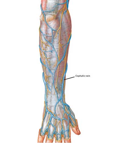
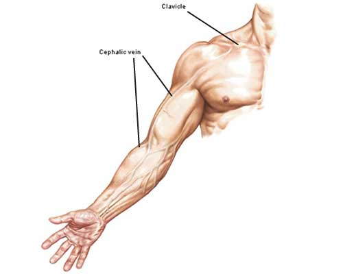
Now, on to our brief anatomy lesson: the cephalic vein. Cephalic is an anatomical term with muddled origins but current usage means “directed toward, or situated on, near or in the head.” Photo A shows the cephalic vein in forearm and arm (Anatomy Lesson #4) so why the name? The name is based on blood flow which is directed upward toward the head, hence cephalic. In the lean and muscular, the vein (they are paired) is visible just under the skin (subcutaneous).

Insert-Photo-A--Figure0401-cephalic-vein-01-KLS-edited-

Photo A

Each cephalic vein forms near the base of the thumb by union of veins of the back of the hand (Photo B). Arteries lie deep and are not visible through the skin. Try this: check the venous pattern on the back of your hand; each pattern is unique.

Figure0463B-cephalic-vein-03-KLS-edited

Photo B

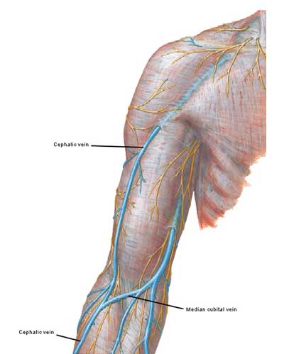
The cephalic vein can be traced up the thumb side of the forearm where it communicates with a vein in front of the elbow (median cubital – the one typically used for a blood draw). Photo C shows the subcutaneous position of the cephalic vein along the lateral borders of forearm and arm but near the armpit(axilla) it disappears. Where did it go?

Figure0462A-cephalic-vein-05-KLS-edited

Photo C

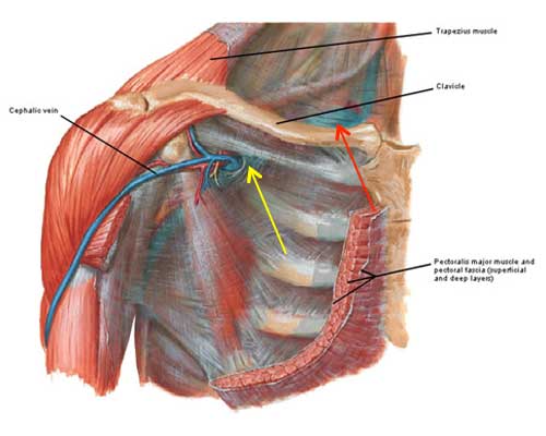
The cephalic vein disappears from view because near the armpit it dives deeply toward the clavicle where it empties blood into the little known axillary vein (Photo D – yellow arrow), a large vein that drains the entire upper limb. Deep to the clavicle, the axillary vein drains into the subclavian vein (Photo D – red arrow). The subclavian vein is an important site for central catheterization because it offers many advantages to both patient and practitioner. Just so you know blood then travels through two even larger veins (not shown in Photo D) before reaching the right side of the heart.

Insert-Photo-D--Figure0411A-cephalic-vein-02-KLS-edited

Photo D

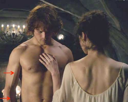
Jamie has agreed once more to hop up on the dissection table; thank you Mr. Bonny Highlander! Several students have asked me to explain Jamie’s arm vein. So here’s Claire playing round and round and round she goes – where she stops everyone knows! Jamie’s cephalic vein is clearly evident in his right forearm and arm (red arrows). Again this vein is best seen in the muscular and lean so Jamie is a perfect model. Try this: with elbow bent, lift one hand and turn the forearm so the palm faces downward (pronation). You will likely find a large vein on the thumb side of the forearm about 2-4 inches (5-10 cm) above the wrist. Trace it upwards as far as you can; this is the cephalic vein.

ep-7-cephalic-vein-02-KLS-edited

This brief lesson is over and the time has come to start the practice practical!

Our first question is from the lead-in to Claire and Frank’s wedding scene which, BTW, is a figment of the writing team’s imagination. Here Frank tells Claire that he doesn’t really give a figment if his parents are waiting to meet them at a restaurant. Let them eat cake!

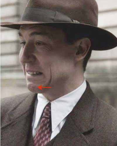
Q #1: Name the paired facial muscle that wrinkles Frank’s chin-skin. (Anatomy Lesson #13)

Insert-image--ep-7-Frank-02-KLs-edited

A #1: Mentalis. Did you get it right? The body, the body, rah, rah, rah, anatomy, anatomy, sis boom bah! (didn’t know there was an official anatomy cheer, did ye?).

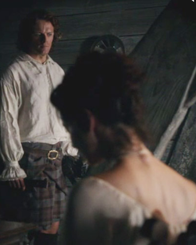
Next, Claire sits with tendrils of her not-dull-at-all brown hair framing her face and curling down her neck. She’s dressed in shift, corset and skirts musing on how life with Frank was like pearls on a string. Whaaaat? Claire, anyone with half a visual cortex can see that is pure fantasy! Frank is a nice man (until episode 8) but it was you that kept team Beauchamp-Randall going. Sorry, canna imagine you as a contented don’s wife. Stick around though you are going to be content verra soon. Happy dance everyone!

Q #2: What shape is the shaft of one curly Claire-hair: flat or round? (Anatomy Lesson #6)

ep-7-vertebrae-prominens-KLS-edited

A #2: flat

The next image uses a quote from Outlander book wherein Herself records:

I took a deep breath. “I have questions,”…”I’d suppose ye do,” he agreed. “I imagine you’re entitled to a bit of curiosity, under the circumstances. What is it ye want to know? “He looked up suddenly, blue eyes bright…”

What’s the matter Claire, did those earnest baby blues make you forget your question? Hahaha, she can barely breathe! Oops, they made me, erm, forget my own question. Ohhhh, here it is:

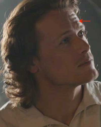
Q #3: Name the muscles lifting Jamie’s eyebrows and wrinkling his forehead skin. (Anatomy Lesson #11)

ep-7-frontalis-01-KLS-edited

A #3: Frontalis

What Claire wonders is “why did you agree to marry me?” Jamie explains he did it to save her from BJR so in gratitude she glides over, sits next to him and leans in for a kiss. Jamie almost makes it to first base but Claire interrupts the kiss with: “Tell me about your family.” Abort mission! Ha, Mistress Beauchamp, ye aren’t fooling Jamie. He’s as bright a lad as ever strode the heather. He kens exactly what ye are doing!

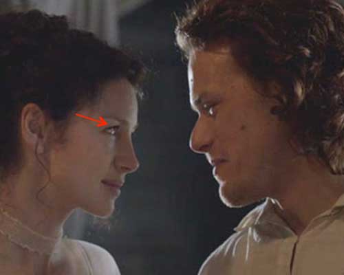
Q #4: Name the muscles lifting Claire’s eyelids. (Anatomy Lesson #14)

Insert-image--ep-7-vermillion-zone-KLS-edited

A #4: Levator palpebrae superioris (lifter of the upper eyelid)

“How many generations back?” Jamie is a Highlander born and bred and also a gentleman so he patiently traces his geneaology back to Adam and Eve for the benefit of nurse Beauchamp. Story telling obviously requires a kilt-hike so we see Jamie knees and thighs…no problema (or problemo if ye are into Schwarzenegger films). Come on lass, get with the program! There’s plenty waiting in line if ye are no willing to do your duty!

Q #5: Name the muscle at the inside of Jamie’s right and left knees. Smooookin’! (Anatomy Lesson #7)

ep-7-vastus-medialis-03-KLS-edited

A #5: Vastus medialis (Aye, it is vast!)

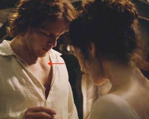
So finally after two days of storytelling and prurient comments from Angus and Rupert they finally get down to business: “To bed or to sleep?” What’s a friend for? Weel, to help Claire with her laces of course – she willna sleep in her corsets tonight. Yippee! What with all the excitement, Jamie’s sark conveniently comes unbuttoned so let’s take a wee keek at his warrior’s neck.

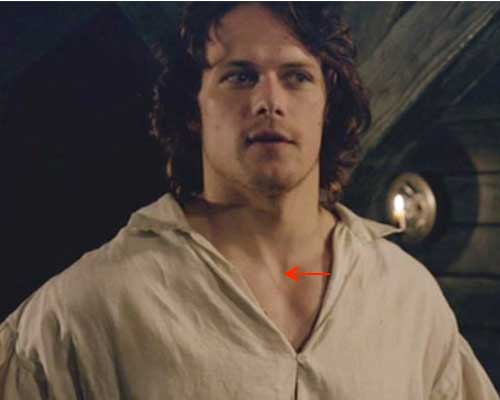
Q #6: Name the midline bony depression at the top of Jamie’s sternum. (Anatomy Lesson #12)

ep-7-jugular-notch-04-KLS-edited

A #6: Jugular notch

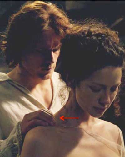
Now, this little ribbon went to market…Ah, no, it never got to the grocery store because it was hung up on its way off Claire’s neck! Who knew that such a wee bit of fluff could be soooo enchanting? As Jamie slowly, sensuously slides that ribbon, he grips the base of her neck (whoa Nelly!)…

Q #7: Name the muscle under Jamie’s right fingers (Anatomy Lesson #2 & Anatomy Lesson #3)

ep-7-ribbon-off-KLS-edited

A #7: Trapezius

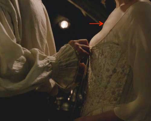
He canna wait to undo the laces of that magnificent corset (Bravo Terry and team)! As he makes his way down the loops, let’s note the bony bump on Claire’s sternum (red arrow)? We all have this bump, a palpable clinical landmark from which ribs are counted; it also serves as a marker for various internal anatomical features that can’t be seen from the surface (e.g. aortic arch). Please find yours or a friend’s…hee hee.

Q #8: Name the bony bump of the sternum (Anatomy Lesson #15)

ep-7-sternal-angle-KLS-edited

A #8: Sternal angle (of Louis)

Next, Claire timidly glances at Jamie with a pleading expression on her face. Clad only in her shift, she’s feeling a wee bit self-conscious under his careful scrutiny. Does he like what he sees?

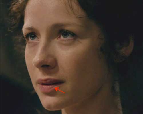
Q #9: Identify the rim around Claire’s luscious lips (Anatomy Lesson #14).

ep-7-vermillion-border-01-KLS-edited

A #9: Vermillion border

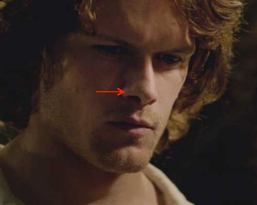
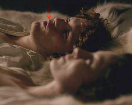
Oh, aye, Jamie likes his bride of astonishing beauty soooo much that his nostrils flare! A wee muscle does this but it has the longest name (5 words) of any in the human body. Take note! This may be on the final exam.

Q #10: Name the muscle that flares each nostril (Anatomy Lesson #11).

ep-7-LLSAN-KLS-edited

A #10: Levator labii superioris alaeque nasi (lifter of the superior lip and nasal ala = LLSAN)

Claire’s no about to let Jamie have his way and not get hers too! Stand still Jamie as she unbuckles yer belt! Bet you weren’t expecting that move from an Oxfordshire widow!

Q #11: Name the glorious mass of muscle on Jamie’s upper chest (Anatomy Lesson #12)

ep-7-pectoralis-major-01-KLS-edited

A #11: Pectoralis major (and they are major!)

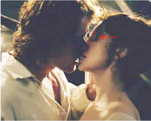
Okay, Jamie, now you can finally kiss Claire without those darned wedding guests oogling the pair of you. Instead, an entire planet hangs on “every minute – every second!” It’s a fab kiss too!

Q #12: Name the muscles closing Claire’s eyelids. (Anatomy Lesson #11).

ep-7-kiss-KLS-edited

A #12: Orbicularis oculi (palpebral parts)

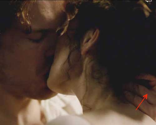
Next, Jamie grips Claire’s neck during that awesome kiss (Q #13, red arrow); that’s how he pulls her up to him. Aye, throughout the books Jamie either grips or bites her neck; it’s that horsey thing again! The next quote from Outlander book records the first time he touches Claire’s neck:

The lad had nice feelings. Instead of calling for help or retreating in confusion, he sat down, gathered me firmly onto his lap with his good arm and sat rocking me gently, muttering soft Gaelic in my ear and smoothing my hair with one hand…but slowly I began to quiet a bit, as Jamie stroked my neck and back…

Q #13: How many cervical vertebrae in Claire’s neck? (Anatomy Lesson #12)

Insert-image--ep-7-kiss-03-KLS-edited

A #13: Seven (Nay, she doesna have any extra vertebrae!)

Again from Outlander book:

“Where did you learn to kiss like that? I said a little breathless. He grinned and pulled me close again. I said I was a virgin, not a monk.”

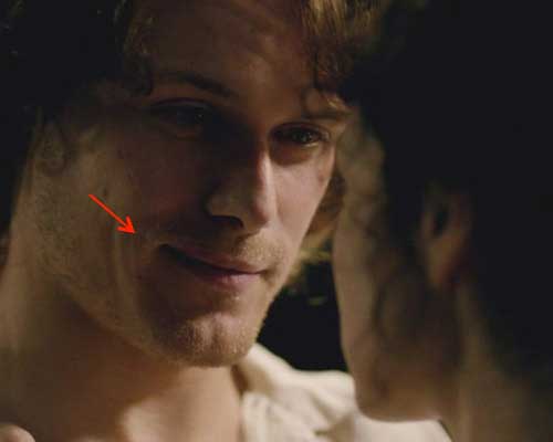
Q #14:

Name one muscle (there are three) that lifts the right side of Jamie’s mouth. (Anatomy Lesson #11 & Anatomy Lesson #13)

ep-7-zygomaticus-major-05-KLS-edited

A #14: zygomaticus major, zygomaticus minor or levator anguli oris

Another fabulous quote from Outlander book. Herself records:

“Was it like you thought IT would be?”…”Almost; I had thought – nay, never mind.” “No, tell me, what did you think?” “I’m no goin’ to tell ye; ye’ll laugh at me.” “I promise not to laugh. Tell me.”…Oh, all right. I didna realize that ye did IT face to face. I thought ye must do IT the back way, like; like horses, ye know.”

Jamie, a wee warning: if Claire promises anything ye must take it with grain of salt. Although she tells Geillis Duncan that a promise was a serious thing in her country too (Starz episode 4, The Gathering), she tends to break her promises!

Q #15: Name the muscle puckering Jamie’s mouth (Anatomy Lesson #11)

ep-7-orbicularis-oris-01-KLS-edited

A #15: Orbicularis oris

“Did ye like IT?” Ahhh Jamie, dinna be daft; ye mistake Claire’s downcast eyes as a NO! Ye don’t ken it yet, but this is one LL for lusty lady. Oh dear, Jamie is totally bummed – mayhap Murtaugh was right and women dinna like IT! Weel, mayhap some leddy should give Murtaugh his own anatomy lesson. Volunteers? Snort!

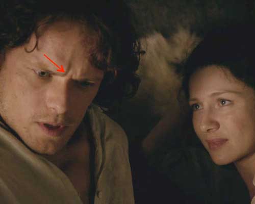
Q #16: Name the muscles wrinkling the skin between Jamie’s eyes. (Anatomy Lesson #11)

ep-7-corrugator-supercilli-01-KLS-edited

A #16: Corrugator supercilii

Next, we hear Claire’s feet pitter patter across the floor: she needs food! She also feels a wee bit unnerved being an adulteress, a bigamist and all: guilt surge because she did enjoy IT (apparently IT doesna have a name).

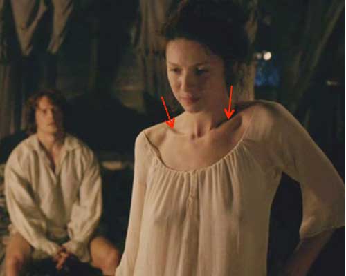
Q #17: Name the stunning bones that peek above the curving neckline of Claire’s shift. (Anatomy Lesson #2 & Anatomy Lesson #3)

ep-7-clavicle-01-KLS-edited

A #17: Clavicles (collar bones)

Ever the gentleman, Jamie descends the stairs for food while the Highlanders rattle the rooftop with crude and ribald jokes. A little chaff rarely bothers Jamie though so he fills a plate with goodies for the pair of them and hightails it back to the bridal suite.

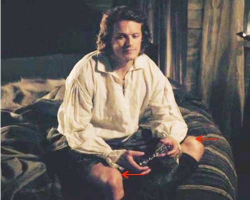
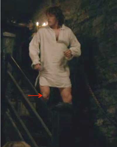
Q #18: Name the bone at the tip of the red arrow. (Anatomy Lesson #7)

Insert-Image--ep-7-patella-KLS-edited

A #18: Patella (knee cap)

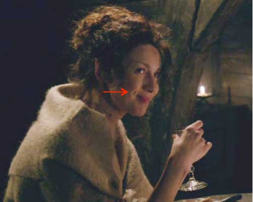
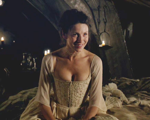
As Jamie offers Claire a chunk of cheese she learns that Dougal was a royal jerkwad in the tap room advising him to never “give a woman too much power!” But, Jamie (aye, he is a bright one) says he is “completely under her power and happy to be there.” Check mate! Claire needs more whisky so off Jamie goes to grab a decanter. After he gracefully pours without spilling a single drop (that man can do anything!) he touches her hair and gets a major rebuff! Ah, Jamie, if only ye only knew her secret ye might ken her push-pull behavior. But, she regrets her snub and favors him with a killer-watt smile! Check out those darling dimples!

Q #19: Name one of three muscle pairs lifting Claire’s lips. Try for a different pair than you gave in question #14 (Anatomy Lesson #11 and Anatomy Lesson #13).

Insert-image--ep-7-zygomaticus-major-04-KLS-edited

A #19: Zygomaticus major, zygomaticus minor or levator anguli oris. Did you answer differently this time? Good job fledglings! Your wings are getting stronger.

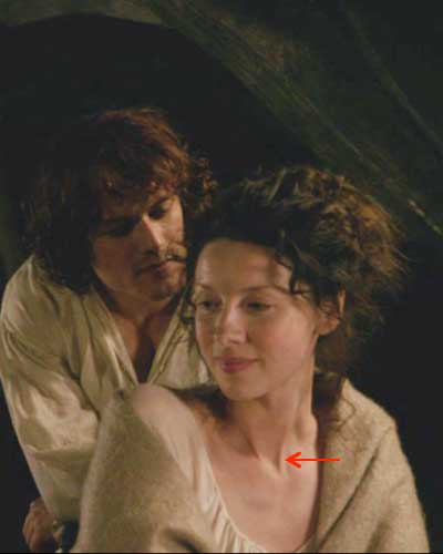
Next, Jamie sidles up with more horse-gentling techniques, comparing her hair to the water in a burn: “dark in the wavy spots and auburn where the sun hits it…“  Whew, the man is determined and she likes those soft, tender words! On, and on he gently coaxes her. “Here’s looking at you kid” isna likely to do for our Claire.

Q #20: Name the muscle in Claire’s neck. (Anatomy Lesson #12)

Insert-image--ep-7-jugular-notch-01-KLS-edited

A #20: Sternocleidomastoid! Yessssss…good job! Remember, these are paired.

That was our last question. Now, the practice practical is over. How did ye do?

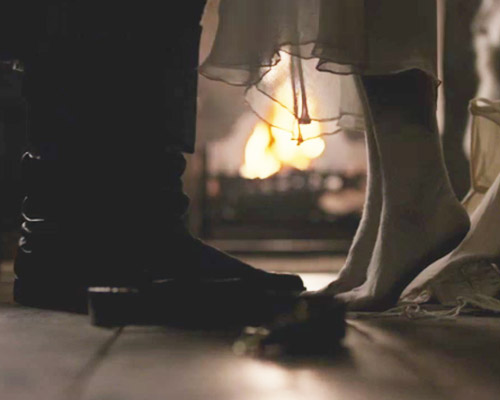
But, we’re not done yet! Time for a little sex, erm, sox talk! As Jamie pulls Claire up for that breath-taking kiss she stands on tippy toes to reach his mouth. Her feet are shrouded in verra fine (lisle cotton?) stockings.

ep-7-stockings-on-03-KLS-edited

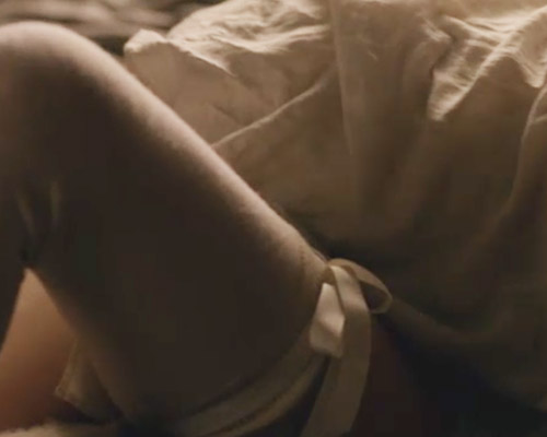
Soon, Claire is on her back baring those lovely cream-colored stockings with matching ribbon garters. Being it’s his first time, Jamie is still clad in a three-yard sark and boots! Puir lad. How do I know the sark (shirt) uses three yards of fabric? Weel, Herself says so in Dragonfly in Amber and that should be good enough for any of us! I’ve said it before and I’ll say it again, if ye aren’t reading the books you are missing the best of the best (no, this isna a paid ad).

“…I had made such sarks for him; I could feel the softness of the fabric…the billowing length of the three full yards it took to make one, the long tails and full sleeves that let the Highland men drop their plaids and sleep or fight with a sark their only garment.”


ep-7-stockings-on-01-KLS-edited

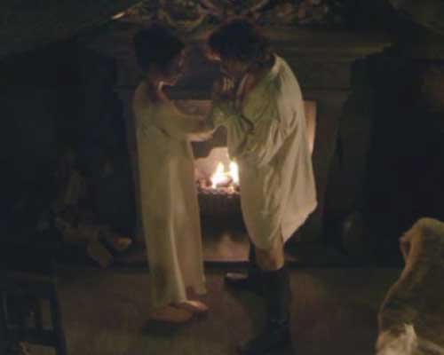
I just canna get those stockings off my mind. The next time we see Claire’s feet, she is barefoot in front of the fireplace as Jamie plants a tender kiss on her wounded wrist. When did Claire take off those lovely leggings? Did she peel them off à la Mrs. Robinson? Did they think we wouldna notice? I want to know…Does this make me professor nitpicker? Tcha, course it does!

ep-7-stockings-on-03-KLS-edited

Alrighty, then! Let’s stop getting our socks knocked off by the Frasers. We’ve all got some studying to do for next week’s midterm which will be next Tuesday, March 31st. Here are links to all of the anatomy lessons for yer studying pleasure: Anatomy Lessons 1-13; Anatomy Lesson 14; Anatomy Lesson 15; Anatomy Lesson 16. The happy news is the midterm exam will cover more of The Wedding (Starz episode 7)!

Until next time…HALLELUJAH, HALLELUJAH, HALLEULJAH, HALLEULJAH…!

A deeply grateful,

Outlander Anatomist

Follow me on Facebook and Twitter!

Image creds: All images of the cast in this post are from Starz episode 7, The Wedding; Netter’s Atlas of Human Anatomy, 4th ed.

Anatomy Lesson 16: “Jamie’s Belly” or “Scottish Six-Pack”

The belly has many synonyms: abdomen, beer belly, breadbasket, girth, gut, guts, insides, maw, middle, midriff, paunch, pot, potbelly, spare tire, stomach, tum, and tummy to name a few. Although not a synonym, there is only one word for Jamie’s belly and that is spectacular! Greetings everyone and welcome to Anatomy Lesson #16: The Anterior Abdominal Wall.

This lesson will explore bones, muscles and skin that comprise the belly along with surface landmarks that you may have observed but didn’t recognized as either important or significant. In this lesson, I use the term anterior abdominal wall or the simpler belly wall and hope that by its end you will enjoy a greater appreciation for its complexity, purpose and design.

Herself refers to the belly many times in her writings, but the verra first time we encounter the term is in Outlander where Claire’s belly rumbles loudly in protest of lack of food (BTW, rumblings or growling by the intestines is due to muscle contraction of the gut wall and is called borborygmi – pronounced BOR-boh-RIG-mee). Our ever gallant hero offers her a strong drink (Starz episode 1, Sassenach):

“A moment later, a hand with a flask came around in front of me again. “Better have a wee nip,” he whispered to me. “It willna fill your belly, but it will make ye forget you’re hungry.” And a number of other things as well, I hoped. I tilted the flask and swallowed.”
image

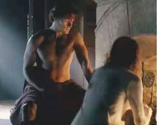
The first time we see Jamie’s belly is Starz episode 2, Castle Leoch after Claire uncovers his back to properly clean and dress his gunshot wound. A number of important surface features are wonderfully demonstrable on Jamie’s belly but in the words of Dougal, we’ll puzzle those out later. One look at Jamie’s bare torso and is it any wonder Claire drops her cleaning cloth? Gah!
image

But the first time we read about Jamie’s belly is in Outlander book as Dougal tells Claire about BJR’s flogging of Jamie. Herself writes:

“His mouth tightened up and he says, ‘I thought this was the young man who only a week past was shouting that he wasn’t afraid to die.Surely a man who’s not afraid to die isn’t afraid of a few lashes?’ and he gives Jamie a poke in the belly wi’ the handle of the whip.”

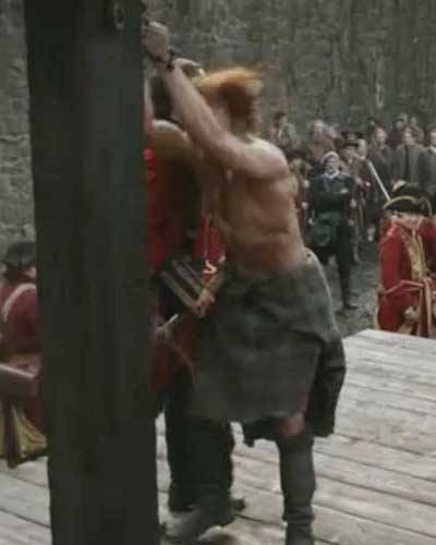
Weel, this looks a wee bit more serious than a poke. The sadistic bastard punches Jamie very hard in the belly with the handle of his cat o’ nine tails (Starz episode 6, The Garrison Commander).

image

Continuing the quote:

“Jamie meets Randall’s eye straight on then, and says,‘No, but I’m afraid I’ll freeze stiff before ye’re done talking.’ ” In otherwords, quit yer yapping BJR and get on with it! Weel, it was a braw speech for which BJR flogs him even harder.

image

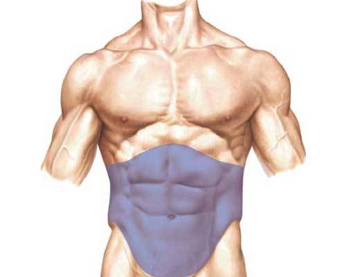
But, let’s leave this sad scene for our anatomy lesson. The abdomen is that part of the trunk between ribs and pelvis (Photo A– blue overlay). Its front and sides are bounded by flexible fibromuscular walls formally known by the lengthy name, anterolateral abdominal wall. As explained earlier, I will use the simpler anterior abdominal wall or belly wall. The posterior (back) abdominal wall is bulkier and less elastic but it doesn’t concern us today.

image

Photo A

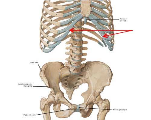
The bony framework for the anterior abdominal wall includes xiphoid process and costal margins and parts of the hip bones: anterior superior iliac spine, iliac crest, pubic symphysis and pubic tubercle.

These bony landmarks are often palpated during physical examination. If you read Anatomy Lesson #15, you have already found xyphoid process and costal margins (Photo B – red arrows).

Try  this: If you can, lie on your back and relax the tummy. At your sides, locate the point  of each hip bone; these are the anterior superior iliac spine or ASIS (Photo B). From the ASIS, press fingers backward along the flared iliac crests. Next, place fingers on the navel and move down to the tops of the pubic bones.  You may feel a midline indentation or pubic symphysis, a disc of specialized cartilage that binds together the paired pubic bones; this disc softens during pregnancy. Run fingers along the top of one pubic bone until you feel a smallish bony knob, one of the paired pubic tubercles.

image

Photo B

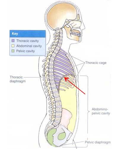
The abdominal walls form a flexible and dynamic container for the abdominopelvic cavity which is more extensive than the abdomen because it reaches from the domed thoracic diaphragm above (Photo C – red arrow and Anatomy Lesson #15) to the sunken pelvic diaphragm (floor) below. The cavity contains most digestive and urogenital organs and is subdivided into upper abdominal cavity (yellow in Photo C) and lower pelvic cavity (Green in Photo C). The abdominopelvic cavity is lined with peritoneum, a serous (wet) membrane that clings to inner walls of the cavity and covers organ surfaces. In males, the peritoneum is a closed sac but in human females the sac has two openings one at the mouth of each uterine tube to allow passage of ova.

image

Photo C

The belly wall may seem simple but it is very complex and critical for our well being. A simple note about orientation before we dissect the belly wall: in anatomy and medicine right (R) and left (L) are defined as the patient’s R and L and not those of the viewer.

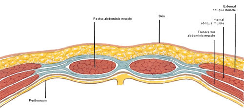
Viewed in cross-section (penny-sliced), the belly wall is composed of several tissue layers. The outer layer is belly skin and the innermost is peritoneum (Photo D). Belly skin bears a number of important landmarks we will discuss shortly. Jamie is our model for that. He is always soooo willing. Thank you Big Ruadh One!

The belly wall includes four pairs of muscles known at the gym as abs. Paired rectus abdominis (RA) muscles flank the midline. Each side has three flat muscles from superficial to deep: external
oblique
(EO), internal oblique (IO) and transversus abdominis (TA).

image

Photo D

The bluish structures in Photo E are fibrous tissues of the belly wall. These include a midline linea alba (Latin meaning white line) and the paired rectus sheaths. Let’s take a closer look.

image

Photo E

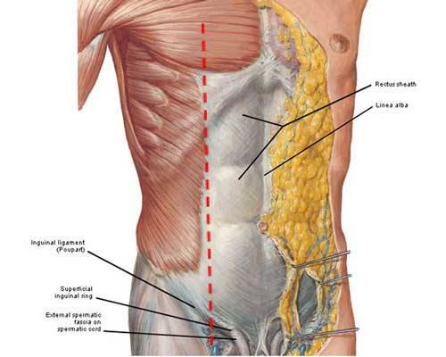
Photo F, shows a superficial dissection of the belly wall exposing linea alba and R rectus sheath. Linea alba is a midline fibrous line. Each rectus sheath is a fibrous envelope extending from ribs above to pubic bone below. The following explains their anatomy: at the mid-clavicular line (Photo F – dashed red line) EO, IO and TA change from muscle to flat fibrous sheets. These sheets envelope each RA muscle as a rectus sheath. In the midline, the sheets meet and decussate (crossover) to form the fibrous linea alba. Amazingly, an incision along linea alba allows an almost bloodless entry into the abdominal cavity as only tiny blood vessels cross the midline.

This is interesting: see the structure labelled spermatic cord (Photo F)? Paired spermatic cords suspend the testes (pl.) in the scrotum and contain structures running to and from these organs. You may think it odd to mention spermatic cords in a belly wall lesson but anatomists teach them here because their layers are derived from the belly wall. Each spermatic cord traverses the inguinal canal (not shown) an oblique channel through the wall and then emerges via its respective superficial inguinal ring.

Each spermatic cord contains a testicular artery the sole blood supply to the testes (This lesson ends with a cautionary tale about the testicular artery. Stay tuned!). Spermatic cords also contain the all-important vas deferens, a small muscular tube for passage of spermatozoa…called the vas deferens because it makes a vast difference if accidentally cut! Um, couldn’t resist a beloved “joke” amongst surgeons.

Lastly, testes do not develop inside the scrotum, they develop inside the abdominal cavity. During the last trimester, they descend through the inguinal canals and exit the superficial inguinal rings to reside within the scrotum. Females also have paired inguinal canals although each contains an unimportant (as far as known) fibrous ligament. Importantly, the inguinal canal is the channel through which passes an  indirect inguinal hernia; more common in males.

image

Photo F

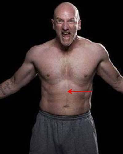
I am breaking my own rule in this lesson by using an image of Graham McTavish because I havna got one of Dougal with his shirt off. Big D is sooo modest in the Starz series. So let’s pretend that this is fierce Dougal (Photo G). The red arrow marks the location of Dougal’s verra fine linea alba. Hopefully this will, erm, satisfy some readers who beg for more Dougal anatomy.

image

Photo G

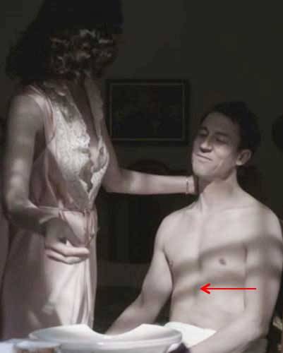
I canna show Dougal-Belly and not show Frank-Belly. Now then Frank, “fair’s fair…take off yours, too.” Ha! Not too many surface features are visible here because Frank is seated, but we can see the location of his linea alba (Photo H):

image

Photo H

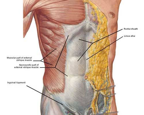
Next, let’s examine muscles of the belly wall, all of which support our abdominal organs. Beginning at the sides, the most superficial pair is the external oblique (EO); each arises from the ribs (Photo I), turns tendinous, helps form the rectus sheath and inserts into linea alba, pubic tubercle and inguinal ligament. Whew! EO muscle fibers are directed downward and towards the midline.

Try this: place hands in the side pockets of jeans or similar trousers; the fingertips point the same direction as EO muscle fibers. Working together, EO muscles flex the trunk and compress the abdominal viscera. Working independently, each EO helps rotate the trunk.

image

Photo I

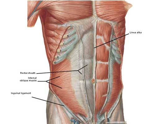
Deep to each EO is an internal oblique (IO) muscle. Each IO arises from thoracolumbar fascia (Anatomy Lesson #10) and iliac crest, turns tendinous, helps form the rectus sheath and inserts into linea alba, pubic tubercle and inguinal ligaments (Photo J). Most IO muscle fibers are directed upward and towards the midline (90° to EO muscle fibers). Working together IO muscles flex the trunk and compress the abdominal viscera. Working separately each IO helps rotate the trunk.

image

Photo J

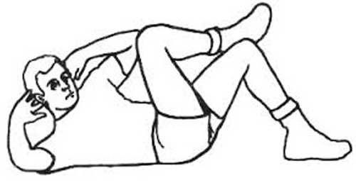
Interestingly, when rotating the trunk as in cross crunches (Photo K) EO of one side works with IO of the opposite side. Here, left EO contracts with right IO to rotate and flex the trunk toward the
right knee. Note: when doing cross crunches, one should reach shoulder/arm pit (not elbow) toward the opposing knee.

image

Photo K

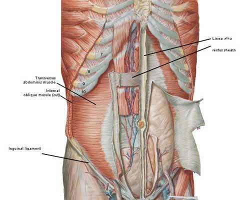
Deep to each IO is transversus abdominis (TA), deepest of the side muscles (Photo L). TAs arise from costal cartilages, thoracolumbar fascia, iliac crests and inguinal ligaments and insert into linea alba and pubic bones. TA muscle fibers are horizontal and like EO and IO give way to a flat tendon that helps form each rectus sheath and linea alba. TA compresses and supports the abdominal viscera. They can be strengthened by doing exercises that tighten the belly wall (e.g. Pilates).

image

Photo L

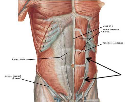
Our final set of abdominal muscles is the paired rectus abdominis (RA); each is a flat vertical muscle extending from pubic bones to costal cartilages (Photo M) and is enveloped by its respective rectus sheath.

RA muscles are separated in the midline by linea alba. The lateral border of each RA is marked by a curved line, the linea semilunaris (Photo M – black arrows).

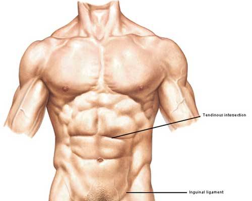
Tendinous Intersections: The grey horizontal bands are tendinous intersections (TIs), fibrous bands that traverse each RA. Most individuals have three TI per RA. The left RA shows three typical TIs and a partial fourth. Acting together RA muscles flex the trunk and compress abdominal viscera.

image

Photo M

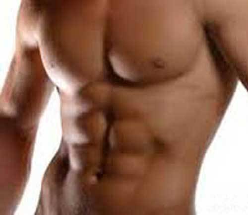
If rectus abdominis muscles are well-toned, they bulge between the tendinous intersections accounting for the six-pack appearance of a muscular anterior abdominal wall. The man shown in Photo N has three pairs of TIs and three RA bulges: one set above the top TI, second set between top and middle TI, third set between middle and bottom TI. The belly below the navel is
not part of a six-pack.

image

Photo N

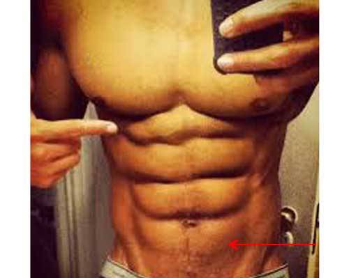
Occasionally, a 4th and 5th pair of TIs appears below the navel resulting in an 8- or even more rarely a 10-pack ab. This man has four pairs of TIs – the 4th appears below the navel (Photo O – red arrow)
resulting in an 8-pack. This is not an anomaly, it just represents individual variation.

image

Photo O

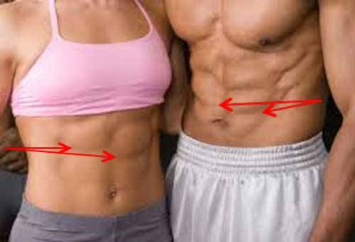
TIs may be asymmetrical such that a pair of bands is offset relative to each other. In the image below (Photo P – red arrows) both woman and man have stair-step TIs. Again, this is individual variation and has no known functional significance.

image

Photo P

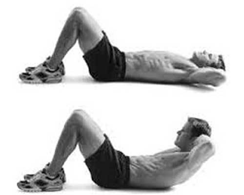
Try this: Would you like your own 6-pack abs? Simple crunches help as they employ EO, IO and RA muscles. Crunches may be done many ways but a common method is to bend the knees with feet hip
distance apart and resting on the floor, elbows to the sides, hands resting lightly behind the head (Photo Q). Lift shoulder blades off the floor and repeat many, many, many times. But, more importantly for protection the lower back (lumbar region) remains on the floor and do not pull on the neck.

image

Photo Q

Now, for surface anatomy: Belly skin bears a number of topographical landmarks which are best observed in the lean and/or physically fit.

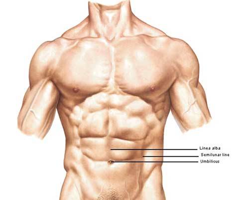
First, a vertical midline groove or furrow overlies the fibrous linea alba (Photo R) marking the medial border of each rectus sheath and its enclosed RA muscle.

Next, the umbilicus, navel or belly button lies near the center of the belly wall at the level of L3
– L4 IV disc (Anatomy Lesson #10). The umbilicus is a midline cicatrix or scar that marks the
attachment site of umbilical cord to body wall during prenatal life. Its size and shape vary considerably and with increased weight or distortion from pregnancy its placement may change. Although most human have an umbilicus, some do not. People may require surgical removal of the umbilicus due to herniation, birth defects, injury and infection or for elective cosmetic reasons.

The umbilicus bears a small opening or umbilical ring. Try this: Lie on your back, place a fingertip in the center of the umbilicus and press down. You may feel the small tight opening of the umbilical ring through which passed the umbilical vessels during intrauterine life.

Two curved vertical skin grooves are also visible on belly skin; lineae semilunares (Latin meaning half-moon lines) mark the lateral border of each rectus sheath and its enclosed RA muscle. These extend from costal cartilages to pubic tubercles.

image

Photo R

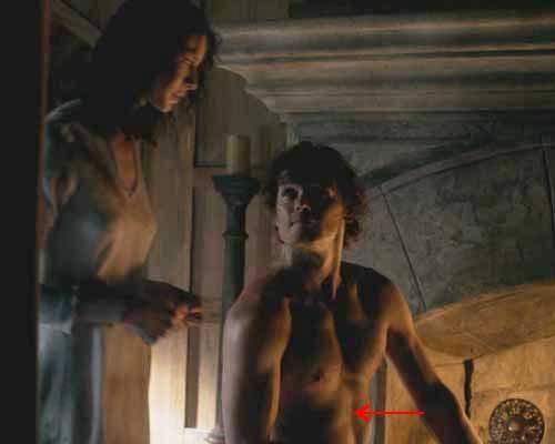
Time for a Jamie-fix! Does Jamie’s belly show these surface features? Oh, aye, to be sure. The red arrow (Starz episode 2, Castle Leoch) points to the vertical furrow overlying linea alba. Ahhhh, now readers, quit looking at Jamie’s chest! I ken what ye are thinking. We are supposed to be
learning the belly wall. Erm, Clarie keep hold of your cleaning cloth!

image

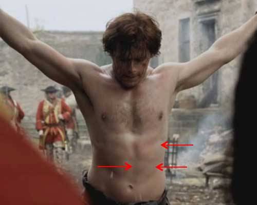
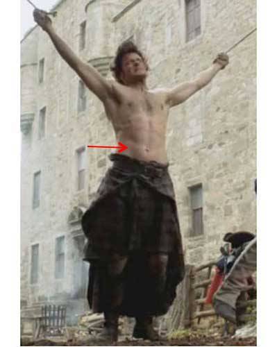
Here Jamie is suspended from ropes as the Mad Man of Fort William beats him the first time (Starz episode 2, Castle Leoch). The red arrows point to medial and lateral margins of the L rectus sheath which is tightened because he is hanging by his arms. Because both rectus sheaths are taut his tendinous intersections and rectus abdominis muscles are flattened. The point is this extended position stretches of his belly wall and its associated structures. No need to ask about his navel, I’ll tell ye: it is a little to the right because his hips are slightly twisted. Jamie has just spit, whether from pain or disgust or both, it’s a verra manly gesture and I love it! Ye can see three drops of spit falling near his navel.

image

Keen observation also reveals Jamie’s L linea semilunaris (red arrow) marking the lateral margin of the same rectus sheath (Starz episode 2, Castle Leoch). Now Jamie, I’m not sure Claire is paying heed to your warning about being English in a place where that’s not a pretty thing to be  –
appears to me that she canna stop looking at your belly wall and neither can we! Yeow!

image

We’ll leave Jamie for a moment but dinna worry he’ll be our model again soon. Two other surface landmarks are pertinent to today’s lesson (Photo S). A toned belly shows three pairs of horizontal skin grooves. Each groove overlies a tendinous intersection of RA muscles. Remember, a well-developed RA bulges between the tendinous intersections causing the much-admired 6-pack abs!

Another pair of skin grooves extends obliquely from ASIS to pubic tubercles; these are the inguinal grooves and each overlies an inguinal ligament, marking the boundary between trunk and lower
limb (Photo S).

Try this: lie on your back and relocate ASIS and pubic tubercle. Press fingers about half way between these bony landmarks and feel a thick tough band, the inguinal ligament. Above this ligament is trunk; below it is lower limb.

image

Photo S

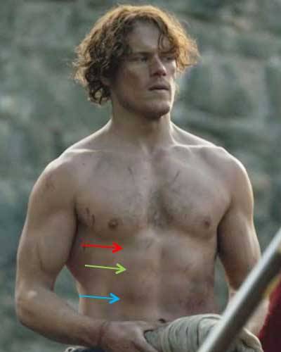
Can we see Jamie’s tendinous intersections and does he have a six-pack? Och, aye, here is the Scottish six-pack in all its glory! Starz episode 6, The Garrison Commander shows his fine abs verra nicely. Arrows point to three pairs of tendinous intersections (TIs). To review: the top pair of TIs (red arrow) lies near the xyphoid process; the bottom TI pair (blue arrow) is near the navel; and the middle TI pair (green arrow) lies between the former two. Remember, in the physically fit, TIs divide each rectus abdominis muscle into bulging segments. Although his lower abdomen is covered by his carefully folded shirt, Jamie does not have a 4th pair of TIs (I’m the anatomist. I’m in chargeso I checked!). Snort!

image

And finally, here is Jamie’s R inguinal groove overlying his R inguinal ligament after being struck by crazy Jack. Because his hips are modestly swathed in plaid we see only the upper half of the inguinal grooves. Wish someone would whip that whip out of Randall’s hands and administer a few smacks to his back.

image

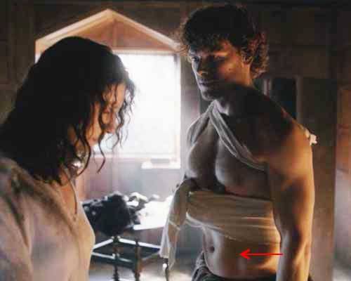
Pop quiz time! Next is an awesome image of Jamie from, well ye all ken where it’s from (Starz episode 7, The Wedding)! And, a lovely quote from the Outlander book:“

‘Because I want to look at you,’ I said. He was beautifully made, with long graceful bones and flat muscles that flowed smoothly from the curves of chest and shoulder to the slight concavities of belly and thigh.”

Woo hoo, are ye paying attention? Slow deeeeeep breaths! All five topographical features of belly
skin
can be seen in this image:

  1. Median groove overlying linea alba.
  2. Lateral grooves overlying lineae semilunaris.
  3. Umbilicus.
  4. Grooves overlying tendinous intersections (There are 6 of them).
  5. Grooves overlying both inguinal ligaments (Only upper parts are visible).

Did ye find them? Would ye like to find them? Ha ha! Hopefully ye all got 100%.

Tcha, Jamie. Didna your mother, Ellen, teach ye not to stare? Gie the puir lass a break! (It’s OK if we stare though.)

image

Sigh. Aye, it’s time to leave Jamie. Bye. Bye.

Now, for the promised true story…some years ago, I had delivered my yearly lecture to first year
medical students on anterior abdominal wall, spermatic cord, etc. A few weeks later, a student shared with me the following harrowing experience. He had just run in the New York Marathon (Photo T – student is not in photo). Weather was warmish and he decided to run wearing only nylon vest and running shorts. After running a few miles, he suddenly crumpled in agonizing pain and
immediately diagnosed his own problem (aye, he remembered the lecture!): as body heat increases, spermatic cords and scrotum relax allowing the testes to move more freely than usual.

His right testis shifted enough during the run to twist its spermatic cord thus strangling the testicular artery and cutting off blood flow. Known as testicular torsion, this is a true surgical emergency that can result in loss of the affected testis if not treated promptly. Paramedics took him to an emergency room where ER docs moved the testis to untwist the spermatic cord and restored its blood supply. Although this may sound slightly frivolous, it is not. Having seen pathology specimens of necrotic (dead) testes following torsion, it is a shocking sight as this organ cannot tolerate loss of its oxygen supply!

I congratulated him on the outcome of quick medical intervention. Needless to say, my future lectures included this prime example of a choice gone wrong (yes, he gave permission – with no identifiers). Men must take precautions to wear appropriate and supportive undergarments especially if a long run is planned. And just to emphasize the point, torsion is the most common cause of testicular loss in adolescent males!

image

Photo T

Okay, you guy readers can start breathing now! The scary story is over.

I hope ye all enjoyed the verra important anterior abdominal wall lesson and appreciate its complex anatomy. Dinna forget – surface features of a muscular belly wall are based on its underlying anatomy. Fare thee well until my next lesson. In the meantime, please take good care of your amazing body!

A deeply grateful,

Outlander Anatomist

You can now follow me on Facebook and Twitter!

Photo creds: Starz, Gray’s Anatomy,
39th ed., Netter’s Atlas
of Human Anatomy, 4th ed., Clinically
Oriented Anatomy, 5th ed., Hollingshead’s
Textbook of Anatomy, 5th ed., Wikipedia, pixshark, workoutnirvana, simplyshredded, the-fitness-motivator, fitnessversusweightloss, build-muscle-101, wikihow, running.es, popworkouts.