
Outlander Anatomy students! Join me tomorrow for the first part of my 2016 Outlander tour of Scotland.
a deeply grateful,
Outlander Anatomist

Human Anatomy taught through the lens of the Outlander books by Diana Gabaldon and the Starz television series

Outlander Anatomy students! Join me tomorrow for the first part of my 2016 Outlander tour of Scotland.
a deeply grateful,
Outlander Anatomist
Greetings fellow anatomy students. Welcome to another lesson, “I have missed your bright eyes and sweet smiles!” This is our first lesson in almost a month, not because I have been lazing about, but because three events have conspired to keep lesson composition at bay.
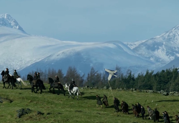
First, I just returned from an Outlander tour of Scotland! Away for almost two weeks, I am still in jet lag but I will be posting my adventures over the summer – I promise!
Second, blueberries are on… plump, dark and sweet! It’s a bumper crop because here, in the Pacific NW, we’ve had more rain than usual. Only 50 plants in my two patches but they are heavy producers and we do our own picking so the process takes time (Image A).
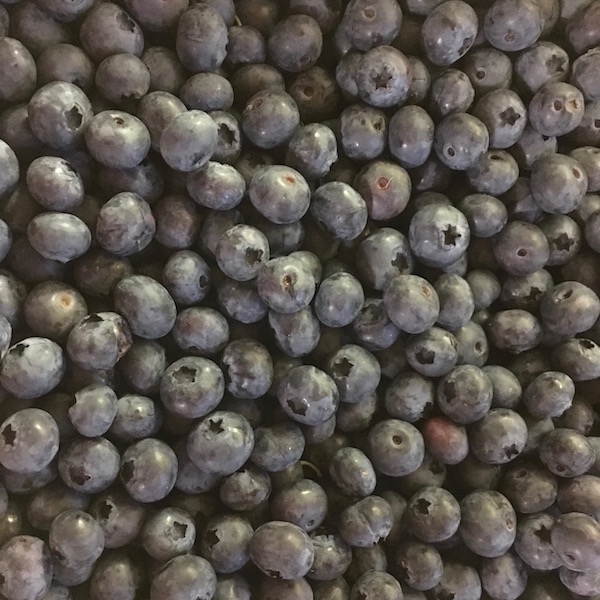
Image A
Third, family members arriving this week for a long-anticipated visit (Starz episode 212, The Hail Mary). Food shopping, fresh sheets, clean house…. yadda, yadda, yadda. You know the drill.
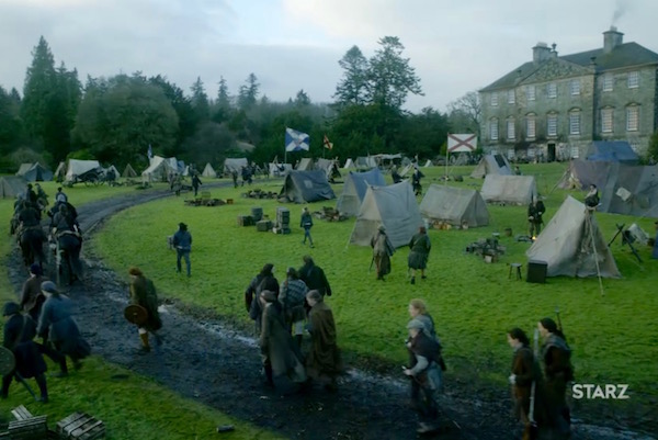
OK prof, enough excuses, let’s get on with the lesson! Today, Anatomy Lesson #41, The Sad Demise of Angus Mhor, explains events leading to his death. This lesson is written in response to a request by long-time reader, Ed, and in deference to our beloved Angus. And, as the lesson confines itself to this single topic and I have lots of other stuff going on (more whining), it will be briefer than usual.
Just one more note before we begin: Angus Mhor (Diana’s spelling) is a character who appears in Outlander and Dragonfly in Amber books. A huge, burly guy, he is Colum’s body-servant. In the Starz series, I don’t recall Angus’ last name being revealed. But, pretty much everyone, including me, assumes Angus Mhor of the book and the series are one and the same.
We find Angus in the thick of it at the Battle of Prestonpans. He fires at a charging British office whose goal is to skewer his pal, Rupert (Starz episode 210, Prestonpans). Angus is having none of it and swiftly dispatches that redcoat to “meet his maker.”
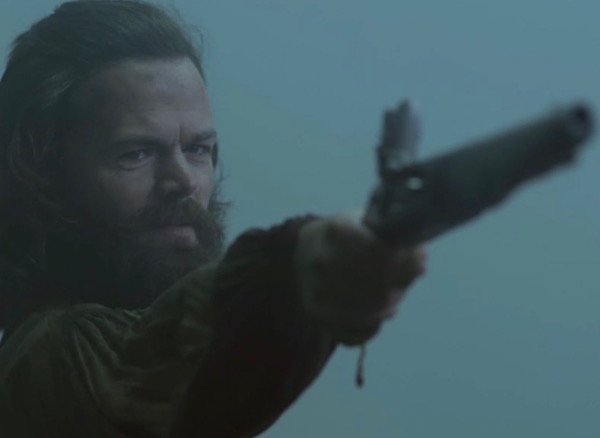
As Rupert staggers to his feet, a cannon blast hurls Angus through the air like “A Leaf on the Wind of All Hallows” (Starz episode 210, Prestonpans)!
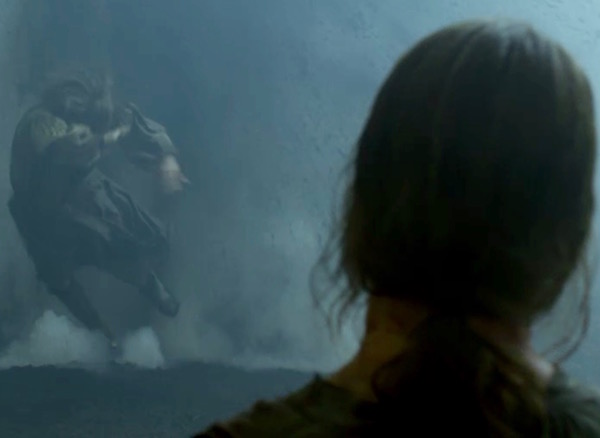
Next, we see puir Angus land with a thud, face down on the cold, hard ground (Starz episode 210, Prestonpans). At this point, his fate is unknown but clearly, the cannon blast dealt him some damage.
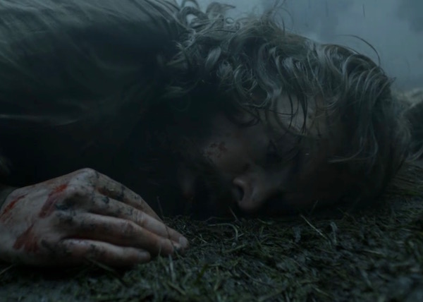
To understand what happened to Angus, we turn to the field of pathology, the study of abnormal anatomy (Anatomy Lesson #35, Outlander Owies! – Part One).
Blast injuries have been with us for a very long time, but they weren’t described as an entity until 1924. And, it wasn’t until World War II that research established their clinical and pathological sequelae (aftermath of disease, condition, or injury). Although we learned about blast injuries in Anatomy Lesson #36, Outlander Owies – Part Deux!, details bear repeating in the context of Angus’ mortal wounds.
Here’s the nitty-gritty: the term, blast, refers to the sound wave produce by an explosion. Blasts also generate a shock (pressure) wave which passes through a victim’s body producing anatomical and physiological injuries. Interestingly, because blast exposure may not produce external injury, internal injuries can pass unnoticed or their severity underestimated. Keep in mind, that the Prestonpans’ cannon blast was sufficient to blow Angus entirely off his feet! As long as explosives are detonated, there will be blast injuries.
Types of Blast Injury: Explosive energy creates four types of multi-system, life-threatening injuries depending on the proximity of the victim to the blast center: the greater the pressure wave and the longer its duration, the more severe the injury:
Image B, is a simple graphic showing the worst injury nearest the epicenter with diminishing effects further from detonation. An entire scientific field exists that studies explosives, associated physics, and the aftermaths. The fields has grown rapidly because blast injuries remain a terribly, timely reality due to global terrorism (think 2013 Boston Marathon).
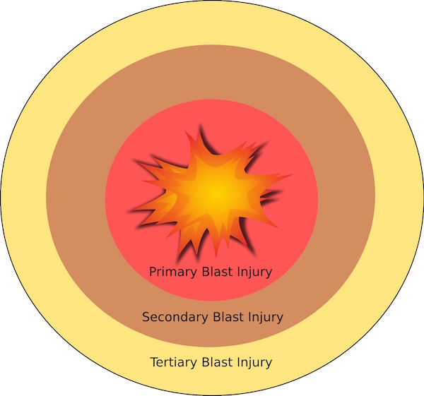
Image B
A more detailed image (Image C) informs us that high explosives are converted to hot gases which create a blast wave of compressed air traveling at supersonic speeds – speeds which can exceed the muzzle velocity of today’s bullets! Now, 18th century cannon may not have created such intense pressure waves, but certainly sufficient to maim and kill.
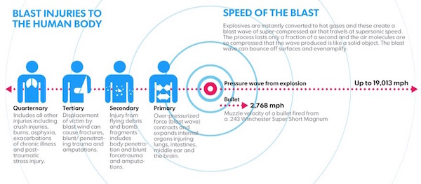
Image C
For today’s lesson, let’s discuss two of the four types of blast injuries: PBIs and TBIs.
PBIs: Gas-containing organs such as lungs, hollow organs of the gastrointestinal (GI) tract, and the middle ear are most vulnerable to blast trauma. Why? Because, as a pressure wave passes through the body, it produces shearing forces at air-tissue interfaces destroying cells, tearing tissues, creating gas emboli (air bubbles), and causing thrombosis (blood clots). PBIs also effect cardiac function.
We cannot cover all such organ injuries associated with blasts, but let’s use lung damage as an example because pulmonary (lung) trauma remains the most commonly fatal PBI. I have not yet posted an anatomy lesson on lung, but bear with me as we explore the effects upon it by a blast wave.
Lung tissue is incredibly delicate. Composed of millions of air sacs (alveoli) with walls a few cells thick, lung tissues are also riddled with capillaries. This design allows inspired oxygen to quickly pass from alveoli into nearby capillaries in exchange for carbon dioxide (a simplified explanation).
Blast injury of the lungs is characterized by bleeding, swelling and bruising. Such changes can be seen by microscopy. The two top photos of Image D show normal lung tissues. The open white spaces are various alveoli, small airways, and blood vessels. Compare and contrast the appearance of the top two photos with the bottom two. The tiny red dots riddling the open spaces of the bottom photos are red blood cells (Anatomy Lesson #37, “Outlander Owies Part 3 – Mars and Scars”). Now, red blood cells are normally confined to blood vessels but here, they are not because the lung tissues have suffered blast injury. Bleeding into air sacs and airways of the lung is the direct result of a pressure wave tearing delicate lung tissues along its path. So, think! If air sacs fill with blood, can there be room for air? If the lungs cannot fill with air, then oxygen is not delivered to the circulatory system. If the circulatory has no oxygen to deliver to the body, then that outcomeis incompatible with human life.
Although hollow organs of the GI tract are not as delicate as lung tissues, PBIs also injure these at air-tissue interfaces. However, compared with lung PBIs, GI injuries, may not present for hours or even days after blast exposure when these are characterized by nausea, vomiting, vomiting blood, etc.
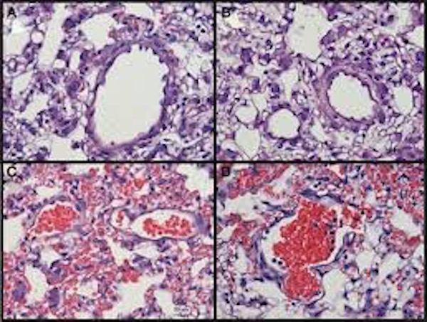
Image D
TBIs: Air displaced by an explosion creates a blast wind that can hurl victims against solid objects, such as the ground. Damage caused by this type of traumatic impact are tertiary blast injuries (TBIs). TBIs may present as some combination of blunt or penetrating trauma, as well as coup contre-coup injuries, basically a fancy term for brain contusion caused when the head is thrown back by a blast and then rebounds.
Back to Outlander! So, our beloved lad is struck by a cannon blast. Then, he lands hard in the bosom of mother earth and loses consciousness. What happened to him? The answer is, our sweet Angus likely suffered both a PBI and a TBI. Back at the make-shift surgery, he presents in succession six red flags (signs) suggesting blast injury.
First Red Flag: The blackened skin of his forehead appears to be a head wound. It could be a PBI, it might be a TBI from the face-plant, or another off-camera battle event may have been the culprit. Look carefully at his forehead as he fires his pistol just before the cannon blast – the hint of a bruise is already present.
Claire is rightly concerned: she checks the wound and his pupils but his “eyes are clear,” a good sign.
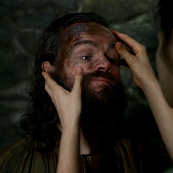
Second Red Flag: Angus is oh, so sweet and docile. His usual cocky strut is long-gone!
Brain injury from the cannon blast must be considered so Claire instructs him to stay awake – by watching the rise and fall of Rupert’s belly. He agrees but says he feels bone weary. Angus is such a feisty rooster, have we ever seen him compliant and willing to follow Claire’s orders?
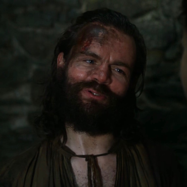
Third Red Flag: Angus pauses to grip his forehead in apparent brain-pain. A possible concussion due to PBI or TBI would explain his discomfort. Claire doesn’t see this action but we do.
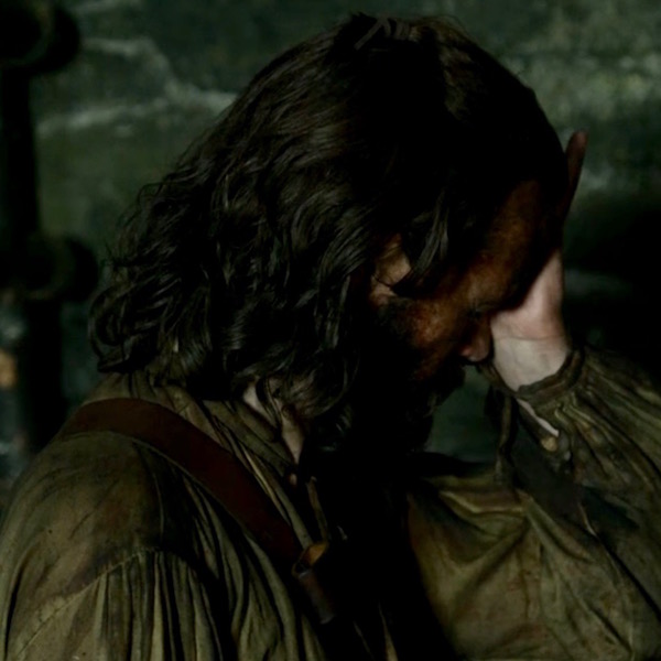
Fourth Red Flag: Angus falls to the floor and Dougal calls for help. Claire and Jamie hasten to his aid. Why does Angus swoon? Drop in blood pressure (due to internal bleeding) seems likely but brain injury or cardiac malfunction could also be factors.
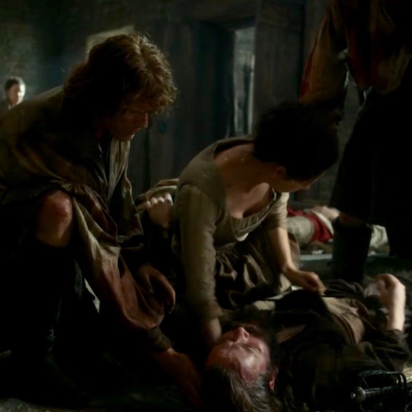
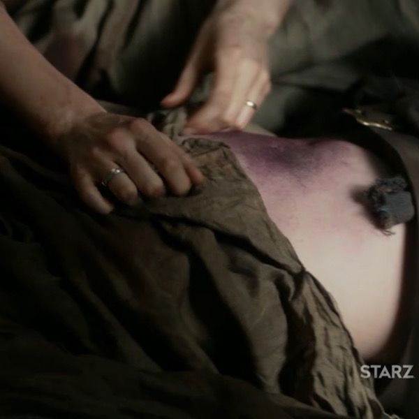
Fifth Red Flag: Claire lifts his sark to reveal a huge contusion (Anatomy Lesson #35, Outlander Owies! – Part One) of the anterior abdominal wall (Anatomy Lesson #16, Jamie’s Belly or Scottish Six-Pack). She cries out that he is bleeding internally.
A wee side note: A belly bruise may or may not indicate internal bleeding. Hemorrhage of abdominal organs can translate to the skin but usually some time passes before presentation. The bruise might also be a TBI of the abdominal wall from being thrown to the ground.
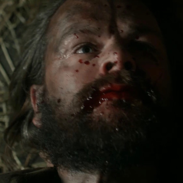
Sixth Red Flag: Blood fills Angus’ mouth, a reliable indicator of internal bleeding (Starz episode 210, Prestonpans). Actually, hemorrhage from any body orifice should set off alarm bells. What is the source of the blood? This is not disclosed in the episode, but I put my money on pulmonary hemorrhage because lung injury is the most common cause death in those that survive a blast and because lung hemorrhage could reach the oral cavity fairly swiftly. Another less likely source of internal hemorrhage is from abdominal GI organs as Claire implies upon seeing the belly contusion; it’s a fair distance for blood from ruptured abdominal organs to reach the oral cavity.
Angus chokes on his own blood as he desperately tries to breathe and to speak but he swiftly expires. Despite all her healing gifts, Claire is helpless. There was really nothing she or anyone could have done in the 18th century to alter the outcome….an outcome as inexorable as Culloden. Needless to say, his end was not an easy one.
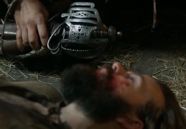
Our wonderful Angus dies 21 September 1745, surrounded by loyal friends (Starz episode 210, Prestonpans). The fabulous Rupert-Angus tag team has left the building. Tears a falling!
The red flags and associated comments are based on my own observations and conjectures. They are also over-simplified for this lesson. So, what really killed Angus? Truly, the only way to answer this question with surety is to perform an autopsy which, of course, isn’t available for Angus. As you viewers of forensic TV shows realize, the autopsy routinely examines organs of the abdominal and thoracic cavities as well as the brain and any other sites that appear abnormal. Gross (macroscopic) observations are made and tissue samples prepared for microscopic examination. Clues from these observations usually yield a likely cause of death.
Autopsies are the turf of anatomical pathologists who are, in my humble opinion, the true brains of the medical profession (no insult intended to other healers). There is an old adage that pathologists are physicians who know everything and do everything … three days late.
Let’s end this lesson by honoring our delightful and unforgettable friend of two Starz seasons.
Ode to Angus Mohr
Two missing teeth and sass to bequeath,
He was loved for his feisty grin.
Rupert’s girth and his own brand of mirth,
Was a brew more potent than gin!
Eyes for Nurse Claire earned Jamie’s fierce glare,
Her kiss put his head in a spin.
Willing to bet, we shall ne’er forget,
This proud warrior of kith and kin.
Our tears are shed with hearts full of dread,
His passing was ever so grim.
Feeling so sad, farewell dearest lad,
Our loss burns like mortal sin.
Angus Mhor, R.I.P.!
Finally, it is only fitting to end this lesson with Stephen Walters singing his own farewell:
A deeply grateful,
Outlander Anatomist.
Photo creds: Starz, Outlander Anatomy (Image A), www.en.wikipedia.org (Image B), www.journal.frontiersin.org (Image D), www.usatoday.com (Image C)
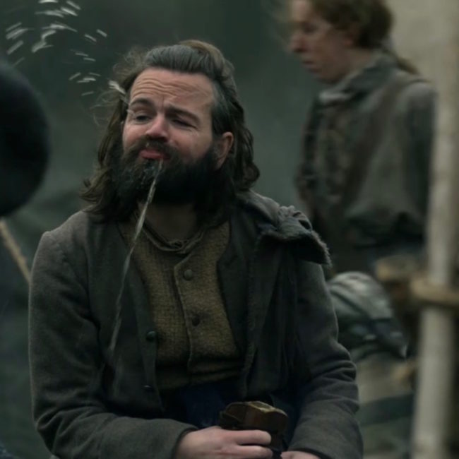
We miss ye already yoo ever lovin’ goof! Join Anatomy Lesson #41 tomorrow, as we bid Angus adieu.
A deeply grateful,
Outlander Anatomist