Anatomy Lesson #49, “Our Liver – The Life Giver!” – GI Tract 6

Hallo, anatomy students! Welcome to Anatomy Lesson #49, Our Liver – The Life giver. We finished our exploration of the GI tract proper (lips through anus), and today, we continue with associated organs of the GI tract. In former lessons, we learned that during embryonic development, various organs arise as outgrowths of the gut tube (Anatomy Lesson # 44, “Terrific Tunnel – GI Tract I”). Such organs retain these connections throughout life, so their products flow via ducts (tubes) into the GI tract proper.

Liver is on our docket today. Liver develops during intrauterine life as an outgrowth of the nascent gut, retaining its attachment via a system of hepatic ducts. And, as the title correctly states, liver is a “giver,” not a “taker.” Liver is the type of “anatomical“ personality we crave in our lives, as it is the seat of many life processes!

As always, this lesson sports Outlander images and quotes about liver. What? Are there really liver quote from our fav books and TV series? Indeed, there are!

Word Origin: Liver, is an Anglo-Saxon word (1325) for liv-er or lifer. Logically, the etymology for liver infers “life,” although the original intent is lost. The name is apt, though, because many vital life functions occur therein. And, our very busy liver has insinuated itself into history, prophecy, character, culture, horror, mythology, and medicine!

History: Greek physician, Claudius Galen (130-200 CE), anointed liver as the principle organ of the body, teaching it as the primary source of blood and lymph (Anatomy Lesson #34, The Amazing Saga of Human Anatomy) – a status enjoyed for 1500 years! After this was disproven, liver fell from grace (sad face), only to have its stature revived in the 19th century by French physiologist, Claude Bernard. Bernard showed the liver serves many vital functions; indeed, we now know of at least 50 ways (TY, Paul Simon), to “love your liver” (Image A). But, counting all known liver functions, its duties approach 500!
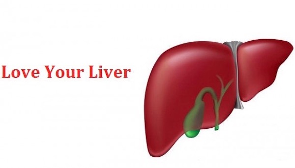
Image A

Prophecy: Ever thought of a liver as a crystal ball? Believe it or not, livers were used for divination. Ancient Babylonians (19th century B.C.) studied sheep livers as a means to foresee the future. Such findings were documented on clay tablets in the shape of a sheep’s liver, complete with surface markings used for prophesying and instruction (Image B).
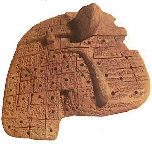
 

Image B

Character: The liver has the dubious honor of being used to symbolize a number of less savory human attributes, such as:

  • lily-livered (cowardice)
  • chopped liver (worthless)
  • white liver (cowardice)
  • hard liver (sensualist)
  • liver-hearted (craven)
  • liver-faced (mean-spirited)
  • liver-lipped (pale and feeble)
  • liver-sick (dropsy = cirrhosis or hepatitis)

Our beloved Outlander provides stellar examples of several such liver characteristics:

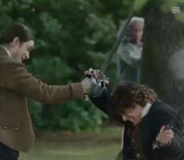
That lily-livered (coward), dastardly Duke (Starz, episode 110, By the Pricking of My Thumbs) peeks from behind a tree, leaving Jamie to battle three grumpy MacDonalds! Mayhap they only got haggis for brekky?

I think that I shall never see,

A more cowardly knave behind a tree!

From the same Outlander episode (Starz, episode 110, By the Pricking of My Thumbs), liver-faced (mean-spirited) Laoghaire spies Claire being arrested for witchcraft. She will happily sling the Heiland fling on Claire’s still-warm ashes!

I think that I shall never see,

A more wicked lass brimming with glee!

Now, please dinna fash, I affront the character, not the gifted and beautiful actress!

And before we leave Character-Assassination by liver, consider the hard-livered (sensualist), Black-Jack. A sociopathic-psychotic-sensualist if ever I saw one (Starz, episode 116, To Ransom a Man’s Soul)!

I think that I shall never see,

A more depraved voluptuary than HE!

Culture: Many cultures offer idioms about the liver. This “pointed” one is an ancient Tibetan Proverb: My enemy’s liver is the sheath of my sword!

Och! Jamie and his men present a perfect metaphor for this dire warning, with raised blades at the Battle of Prestonpans (Starz episode 210, Prestonpans). Redcoat livers: take care and beware – your livers willna be living long!

I think that I shall never see

a more deadly blade than…Owie!

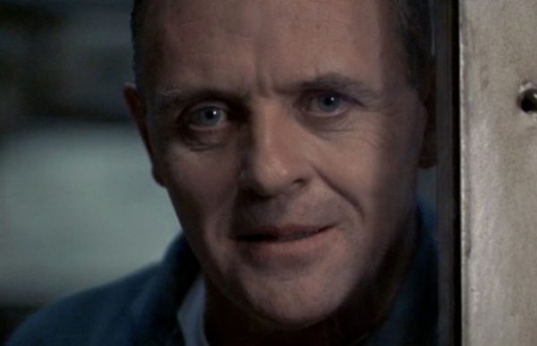
Horror: The liver appears in a surprising number of “settings and upsettings” to convey horror!  Jonathan Demme’s 1991 horror film, The Silence of the Lambs offers a perfect example:

Check out this link to YouTube for a peek at this creepy scene!

Liver, fava beans, and a nice Chianti? Yuck. Well, maybe the nice Chianti. Hilarious Hannibal – the life of the party!

Anthony Hopkins as Dr. Hannibal “The Cannibal” Lecter in the 1991 film “Silence of the Lambs.” Photo Courtesy: MGM Home Entertainment

Mythology: Let’s not forget myths! Liver was immortalized in Theogony, a poem by Hesiod (8th – 7th century BCE), wherein the fate of the Titan, Prometheus (no relation to Ridley Scott), is recorded. Much to Zeus’s fury, Prometheus unwisely granted humans the gift of fire and the skill of metalworking. Zeus was so pissed, he chained Prometheus to a mountain rock. Immortalized in this stark painting (Image C), each day, Zeus sent an eagle to eat Prometheus’ liver and every night, his liver regenerated only to be devoured the next day. Eternal retribution (Prometheus eventually escapes)! Lesson learned: Yo mama always said, “dinna play with fire!”

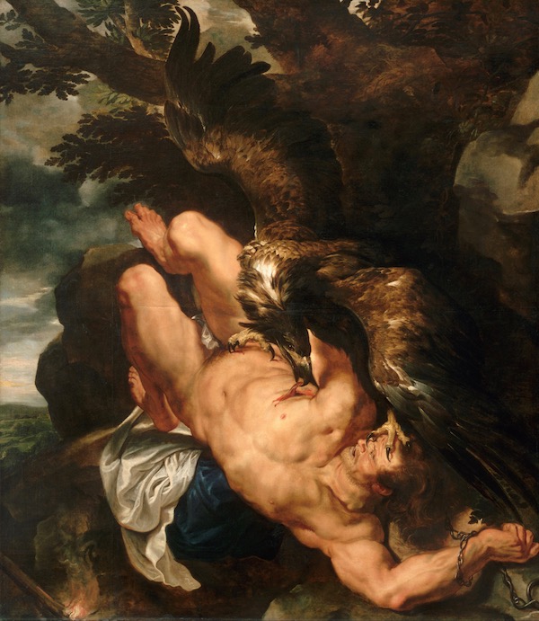
Image C by Ruben and Snyders

 Medicine: Many cultures have used liver as medicaments. Even today, cod liver oil is taken to lower high triglycerides and high blood pressure, and to treat some symptoms of diabetic-related kidney disease (www.webmd.com).

An 1899 book titled “Animal Simples,” by W. T. Fernie, M.D., states:

Dr. Heffinger advocates the taking of crocodile liver oil for phthisis; which almost supersedes the use of cod-liver oil in Peru; being more nourishing and palatable.

I dinna ken Dr. Heffinger, but I assume and presume he was a renown practitioner of 19th century medical arts. Oh, and phthisis is an old term for pulmonary tuberculosis, although liver oil likely had zero effect on this diabolical disease.

We encounter crocodile oil at Master Raymond’s Paris apothecary shop (Starz, episode 204 La Dame Blanche)! This “little shop of horrors” (snort!) displays pretty much every oddity in gay Paree! There’s even a quote from Dragonfly in Amber book:

“My crocodile ? Oh, to be sure, madonna. Gives the customers confidence.” He jerked his head toward the shelf that ran along the wall just above eye height… Three of the jars closest to me were labeled in Latin, which I translated with some difficulty—crocodile’s blood, and the liver and bile of the same beast, presumably the one swinging sinisterly overhead in the draft from the main shop

“never smile at a crocodile…..”

Gross Anatomy: OK, enough  socializing – time for gross anatomy. Yay! Here, gross means visible with the naked eye (Anatomy Lesson #34, The Amazing Saga of Human Anatomy); it does not mean yucky!

Location: Where, oh where, does the liver dwell? It lives in the abdominal cavity along with most of the GI tract. Its location is best defined by dividing the abdominal wall/cavity into imaginary quadrants (Image D); vertical and horizontal lines intersect at the umbilicus (Anatomy Lesson #16, Jamie’s Belly or Scottish Six-Pack), dividing the area into RUQ, LUQ, RLQ, and LLQ.

Most of the liver resides in the RUQ although some extends into the LUQ. There’s another anatomical method dividing abdominal cavity and wall into 9 segments but the quadrant system works best for today.
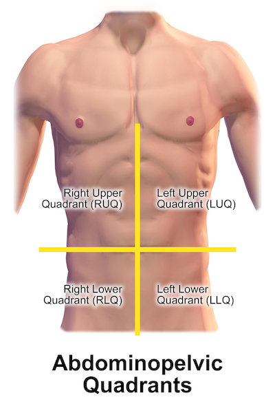
Image D

Respiratory Diaphragm: What does respiratory diaphragm have to do with liver? Lots! Anatomy Lesson #15, Crouching Grants – Hidden Dagger, taught that the respiratory diaphragm is parachute-shaped, doming upwards such that it is largely surrounded by rib cage, sternum, and vertebrae (Image E).
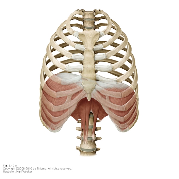
Image E

Anchorage: Liver abuts the respiratory diaphragm (Image F – hooks) but it doesn’t flop around in the RUQ. Rather, it is anchored in place by membranes (Image F – red arrows), layers of peritoneum which bind liver tightly to the diaphragm.
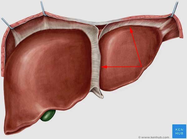
Image F liver

Ribs and Sternum: Because the liver abuts the respiratory diaphragm, it too, enjoys the immediate and privileged protection of sternum, ribs and vertebrae (Image G). As such, a normal liver is challenging for a practitioner to palpate. To do so, a patient lies on the back and inspires deeply; the practitioner presses fingers under the right costal margin and, if skilled, may be able to feel the margin of the liver slip under the fingers. Normal liver is soft, smooth and slightly tender to palpation. An easily palpated liver is usually associated with one of several issues, the most dire being hepatitis or neoplasia (tumor).
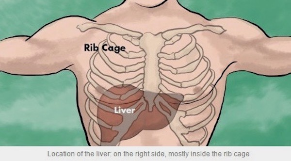
Image G

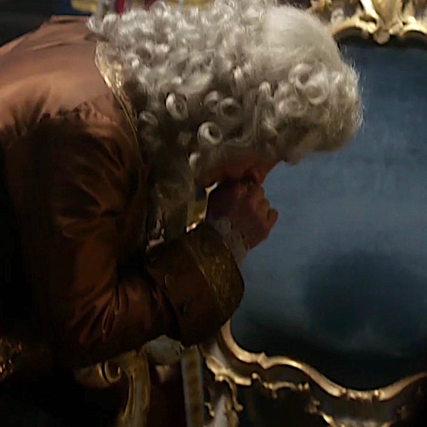
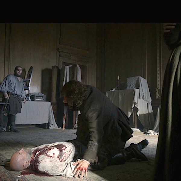
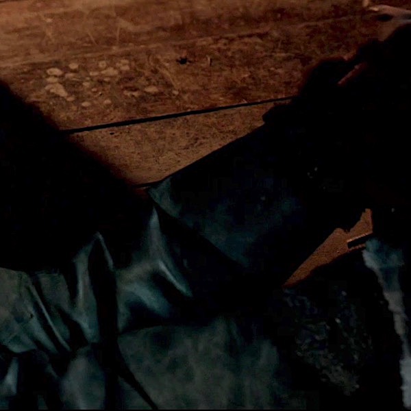
Forensics: It’s Outlander Time. Yay! Ever wonder where Dougal met his Demise-by-Dirk (Starz, episode 213, Dragonfly in Amber)? No, not in the attic! I mean,  where was he stabbed? Did you think Claire and Jamie got him in the heart? Or, mayhap a lung? Forensic caps on!

Most likely, Dougal was stabbed in the liver! Yep. If ye look carefully, his wound is to the right of midline and above the costal margin. This means the dirk likely entered his RUQ, penetrating the right dome of respiratory diaphragm and piercing the right side of liver (Starz, episode 213, Dragonfly in Amber). No, Rupert, Jamie didn’t do the deed; or rather he didn’t do it alone!  It took four hands to finish Dougal off by Dirk-Deed! Dougal, man, we miss your sass!

As the heart lies mostly to the left of midline and right lower lung is to the right of the wound, neither of these organs were likely pierced. Good job, anatomy sleuths!

Physical Attributes: The adult liver is wedge-shaped, dense, and meaty, weighing 3-3.5 lb. Liver is our largest internal organ and the largest gland of the body, superseded in size only by our body’s largest organ, the skin (Anatomy Lesson #5, Claire’s Skin – Opals, Ivory and White Velvet). Ken the difference? Bonny!

Liver is solid in texture and deep red-purple-brown in color (Image H). Although liver mass constitutes only 2.5% of total body weight, the liver receives a whopping 25% of the cardiac output. Two and one-half pints of blood flow through a normal liver every minute of our lives!
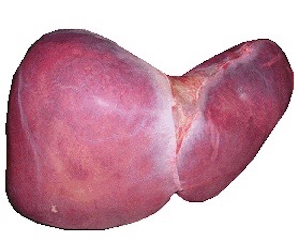
Image H

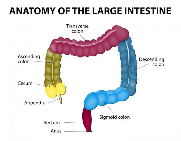
Impressions: Front, back, and top surfaces of liver are relatively smooth; flip liver up-side-down, and its under-belly bears ridges and valleys. The depressions, named impressions, are created by contact with adjacent organs including stomach (gastric), duodenum, adrenal (suprarenal) gland, kidney (renal), and colon (Image I). Gallbladder also leaves an impression, but this is visible only after its removal. Ergo, liver is centrally-located in the upper abdominal cavity with many important organ contacts.
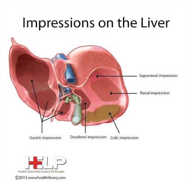
Image I

Anatomical Liver Lobes: Anatomists used landmarks to divide the liver into four anatomical lobes: right, left, caudate and quadrate (Image J). The large, right lobe (tan) is separated from the smaller left (yellow) by one of the membranes mentioned above, the falciform ligament. Only by viewing the undersurface are caudate (blue) and quadrate (green) lobes visible; caudate is nearer the spine and quadrate lies adjacent to gallbladder.
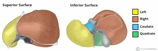
Image J

Blood Supply: Most organs receive blood via an artery and are drained by a vein. Not the liver! It is unique because it is the only organ receiving blood from both an artery and a vein; the hepatic vein delivers nutrient-rich blood derived from food digested in the gut, and the hepatic artery supplies oxygen-rich blood via the aorta. Three hepatic veins drain the liver (Image K).

Most organs are variations of a scarlet color because they receive oxygenated blood. Liver is a red-purple color because 75% of its blood supply is deoxygenated, nutrient-rich blood from the hepatic vein.

A dear long-time friend had a bit of trouble last year. Tests revealed that she had lost the blood supply to a segment of her liver with subsequent death of said segment! Cause unknown. She has given me permission to share this experience because it is unusual given she lacks any history of typical liver disorders or diseases. This is a wonderful example, though, of how dependent the liver is upon an intact blood supply!
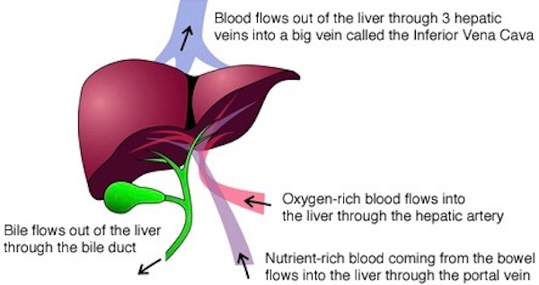
Image K

Clinical Liver Segments: The next image looks scary, but bear with me here! Clinicians divide the liver into eight separate segments, numbered 1-8 or I-VIII (Image L); segment I (1) lies deep to IVa (4a) and IVb (4b), so it is not visible. Segments II and III lie to the left of the falciform ligament; all other segments lie to the right. Segmental divisions are determined by vascular supply and bile duct branching (see below) and are of great clinical import!
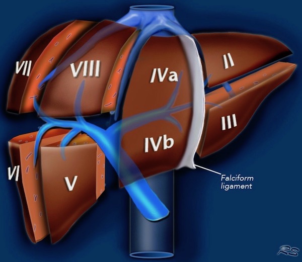
Image L

Clinical Liver Segments (cont.): Let’s say a cancer is found in liver segment IVb (4b). A competent surgeon determines the probable boundaries of IVb and resects only that segment, sparing remaining liver segments and minimizing blood loss. The surgeon performing these highly specialized surgeries must be thoroughly familiar with the segmental design as defined in Couinaud’s system (Image M).

Medical students have difficulty memorizing liver segments, but this fist mnemonic offers a useful visual. Note that the thumbnail (seen in ghost) represents the deep-lying, segment I.
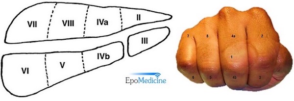
Image M

Outlandish Lobes: Speaking of liver lobes… like you, I was deeply impressed by Master Raymond’s ministrations to Claire (and her liver) after her loss of Faith-faith (Starz episode 207, Faith). Her liver and other organs were infected; she probably had puerperal fever, a post-partum infection of the uterus that, if untreated, usually becomes systemic. Serious-Sobbing here (Dragonfly in Amber book)!

An odd feeling of warmth now emanated from those broad, square, workman’s hands. They moved with painstaking slowness over my body, and I could feel the tiny deaths of the bacteria that inhabited my blood, small explosions as each scintilla of infection disappeared. I could feel each interior organ, complete and three-dimensional, and see it as well, as though it sat on a table before me. There the hollow-walled stomach, here the lobed solidness of my liver, and each convolution and twist of intestine, turned in and on and around itself, neatly packed in the shining web of its mesentery membrane.

Bile Ducts and Gallbladder: Liver enjoys a robust working relationship with gallbladder and bile ducts. Why? Because, liver cells continuously produce bile, a dark green to yellowish-brown fluid. Bile is transported by a converging system of bile ducts from liver to gallbladder, a bag-like structure which stores and concentrates bile (Image N). Bile is then released in a controlled fashion after chyme (Anatomy Lesson #46, Splendid Stomach, Wobbly Wame) enters the duodenum. In folks lacking a gallbladder, bile ducts continuously release bile into the duodenum.

Bile: Two important purposes are served by bile:

  • Digestive aid: After eating a meal (especially high in fat), bile is released from gallbladder into duodenum (Anatomy Lesson #47, Brave Bowels – Gurgly Gut!) where it aids in lipid digestion by fats emulsification.
  • Excretory product: Specialized liver (Kupffer) cells devour effete red blood cells, breaking down hemoglobin into bilirubin, a waste component of bile that is eliminated with the stool.

Bile enjoys a long history in western medicine. From 500 B.C. to the early 19th century, standard practice adhered to a four humor system: black bile, yellow bile (liver bile), phlegm, and blood. Excesses of black and yellow biles were thought to produce depression and aggression and were treated accordingly (sometimes with phlebotomy). If only physiology/psychology were that simple!
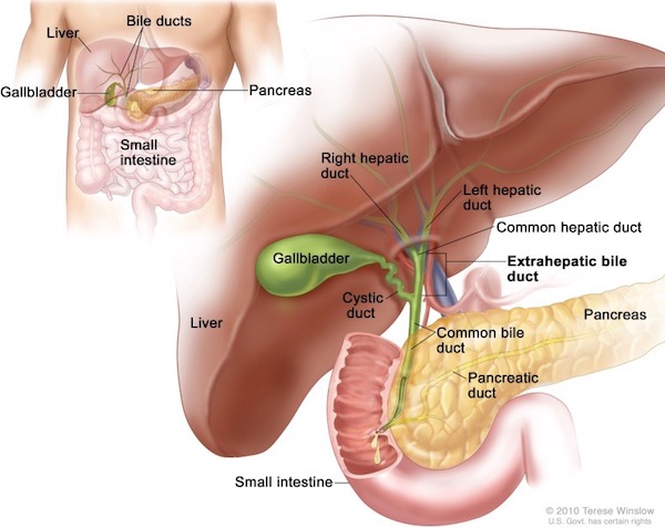
Image N

Hepatocytes: The hepatocyte is the main functioning cell of the liver (there are other cell types, too). It’s prefix derives from the Greek word, hepar, meaning liver. Microscopically, the hepatocyte (image O – arrows point to hepatocyte cell membranes) is a large, round, bland-looking cell, but what a power-house it is!

Hepatocytes are arranged into 1-2.5mm cylinders, each known as a liver lobule. I will not teach liver lobule structure here as it is complex and difficult to understand for students lacking histology training – beyond the scope of this anatomy lesson. Suffice it to say that the liver is packed with millions of these tiny liver lobules. Their designs allows hepatocytes to have immediate access to blood vessels and bile channels. This allows ready access to substances in the blood stream and to release bile into bile channels for transport to the gallbladder.
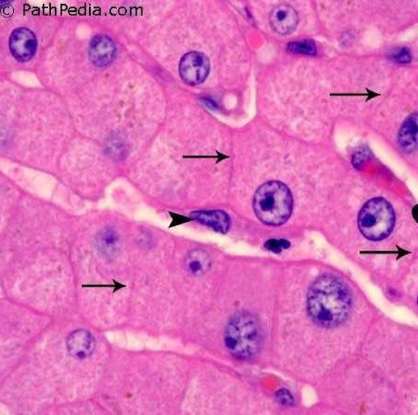
Image O

Image P is a nice graphic summarizing a few liver functions – most are self-explanatory. Hepatocytes are responsible for almost all the ∼500 liver functions. Just a note about the Kupffer cell (Image O – arrowhead). a phagocyte, meaning “eating cell.” Kupffer cells ingest effete red blood cells (and other things), turning hemoglobin into bilirubin, a component of bile. Just consider: maintaining such a huge organ would be wasteful if it weren’t highly important – it wears many hats!
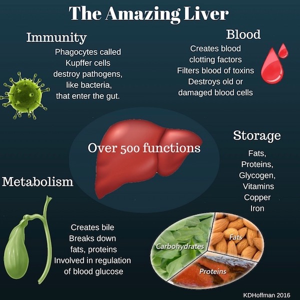
Image P

Liver Diseases: Liver is subject to a staggering array of diseases – from genetic disorders, autoimmunities, viral hepatitis, and parasitic liver flukes to cancer, chronic alcohol disease, poisoning, drug abuse, excessive use of over-the-counter pain relievers, etc. Although liver has a tremendous capacity to regenerate, long-term damage may result in the formation of scar tissue. A scarred liver is characteristically bumpy, a condition known as cirrhosis (Image Q). In the 18th century, Claire would have said the patient with a cirrhotic liver was liver-sick and cirrhotic livers were termed hobnailed livers. Just so you ken, hobnails are a short, heavy-headed nails used to reinforce the soles of boots. Bless Diana’s heart, she even wrote about cirrhotic livers (Dragonfly in Amber book)!

I knelt by the small traveling chest, unfolding the green velvet. Kneeling next to me, Jamie flipped back the lid of my medicine box, studying the layers of bottles and boxes and bits of gauze-wrapped herbs. “Have ye got anything in here for a verra vicious headache, Sassenach?” I peered over his shoulder, then reached in and touched one bottle. “Horehound might help, though it’s not the best. And willow-bark tea with sow fennel works fairly well, but it takes some time to brew. Tell you what—why don’t I make you up a recipe for hobnailed liver? Wonderful hangover cure.”

Liver dialysis can be used as a stop-gap treatment, but liver transplant is the only current option for complete liver failure. If all goes well, the transplant regenerates much of the original liver mass.
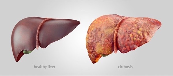
Image Q

Leaving that unhappy liver-note, let’s end on a happier liver quote!

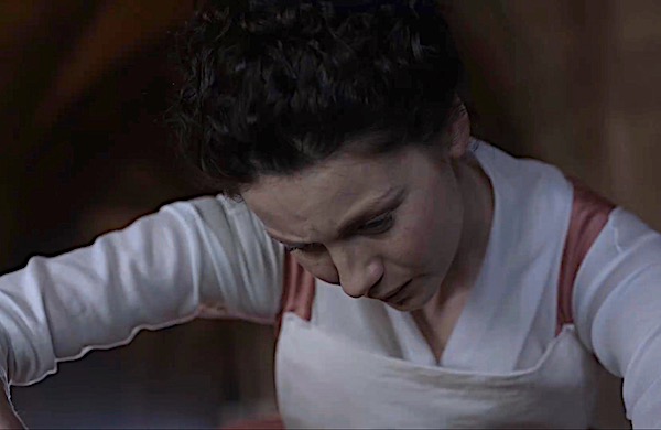
Recall Starz, episode 105, Rent? One of my favs.

Intruder alert! With a massive candle stick, Claire goes after someone fussing outside her door!

Dashing outside, she encounters Jamie, sprawled on the filthy floor. “What are you doing here?” she demands (Outlander book), after stepping on his RUQ. Watch out lovely, lively liver! Crunch!

“What are you doing here?” I asked accusingly. At the same time Jamie asked, in a similarly accusatory tone, “How much do ye weigh, Sassenach?” Still a bit addled, I actually replied “Nine stone,” before thinking to ask “Why?” “Ye nearly crushed my liver,” he answered, gingerly prodding the affected area. “Not to mention scaring living hell out of me.”

Nurse Claire, in the hallway with a candlestick? <G> She almost brains Master Fraser! And her weight? What gently-bred Scottish lad asks a lass how much she weighs? Geez, at 126 lbs, Claire is a just wee thing!

And, Jamie, you drink Scotch whiskey so we all ken your liver can take a trashing! Uh oh, we’ve seen that look before. Run, Claire, run! Hah!

So, the liver may look boring, but is a bundle of life: 500 duties it performs to keep our bodies healthy and happy. We literally live in our liver!

Let’s end this lively liver lesson with a great quote from American physician and psychologist, William James:

Is Life worth living? It all depends on the liver.

And, from my own pen:

Liver the giver

makes us shiver and quiver

bound to deliver

It’s a major caregiver!

So, Love your Liver!

A deeply grateful,

Outlander Anatomist

Photo Creds: Starz, www.techmeanatomy.info (Image J), www.arthistorynewsreport.blogspot.com (Image C), www.cojs.org (Image B), www.commons.wikimedia.org (Image H), www.diets-usa.com (Image A), www.epomedicine.com (Image M), www.kenhub.com (Image F), www.medhealthdaily.com (Image G), www.medvizor.com (Image P), www.nature.com (Image L), www.ncbi.nlm.nih.gov (Image N), www.pathpedia.com (Image O), www.pinterest.com (Image I), www.skylerzarndt.com (Image E), www.vaxy.com (Image Q), www.wikipedia.org (Image D), www.24nurse.com (Image K)

Fun Fact: Premolar or Molar?

Anatomy def. – premolar: One of 8 transitional teeth (2 per quadrant) seated between canines and molars. Premolars are also known as bicuspids because they typically bear two cusps or points (sometimes three).

Anatomy def. – molar: One of 12 large, flat teeth (3 per quadrant) at the back of the mouth used for grinding and chewing. Molars typically have 4 – 5 cusps.

Outlander def: One of many “owie” teeth that Claire yanks from the mouths of impatient – outpatients! Wicked pliers!

Learn about premolars, molars and all the rest in Anatomy Lesson #26, “Jamie’s Chin – Manly Mentus.”

This FF title poses a question: is the extracted tooth shown in Starz ep 211, “Vengeance Is Mine,” a premolar or molar? Time to think! Well, the tooth has two roots (cusps aren’t visible): each first upper (maxillary) premolar and all lower (mandibular) molars have two roots. Otherwise, all upper molars have three roots, and all other teeth have one. Ergo, Claire is pulling either a first upper premolar tooth or a lower molar (see below). Angle of the pliers and tilt of the head implies she is gripping an upper tooth, so I vote for a first maxillary premolar. See how anatomy works? Darned interesting stuff!

See Claire pull the “hen’s” tooth in Starz ep 211, “Vengeance Is Mine,” written by our very own, Diana Gabaldon. Rupert stands as witness even though his belly was just stitched up. Ouch and och!

Read about teeth in Diana’s first big book, Outlander. Although not filmed, the following scene illustrates our Sassynurse’s versatility as she reseats Ned Gowan’s tooth in its socket. Anatomically thrifty!

Ned Gowan was bright-eyed and fizzing with vitality, apparently so intoxicated with the thrill of the fight as hardly to notice the tooth that had been knocked out by an ill-aimed dagger hilt. He had, however, retained sufficient presence of mind to keep it carefully held under his tongue. “Just on the off-chance, d’ye see,” he explained, spitting it into the palm of his hand. The root was not broken, and the socket still bled slightly, so I took the chance and pressed the tooth firmly back into place. The little man went quite white, but didn’t utter a sound. He gratefully swished his mouth with whisky for disinfectant purposes, though, and thriftily swallowed it.

Go Claire – go Ned!

A deeply grateful,

Outlander Anatomist

Anatomy Lesson #48: The Big Guy! – G.I. Tract 5

By Dr. Karmen L. Schmidt

Greetings fellow travelers! Welcome to today’s Anatomy Lesson #48, “The Big Guy,” better known as large intestine. Much is known about large intestine but today’s lesson covers only a tad; otherwise, the lesson would rival one of Diana’s big books! <g>  This topic may prove uncomfortable for some, but I hope you lasso any reservations and read this fascinating stuff. For those not sure, Jamie appears to give a wee warning before the most – ahem – personal bits. The good news: Outlander quotes, scenes, and book spoilers await. Yay!

Last lesson, Anatomy Lesson #47, “Brave Bowels – Gurgly Gut!” – GI Tract, Part 4, we learned that small intestine ends after ileum, the site where large intestine begins.

Divisions: As always, anatomical descriptions and definitions are a must! Large intestine is shorter (5 ft) than small intestine (20 ft) but its diameter is larger (3”) compared with the little guy (1-2”), hence the name. Large intestine is divided into the following regions (Image A):

  • vermiform appendix (cream & green)
  • cecum (yellow)
  • colon
    • ascending colon (green)
    • transverse colon (mauve)
    • descending colon (dark blue)
    • sigmoid colon (light blue)
  • rectum (purple)
  • anal canal (not labelled)

Image A

Other Features: Large intestine also exhibits three other important anatomical features (Image B):

  • ileocecal valve – opening between ileum and cecum
  • right colic (hepatic) flexure – near the right lobe of liver
  • left colic (splenic) flexure – near spleen

Image B

Location: Large intestine lies in the abdominal cavity partially covered by small intestine. Removing small intestine requires cutting the root of the mesentery (Image C – red arrow) – this mesentery suspends small intestine from the posterior abdominal wall (Anatomy Lesson #47, “Brave Bowels – Gurgly Gut!” – GI Tract, Part 4).

Now, lifting greater omentum and transverse colon reveals all parts of the large intestine, except anal canal (Image C).  The mesentery (yellow in Image C) envelopes most of large intestine, securing the parts in their respective positions and containing blood and nerve supplies. Appendix, cecum, and ascending colon typically sit on the right side of the abdominal cavity – descending and sigmoid colons are on the left. Transverse colon hangs between right and left colic flexures. Rectum lies in the midline of the pelvic cavity.

Understand that exceptions to this arrangement do occur. One rare congenital condition, situs inversus totalis, happens once in every 10,000 people. Here, all abdominal and chest organs are flipped 180°. So, for example, appendix lies in lower- left abdominal cavity. Competent health practitioners must keep such rarities in mind while assessing patients.


Image C

Now, onto specific parts of large intestine.

Cecum: The term, cecum or caecum (British spelling), comes from the Latin caecus, meaning blind: not as in sightless, but as in cul-de-sac. The cecum is an enlarged pouch typically located on the right side of the abdominal cavity and is the first part of large intestine. Both appendix and ileum open into the cecum (Image D).

Cecum and ileum are separated by an ileocecal valve, a slit-like opening in the cecal wall. The valve contains a sphincter of smooth muscle, opening to admit chyme from the ileum, and closing to prevent back flow. Thus, cecum serves as a receptacle for fluid chyme.


Image D

History Detour: Almost simultaneously, the ileocecal valve was described by Dutch physician, Nicholaes Tulp, and by Swiss botanist, Gaspar Bauhin. Depending on which scientist one intends to honor, the iliocecal valve is also either the Tulp valve or the Bauhin valve. Yep, the way the world works!

In case you forgot or haven’t read Anatomy Lesson #34, “The Amazing Saga of Human Anatomy, Tulp is the anatomical demonstrator in Rembrandt’s 1632 painting (Image E), “The Anatomy Lesson of Dr. Nicolaes Tulp.” Several modern anatomists have performed dissections trying to recreate the left forearm tendons as depicted in the painting, but to no avail; the painting is anatomically incorrect, which matters ZIP because it is priceless!


Image E

Pretty cool, eh? Now, back to the lesson!

Vermiform Appendix: The appendix is more accurately termed vermiform appendix, cecal appendix, vermix, or vermiform process. Why use one name when four will do, hah! To improve student vocabulary? No, not really. Adding a second word provides specificity because the body has other appendices (pl.) including those associated with ovary, testis, and colon. Thus, vermiform helps verify which appendix is intended.

I prefer the term vermiform appendix. Why? Vermiform is from the Latin vermes meaning “worms” + formes meaning “shape.” Appendix refers to a process or projection. Voila! A normal vermiform appendix is shaped very much like a long, pink worm!

Enough with word roots. Vermiform appendix is a blind-ended, worm-like extension projecting from the cecum and usually located in the lower right abdomen (Image F). It is suspended by a mesoappendix, a fold of mesentery that also provides a conduit for blood vessels and autonomic nerves.


Image F

Appendiceal Location: Illustrations typically place vermiform appendix in the lower right abdominal position, as mentioned above. But, it may assume one of about eight positions relative to the cecum (Image G). Or, a cecum may ride high in the upper right abdominal cavity carrying the vermiform with it, or even shifted to the left side as in situs inversus (Image G). Vermiform appendix may also drift into an inguinal hernia (Image G – red arrow). Interestingly, some alternate appendiceal positions are more prevalent among some populations and cultural groups. Such positional variations can present confusing signs and symptoms and pose diagnostic challenges for practitioners. Truth!

Image G

Function #1: The function of vermiform appendix is unclear. Many anatomists consider it a useless relic of an evolutionary past. But, recent studies suggest it acts as a reservoir for good bacteria. After severe diarrhea, the vermiform appendix apparently “reboots” the large intestine by repopulating it with normal bacterial flora (Image H).

I always taught, and still firmly believe, the body doesn’t have useless organs. I reason that if a body part appears useless, it is our own ignorance that impedes understanding. I must confess, though, one savvy student stumped me with the question: “why do men have nipples?” I had no answer then, but I do now  – subject for another lesson. Understand, the half-life of medical facts is about 5 years, so new and/or revised info emerges daily! Stay tuned! Hee, hee.

Image H

Function #2: Vermiform appendix also serves as a lymphoid organ. Remember the tonsils (Anatomy Lesson #45 “Tremendous Tube” – GI Tract, Part 2) and Peyer’s patches of ileum (Anatomy Lesson #47, “Brave Bowels – Gurgly Gut!” – GI Tract, Part 4)? Moving to a microscopic image, these are aggregates of lymphoid nodules (spheres) containing lymphocytes and other cells functioning in immune responses. Turns out the wall of vermiform appendix is loaded with lymphoid nodules (Image I – blue circles with pale centers) which constantly survey luminal proteins to determine which ones are threatening and require an immune response. Cross my heart!

Now, you may argue that vermiform appendix is of dubious value because many people survive without one. My response: well, people can also live without a leg but that doesn’t mean it isn’t useful! Aye?


Image I

Appendicitis: As we learned in Anatomy Lesson #37, “Outlander Owies Part 3 – Mars and Scars,” any anatomical word bearing the suffix “-itis” means inflammation of…  Thus, appendicitis means inflammation of the appendix, a condition accompanied by swelling, redness, pain, heat, and loss of function.

Book Spoiler #1: The following quote is from Diana’ sixth big book, “A Breath of Snow and Ashes.” You might skip if you haven’t read it, although the quote doesn’t reveal any plot twists. How did prescient Diana know this lesson needed a quote about appendicitis? One of the great mysteries! <G>

The book describes Surgeon Claire treating a young boy with acute appendicitis. I borrow shamelessly from Starz episode 203, Useful Occupations and Deceptions, to illustrate the quote:

I put a thumb in his navel, my little finger on his right hipbone, and pressed his abdomen sharply with my middle finger, wondering for a second as I did so whether McBurney had yet discovered and named this diagnostic spot. Pain in McBurney’s Spot was a specific diagnostic symptom for acute appendicitis. I pressed Aidan’s stomach there, then I released the pressure, he screamed, arched up off the table, and doubled up like a jackknife. A hot appendix for sure. I’d known I’d encounter one sometime…. No doubt about it, and no choice; if the appendix wasn’t removed, it would rupture.

McBurney’s Point: Sounds like a feature of the Scottish coastline <g>, but, its not! McBurney’s Point is the name for a spot on right abdomen that is two-thirds the distance from umbilicus to ASIS, the anterior superior iliac spine of the right hip bone (Anatomy Lesson #39, Anatomy Lesson #39 “Dem Bones – Human Skeleton”). This point roughly corresponds to the most common site where appendix attaches to cecum (Image J). Warning: some descriptions define the point as one-third distance from ASIS to umbilicus. What gives? Going the opposite direction, the latter statement is also true. Do the math!

Try This: Stand before a mirror. Place right fingers over the most prominent bony knob of right hip bone. You know the one – it bumps into corners of tables? Ouch! Now, place right little finger on right ASIS. Place right thumb in navel. Estimate 2/3 distance from thumb to little finger or estimate 1/3 distance from little finger to thumb – this is McBurney’s point! If this is too tough, estimate the halfway point – it will be close. This is likely where the base of your vermiform appendix (if you still have one, of course!) attaches to cecum.


Image J

Back to Appendicitis: And in answer to Claire’s musing, no, McBurney’s point was not described in the 18th century. Who was McBurney? Charles McBurney was an American surgeon who, in 1889, published the relationship between acute appendicitis and McBurney’s point, the typical site of greatest tenderness. Understand, this relationship is not 100% reliable because of appendiceal positional variations and because other abdominal processes may produce tenderness at this site.

Nevertheless, McBurney’s point is significant in three ways:

  • marks the most common site where appendix attaches to cecum.
  • intense tenderness is consistent with acute appendicitis and possible progression toward rupture.
  • most open appendectomies (not by laparoscopy) are performed at McBurney’s point.

Back to Dr. Claire and her spot-on description of anatomy and acute appendicitis!

“All right,” I said out loud, triumphant. “Got you!” Very carefully, I hooked a finger under the curve of the cecum and pulled a section of it up through the wound, the inflamed appendix sticking out from it like an angry fat worm, purple with inflammation. “Ligature.” I had it now. I could see the membrane down the side of the appendix and the blood vessels feeding it. Those had to be tied off first; then I could tie off the appendix itself and cut it away. Difficult only because of the small size, but no real problem … “Forceps.” I pulled the purse-string stitch tight, and taking the forceps, poked the tied-off stump of the appendix neatly up into the cecum. I pressed this firmly back into his belly and took a breath.

BTW, the membrane mentioned in the quote is the mesoappendix but you likely figured that out, already. Image K shows what Aiden’s appendix might have looked like after extraction (sans blue terrycloth).


Image K

Moving on…..

Ascending, Transverse and Descending Colons: Most of the length of large intestine is composed of ascending, transverse and descending colon. These parts can be distinguished easily from small intestine because their diameter is larger and they possess these unique features (Image L):

  • epiploic appendices – fatty globs dangling from the outer surface of colon (even in thin folks)
  • haustra – wall pouches giving the surface a saccular (sack-like) appearance
  • taenia coli – meaning “worms of the colon”, these longitudinal bands of smooth muscle travel the length of the colon (think of them like purse stings holding the wall into haustra)

The mucosa (internal lining) of the colon has no villi, the finger-like extensions riddling the mucosa of small intestine. It does exhibit semilunar folds, transverse folds of mucosa that partially encircle the lumen to increase surface area and support stool (feces). The wall of the colon also contains smooth muscle layers which contract to move stool through the lumen.

Colon Functions:

  • Absorbs water from luminal contents converting them into more solid stool.
  • Saccular design accommodates luminal mass as it solidifies.
  • Houses massive numbers of bacteria which breakdown food matter.
  • Absorbs nutrients from digestion by colonic bacteria and leftovers from small intestine digestion.
  • Releases mucus (protein with attached CHO groups) into the lumen making contents slippery.


Image L

Sigmoid Colon: The sigmoid connects descending colon with rectum. Sigmoid is so named because it is generally S-shaped, although it varies considerably in shape and length! Some of variations are shown in the next two images. Left side of Image M shows a typical sigmoid; right side shows a short, straight sigmoid angling into the pelvis.


Image M

Left side of Image N shows a long sigmoid with a large loop shifted to the right; the right side of the image shows a long sigmoid ascending high into the abdomen. The point is, normal sigmoids assume different shapes, lengths, and positions.

The function of the sigmoid is to accumulate fecal waste until it is ready to leave the body.

This reminds me of a fav saying by surgeons I have worked with: “The good news: there is a light at the end of the tunnel. The bad news: it is a sigmoidoscope!”

Image N

Heeeeeere’s Jamie (Image O) to give you a wee bit of warning! Next up are rectum and anal canal. If these topics cause you discomfort, skip to Jamie and Claire laughing, just before the pelvic floor!

Image O

Rectum: The next organ is rectum, the segment between sigmoid colon and anal canal. At its start, the diameters of sigmoid and rectum are the same, but rectum enlarges as it descends into the pelvis (Image P). Many sources define rectum as extending all the way to the anus, but this is not so; rectum converts to anal canal as it passes through the pelvic floor (Image P – black arrow) and anal canal, being a separate part, ends at the anus.

Rectum has no taenia coli or epiploic appendices and it lies in the pelvic cavity, making it easily distinguished from sigmoid.  Otherwise, its wall contains smooth muscle and a mucosal lining very much like the rest of large intestine.

The rectum serves as a storage receptacle for feces awaiting release via the anal canal. Rectal valves (Image P) are folds of mucosa that help support stool. During elimination, smooth muscle of the rectal wall contracts to aid defecation. And, rectum releases loads of mucus to aid the movement of stool.

Image P

Och! Here is public proof that many mistakenly believe the rectum reaches all the way to the anus; the so-called rectum bar in Vienna (Image Q) is anatomically incorrect. Who on god’s green earth considered this a viable idea? Would you sample refreshment from such an edifice? Snort!

Image Q

Anal Canal:  Last stop! The final segment of large intestine is anal canal (Image R). This complicated structure is pretty short, being only about 1” – 1.5” in length. It begins as rectum traverses the pelvic floor (see below) and ends at the anal verge (anal opening). Its orientation is not vertical, as it is directed downward and backward.

Anal canal is lined with mucosa turning to skin near the anal verge. Its wall contains an internal anal sphincter, smooth muscle over which we exert no voluntary control. Outside the internal sphincter is a sturdy layer of skeletal muscle forming an external anal sphincter, which is controlled voluntarily. Yay!

Normally, both sphincters are tonically contracted but relax during elimination. Psst….a child learns to control the external sphincter during potty training.  In most children, this cannot occur until about 18 months due to nerve immaturity.  A younger child can be taught to alert the parent when defecation is eminent, but he/she cannot voluntarily control the process.


Image R

Here they are, the cuties! Everybody make it through as far as the pelvic floor?

Pelvic Floor: Although not part of large intestine, we must discuss this area because fecal (and urinary) continence requires a healthy pelvic floor. The pelvic floor is formed by the fusion of right and left levator ani muscles forming a muscular diaphragm (we have more than one) that hangs from the bony pelvis much like a hammock (Image S). The floor is pierced by two midline openings in the male, one for urethra and one for anal canal. The female has three midline openings, two the same as a male, plus a middle opening for the vagina (Image S). Each of these openings is supported by levator ani muscles. Although the pelvic floor appears spacious in Image S, in reality it is about the size of half a grapefruit. Very tight squeeze, indeed!

The Way Things Work: A subpart of each levator ani, the puborectalis muscle (Image S) passes backward from each pubic bone to fuse behind the rectum. Forward pull by these paired muscles creates a flexure causing the anal canal to drop downwards and backwards, and thereby closing the channel between rectum and anal canal. During elimination, puborectalis muscles, internal anal sphincter, and external sphincter relax allowing the passage of stool; otherwise, all of these muscles normally remain contracted.


Image S

Blood Supply: Like small intestine, large intestine requires a huge blood supply (Image T). Arteries arise from the aorta and its branches supplying all parts of large intestine, from appendix through anal canal. Major arteries also join to form a marginal artery that follows the contour of the large bowel (Image T). Veins follow similar routes.

This prolific blood supply is used for transport of absorbed nutrients, but also provides a huge oxygen supply because bowels are excruciatingly sensitive to oxygen loss. This is why a twisted bowel, bowel loop caught (incarcerated) in a hernia, or other bowel obstruction is of high concern as such events rob bowels of their blood supply and the accompanying cargo of oxygen –  these conditions present a medical emergency which must be quickly addressed to avoid bowel death! And, yes, bowel obstructions occur in both large and small intestines. BTW, the red arrow of Image T points to the pelvic floor separating rectum from anal canal.

Book Spoiler #2: From Diana’s third book, Voyager, we read about a patient with a strangulated bowel, caught in a hernia. Too late, the bowel is dead, and Claire is none too happy about it. Again, this quote contains no plot twists, so it might be safe for you to read.

The cause of death was more than obvious: a strangulated hernia. The loop of twisted, gangrenous bowel protruded from one side of the belly, the stretched skin over it already tinged with green, though the body itself was still nearly as warm as life. An expression of agony was fixed on the broad features, and the limbs were still contorted, giving an unfortunately accurate witness to just what sort of death it had been. “Why did you wait?”


Image T

Book Quote:  Now, let’s apply some of knowledge gleaned from this lesson. Diana’s second big book, Dragonfly in Amber, describes a horrifying scene wherein Mr. Forez describes to Jamie, the disembowelment of criminals! He hopes to dissuade Big Red from engaging in various occupations and deceptions, no matter how useful. As this scene wasn’t included in the Starz episodes, I’ve used a substitute image of the King’s hangman at L’Hôpital des Anges (Starz episode 204, La Dame Blanche)!

“Just there,” he said, almost dreamily. “At the base of the breastbone. And quickly, to the crest of the groin…  —and the letter opener flashed to one side and then the other, quick and delicate as the zigzag flight of a hummingbird—“following the arch of the ribs. You must not cut deeply, for you do not wish to puncture the sac which encloses the entrails. Still, you must get through skin, fat, and muscle, and do it with one stroke. This,” he said with satisfaction… “is artistry.” .. “After that, it is a matter of speed and some dexterity, but if you have been exact in your methods, it will present little difficulty. The entrails are sealed within a membrane, you see, resembling a bag. If you have not severed this by accident, it is a simple matter, needing only a little strength, to force your hands beneath the muscular layer and pull free the entire mass. A quick cut at stomach and anus”—he glanced disparagingly at the letter opener—“and then the entrails may be thrown upon the fire.

Mr. Forez’s description of disembowelment has a few anatomical problems. Scout’s honor. Here’s why:

  • The sac (peritoneum) sealing the entrails also hugs the body wall; once a knife pierces the body wall, too late, that sac has already been severed.
  • Severing skin, fat, muscle and peritoneum in one stroke is nigh to impossible, waaaay too tough! Sawing, maybe!
  • There isn’t a muscle layer under the bag of entrails.
  • A quick cut at stomach isn’t possible as it is covered by greater omentum and transverse colon and tethered by membraneous ligaments. Cannot be reach quickly or severed easily.
  • A quick cut at anus is even less likely because it lies below the pelvic floor – this tough fibro-muscular layer is very hard to access and dissect in such a confined space!
  • If the gut from stomach through anus were removed, the victim would immediately go into shock and rapidly exsanguinate from the severed blood vessels.

Truth: Evisceration was designed to keep a convict alive long enough to ensure maximum suffering! So, typically, a short segment of small bowel was pulled through a belly incision, cut out, and thrown on the fire or given to dogs to eat. Thus, the still living victim could gaze with horror at the fate of his own entrails before finally going into shock from blood loss. Vicious prospect. Who thought this one up????

My conclusion for the boo-boos in Mr. Forez’s gory tale is that first, he is not an anatomist, and, second, he deliberately embellished his gruesome story with horrific inaccuracies to halt Jamie’s seditious activities. Of course, this threat didn’t work! Jamie is one stubborn Scot, aye?

So, we now know the structure and functions of the large intestine. Let’s take a few moments to discuss what happens inside the dark tube.

Good Bacteria: We generally think of bacteria as being bad, but in a normal gut, most are good (Image U). Although stomach and small intestine contain relative few species of bacteria, numbers in the large intestine are staggering! More than 1,000 different species of bacteria inhabit the normal large intestine, collectively, that’s trillions of bacteria! Here’s an astonishing factoid: dried feces is 60% bacteria. Yowser!


Image U

Microbiota: Gut flora, collectively known as the microbiota, is a complex community of organisms living mostly in large intestine. Interestingly, our gut flora is established by one to two years of age. Because the gut and its bacteria co-developed, our gut becomes tolerant to, and even supportive of, gut flora. These organisms aren’t just commensal, they are mutualistic, meaning both they and we benefit. Here is what they do for us:

  • Ferment dietary fiber (cellulose) into molecules which we can absorb.
  • Synthesize Vitamins B and K.
  • Probably act as an endocrine organ.
  • May protect against inflammatory and autoimmune conditions.
  • Fill ecological niches that that might otherwise be subject to hostile takeover by pathogenic organisms.

Image V presents a marvelous visual of gut bacteria, a scanning electron micrograph (SEM). It would be great fun if gut bacteria really did come in such marvelous colors, but, no, these are computer generated. The woody-looking slab isn’t for the fireplace; it is a wee shard of dietary fiber!


Image V, SEM by Martin Oeggerli

Western Diet: Now, we could get embroiled in a messy discussion about diet as there is a gut-full of claims floating around in the ethernet. I don’t mean to be a harpist about the western diet (Image W), but 90% of Americans do not know there is a link between diet and cancer!

To the point: Colorectal cancer (bowel cancer) is the second highest cause of cancer deaths, both in the US and in Great Britain!  Americans are 13 times more likely to develop bowel cancer than Africans. Thus, in a recent scholarly study, 30 healthy Africans were fed an American diet (high fat, low fiber) and 30 Americans ate an African diet (high fiber, low fat). After two weeks, two weeks I say!, all subjects received a colonoscopy and various tests. Americans on the African diet experienced decreased inflammation and increased production of a fatty acid proven to protect against colon cancer. Africans on the American diet showed increased inflammation and changes that precede the development of cancerous colon cells. This short term study underscores the speed with which an adverse relationship between poor diet and poor colonic health may develop.

Hopefully, you all realize that dietary fiber (roughage) is needed for the large intestine to do it’s job (hee, hee). Fiber helps prevent “traffic jams!” Eating lots of whole grains, legumes, fruits, and veggies aids elimination. Too little dietary fiber coupled with too much fat is a fine recipe for “road blocks!”


Image W

Now, time for some potty talk! Outlander provides, as always…Yep, Diana’s books contain a number of references to beautiful bowels and their dubious duties (Dragonfly in Amber book).

“A verra wearisome business it was, too,” he added, bending over and setting his hands on the floor to stretch the muscles of his legs.

What is wearisome, and whom is Jamie discussing? Why, King Louis of France, of course (Starz episode 202, Not in Scotland Anymore). Annalise de Marillac does Jamie the dubious honor of hustling him off to observe King Louis perform his morning “rituals”  – Murtagh follows, at Claire’s command! The puir man must poop before the entire world. Imagine!

Prof. Jamie explains further in Dragonfly in Amber book:

Took forever; the man’s tight as an owl.” “Tight as an owl?” I asked, amused at the simile. “Constipated, do you mean?” “Aye, costive. And no wonder, the things they eat at Court,” he added censoriously, stretching backward. “Terrible diet, all cream and butter. He should eat parritch every morning for breakfast—that’d take care of it. Verra good for the bowels, ye ken.

This puir King was famously costive because his fat-rich/low-fiber diet resulted in chronic constipation. And, constant straining can balloon the walls of veins draining the anal canal, resulting in hemorrhoids (Starz episode 202, Not in Scotland Anymore). Truth be told, Lionel Lingelser is one fearless thespian! Hope he was well-paid for this scene; check out the rivulets of sweat streaking his forehead. Oooooh, straining!

Our two “gently-bred” Scots are frankly appalled but they observe the theatrics with manly composure leading Jamie to prescribe peasant parritch for breakfast each morn (Starz episode 202, Not in Scotland Anymore)!

Who do you think has the worst job? King Louis who must master his bowels before the known universe, or the court physician who must verify his success – or lack thereof (Starz episode 202, Not in Scotland Anymore)? Hard to say, hah!

Thus “ends” the saga of the GI Tract from lips through anus –  it took 5 lessons to cover this tremendous tube!

Let’s close this lesson with a few jokes about large intestine and its duties…. dumb but not too offensive, I hope!

  • Have you seen that new movie, Constipated? … It’s not come out yet!
  • Have you seen its sequel, Diarrhea? … It leaked, so they had to release it early!
  • Did you know that diarrhea is hereditary? … It runs in your genes!
  • I ate four cans of alphabet soup yesterday. Then I had the biggest vowel movement ever!
  • People who tell you that they are constipated are full of crap. <G>

The body is sooo cool! Thus ends the fascinating tale of our large intestine, “The big Guy!” Next lesson, liver and its cohorts!

A deeply grateful,

Outlander Anatomist

Karmen L. Schmidt, Ph.D.

PIcture creds: Starz, Atlas of Human Anatomy, Frank H. Netter, M.D., 4th ed. (Images C, D, E, F, G, I, L, M, N, S, T), Medicine Perspectives in History and Art, Robert E. Greenspan, M.D., 2006 (Image E), www.care2.com (Image O), www.enwiki.org (Image H; Image S), www.findchart.com (Image B). www.icanrunaminute.com (Image J), www.lydiaskindfoods.com (Image W), www.mayoclinic.org (Image R), www.medicalnewstoday.com (Image A), www.motherjones.com (Image U), www.ngm.nationalgeographic.com (Image V), www.researchgate.net (Image K), www.sites.google.com (Image P), www.walyou.com (image Q)