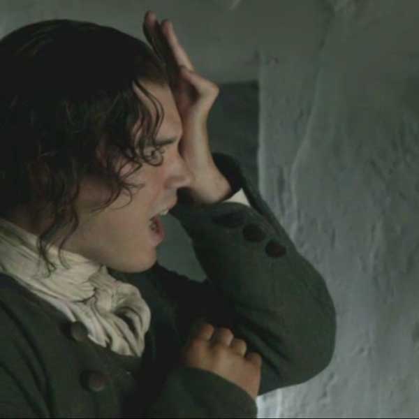
“Och! Willie just took it in the eye! Puir lad! See your way to class tomorrow for Anatomy Lesson #33 – The Eye, Part 5!
A deeply grateful,
Outlander Anatomist

Human Anatomy taught through the lens of the Outlander books by Diana Gabaldon and the Starz television series
“Och! Willie just took it in the eye! Puir lad! See your way to class tomorrow for Anatomy Lesson #33 – The Eye, Part 5!
A deeply grateful,
Outlander Anatomist
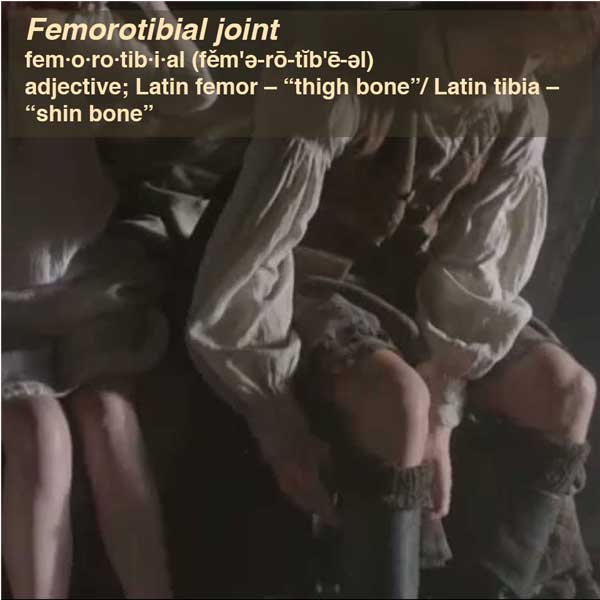
Anatomy def: Articulation between two condyles of femur and superior surface of tibia.
Outlander def: Darling and heart-stopping knee joints! ?the femoral bone’s connected to the tibial bone…?
Learn about the femorotibial joint in Anatomy Lesson #7, “Jamie’s Thighs” or “Ode to Joy.”
See these cute femorotibial joints in after-morning bliss, Starz episode, 7, The Wedding.
Read about Jamie and Claire’s knees in Outlander book:
A cool shadow fell over my heated face and a large pair of hands took firm hold of mine and pulled me to my feet. Jamie took my place on the log, and patted his knee invitingly. “Sit,” he said.
The bed creaked with a shifting of weight and I felt my knees being nudged further apart. “Not as dead as you look, I hope?”
A deeply grateful,
Outlander Anatomist
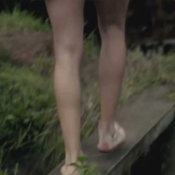
Gastrocnemius gas·troc·ne·mi·us ˌɡastrō(k)ˈnēmēəs/, noun: gastrocnemius; plural noun: gastrocnemii
Anatomy def: the chief muscle of the calf of the leg, it flexes (bends) the knee and ankle. Two heads arise from the femur and attach to the heel bone (calcaneus) by the single Achilles tendon. From Greek gastēr, gastr- ‘stomach’ + knēmē ‘leg. meaning stomach of the leg due to the bulging shape of the calf.
Learn about the gastrocnemius in Anatomy Lesson #27, “Colum’s Legs and Other Things Too.”
See Jamie’s gastrocnemius muscles as he crosses a plank bridge at the Lallybroch millstream, Starz episode 112, Lallybroch.
Read about Jamie’s legs in Outlander book:
I sat up, admiring the long legs, with the smooth line of muscling that indented the thigh from hip to knee, and another that ran from knee to long, elegant foot. The bottoms of his feet were smooth and pink, slightly callused from going barefoot.
A deeply grateful,
Outlander Anatomist