If you are like me, you binge watched Men in Kilts as soon as it became available on Starz. What a delightful respite after a very looong Droughlander! At least we can rewatch as we wait and wait and wait for season six of Outlander! 😉
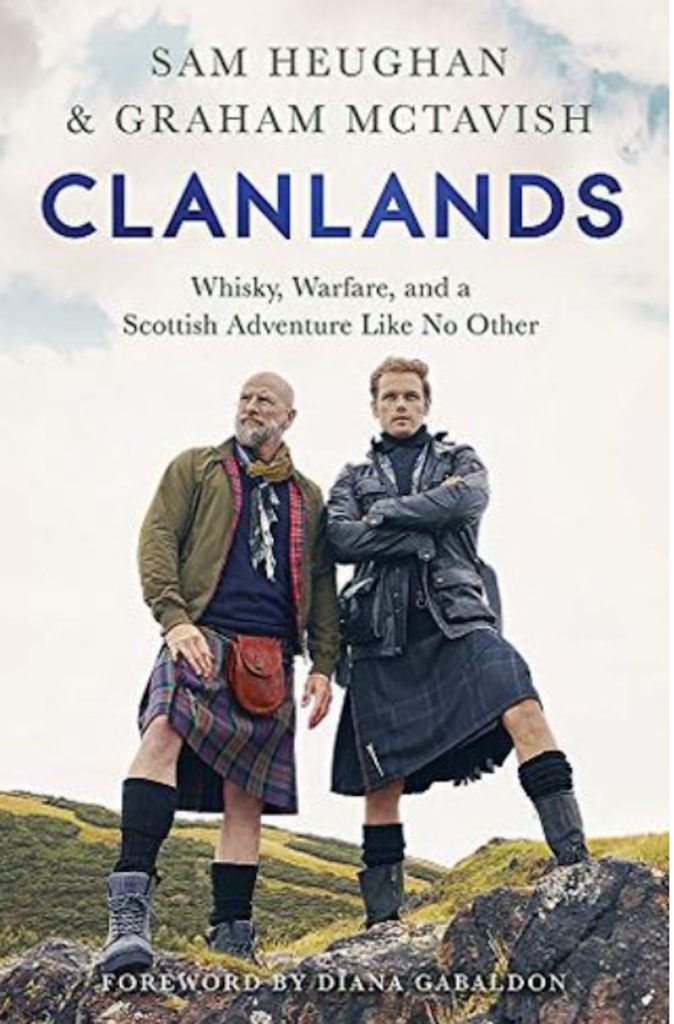
Also, I immediately snagged hard copy and digital versions of Clanlands, the book written by these two inventive lads. And, I listened to the audible version, the official title of the books being: Clanlands: Whisky, Warfare, and a Scottish Adventure Like No Other.
Basically, TV series and various book forms offer a splendid record of one big, fabulous road trip taken by Sam Heughan and Graham McTavish, stars of the beloved Outlander TV series. And, in an interesting switch-up, the books, the show, and the audible versions are not precisely the same. So, if you are able, I recommend all three forms for conspicuous consumption! 😋
As if reading about this Scottish pair isn’t sufficient to pique ones interest, the book forward is written by Herself, Diana Gabaldon! With typical wit and charm, her first words are:
“Well, in The Beginning…there was a man in a kilt.” 😍
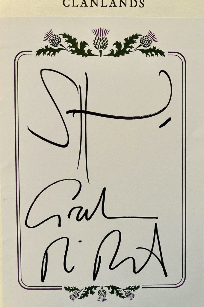
Now, I don’t want to crow (well, maybe a little), but I snagged an autographed version of the hard copy of Clanlands. 🤗
Yes, I know. The “autograph” consists of Sam’s initials and Graham’s… what? There’s maybe a “G” in there, but can the last line possibly read as McTavish??? 🤷♀️
The book of their Scottish odyssey begins with this prophetic promise:
“The story of two men who know nothing”
Sam and Graham
Followed by:
“It’s a dangerous business, Graham, going out your door. You step onto the road, and if you don’t keep your feet, there’s no knowing where you might be swept off to.”
Sam Heughan on behalf of
J.R.R. Tolkien, The Lord of the Rings
Annnnd….. it goes downhill from there.
Seriously, they begin an arduous week-long journey “to find the real Scotland and what it means to be Scottish,” while sequestered in a Fiat Auto-Roller Clan-Van sporting a gear shift! Now, I wonder who came up with that Clan-Plan? 🤔
It had been years since Sam used a gear shift. So, Graham-wise patiently reviewed the ABC mnemonic for the three foot pedals: A = acceleration; B = brake; C= clutch. A discussion then ensues – is the ABC from left to right or right to left? Right to left assures Graham.
“Ka-chunk wheeeeeeeeeeeerrrrrrrrrt ka-chunk”
And, They’re off! 😱
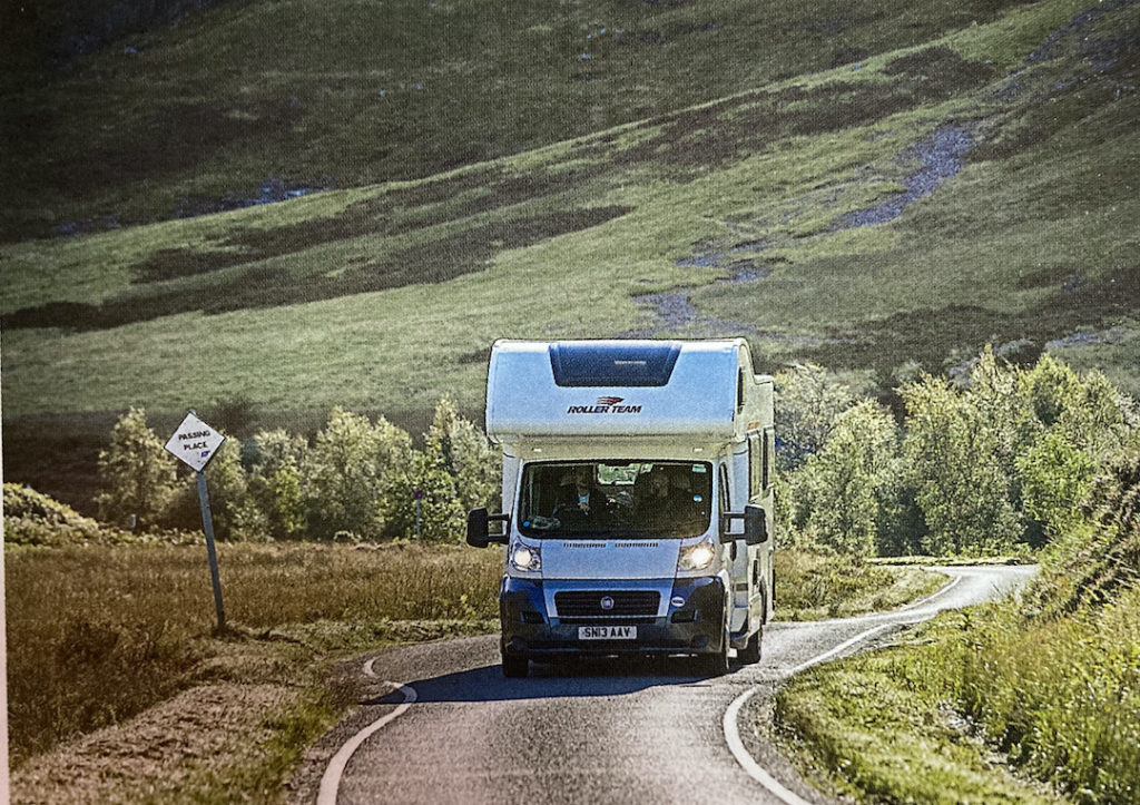
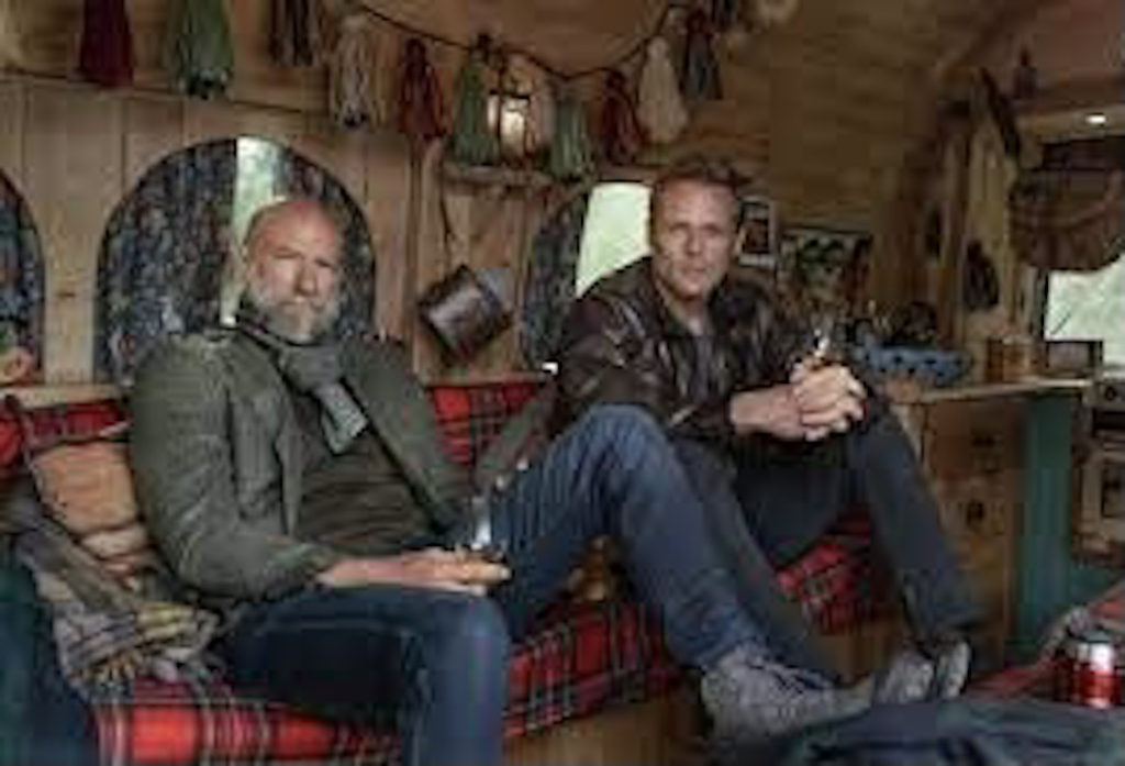
The camper looks rather typical from the outside, but was ever so exotic on the inside. Looks a wee bit crowded for these two rather largish Scots!
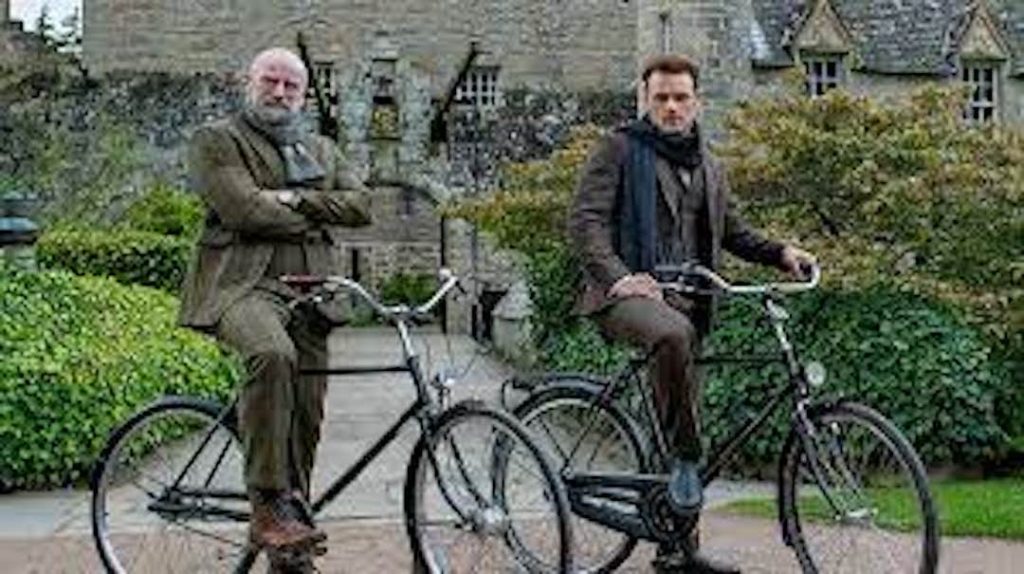
Between camper van excursions the blokes also travel by bike.
Look ma – No hands! 🙌🏻
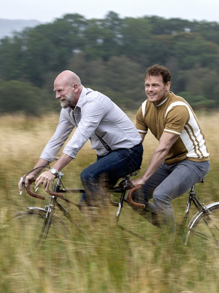
They also toured by tandem bike, ending with the slightly naughty, but apropos ditty, “Shovellin’ Coal” by Tony Pranses!
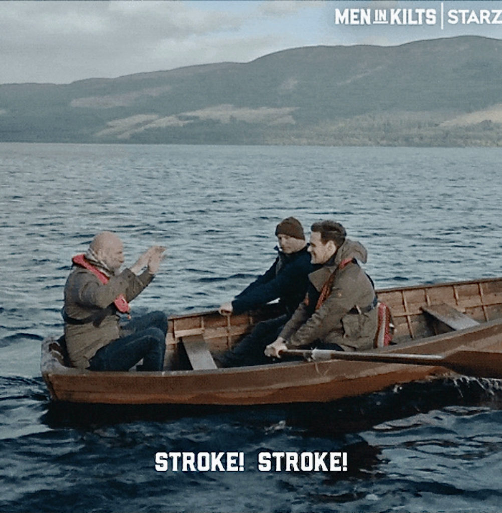
Travel by kayak. Well, below is not a kayak – more like a rowboat. Notice Graham relaxes as “coxswain” while Sam and Gary Lewis (aka Colum McKenzie) work! Row, row, row your boat! 🚣🏼
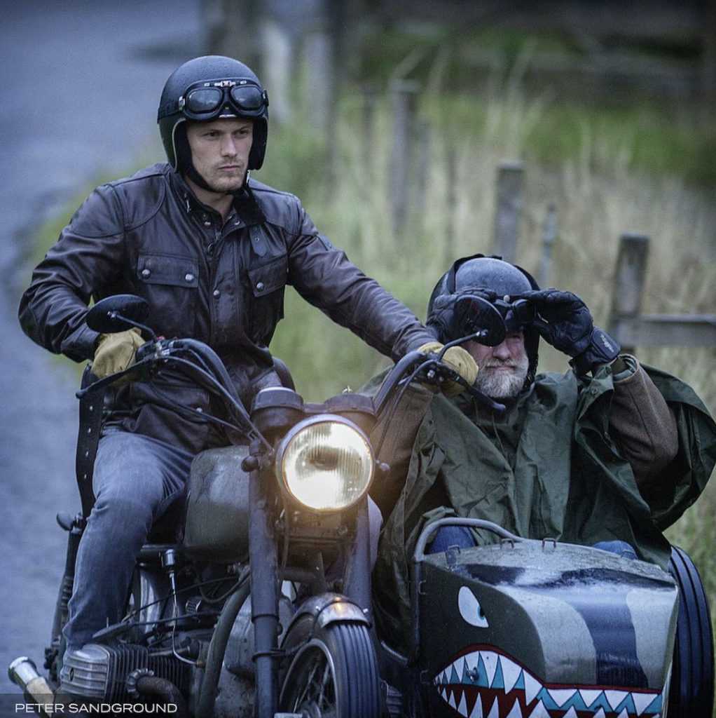
And, the most notable mode of transport: a motorbike with sidecar! Guess who’s driving “Miss Daisy?”
(Sam looks grim; Graham appears worrit)
Their adventure took them to the Highlands of Scotland where they scampered up hills.
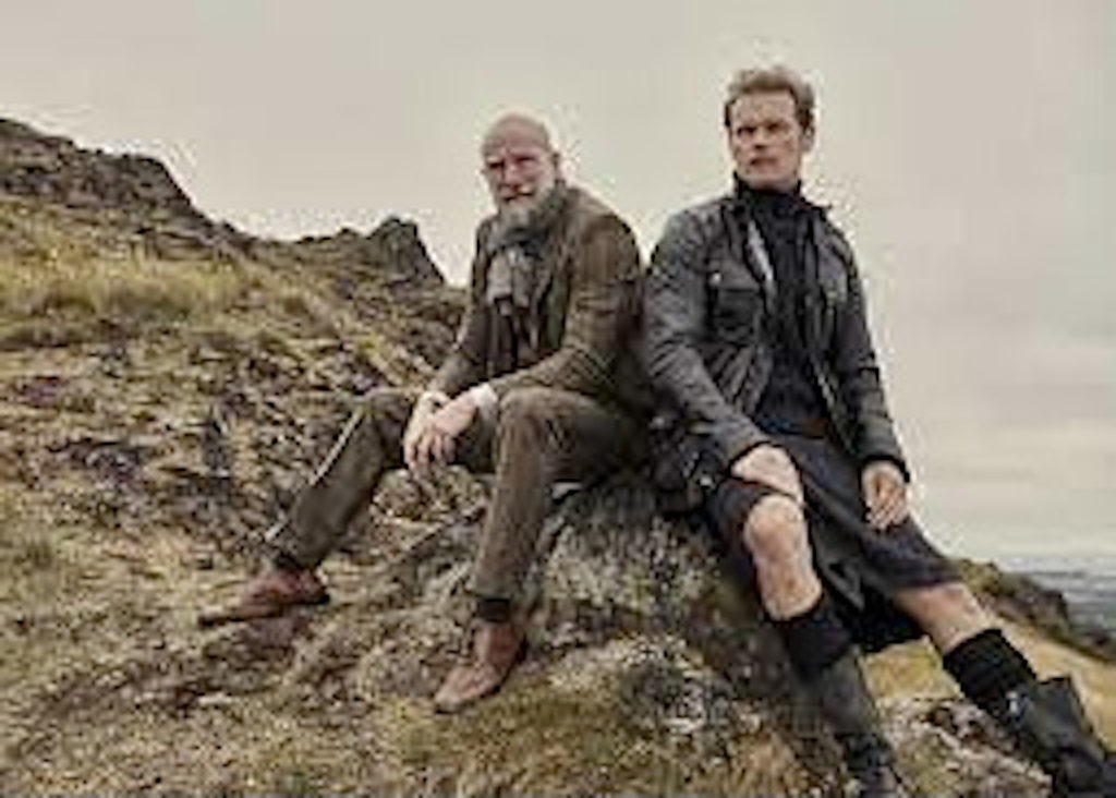
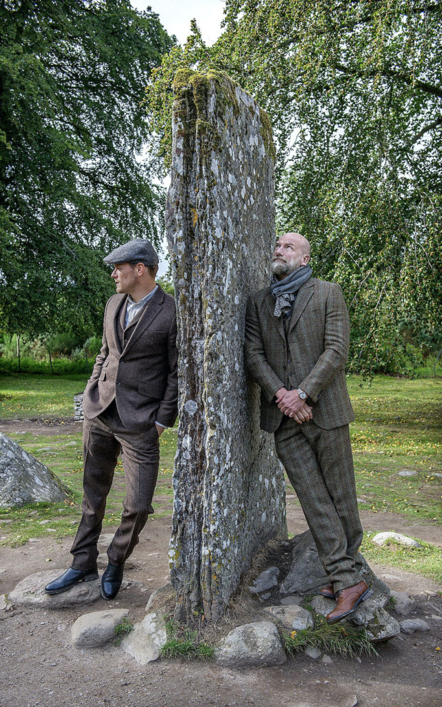
And, gazed on magestic vistas! (These guys own a lot of clothes!)
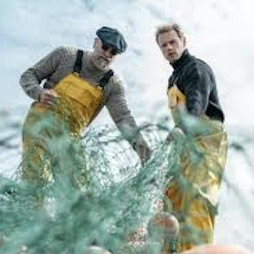
They pulled fishing nets from the sea. And, why wouldn’t they? Scotland has over 6,000 miles of coastline!!!
Hum…. wonder if they had fishing licenses? (Nice shades, Graham!) 😎
They marched with a bagpipe band, possibly the Inverness Youth Pipe Band, as described in Clanlands:
‘Scotland the Brave’ is blasting from the next room as the Inverness Youth Pipe Band warm up, the base drum reverberating off the walls.”
Clearly, Graham and Sam are pumped by the pipes! Bagpipes, that is. 😉
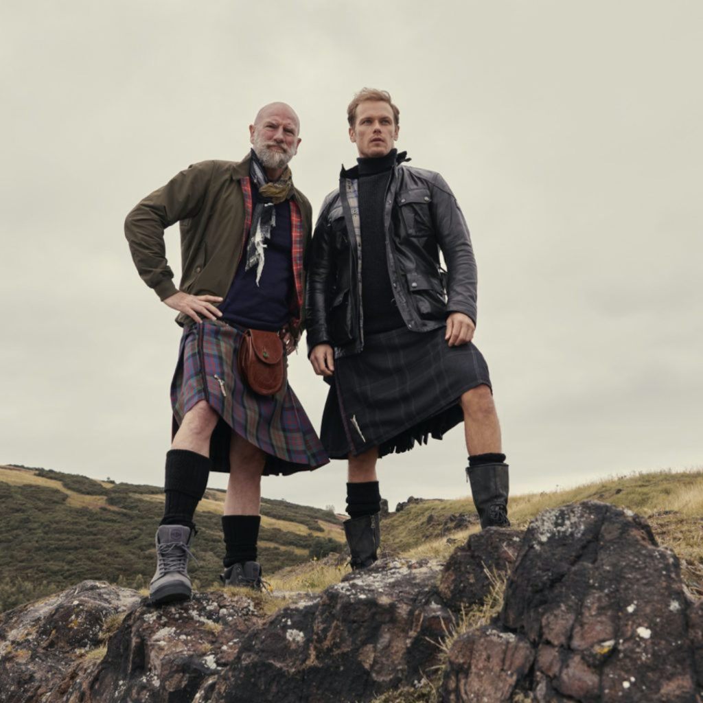
Dressed in snappy threads, they visit Clava Cairn, a type of Bronze Age circular chamber tomb near Inverness. (Like Jamie and Claire, they are separated by 202 years 😜)
Btw, I was privileged to tour this amazing historical site in 2016. Please go if you get the chance. You won’t regret it.
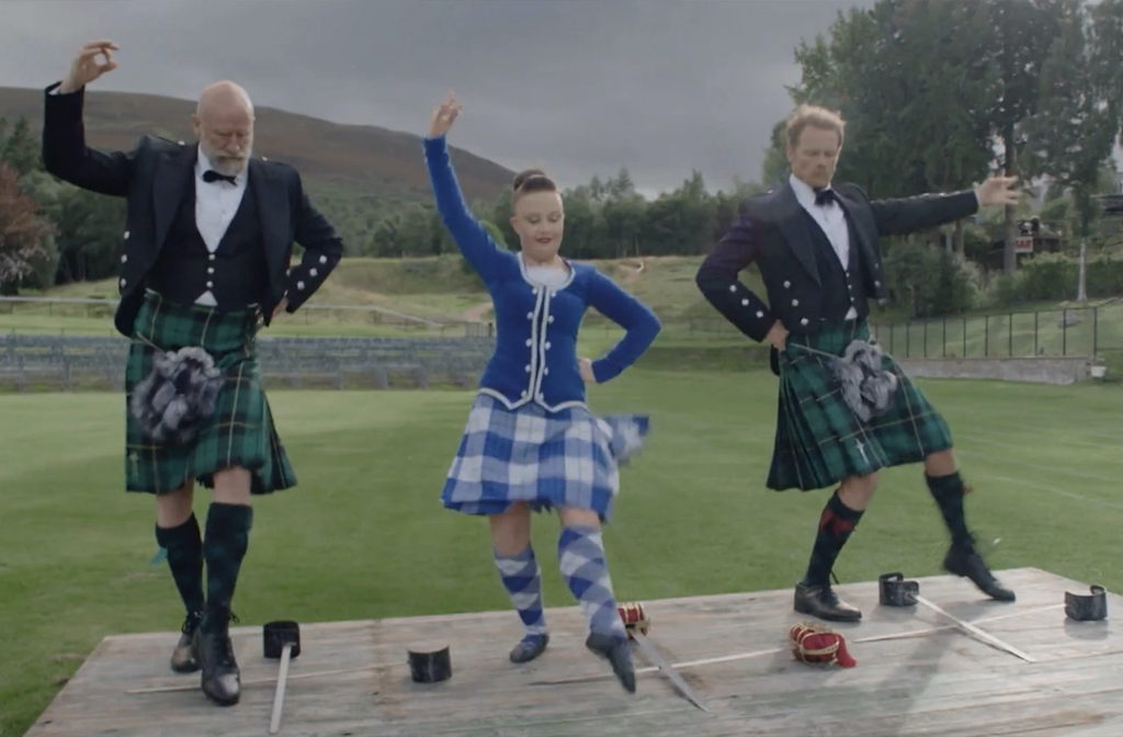
The lads even put on Sunday best to attempt the Scottish sword dance.
Not sure what in the world Sam is doing, but Lord of the Dance, it is NOT! Tiptoeing through the tulips, perhaps? 😂
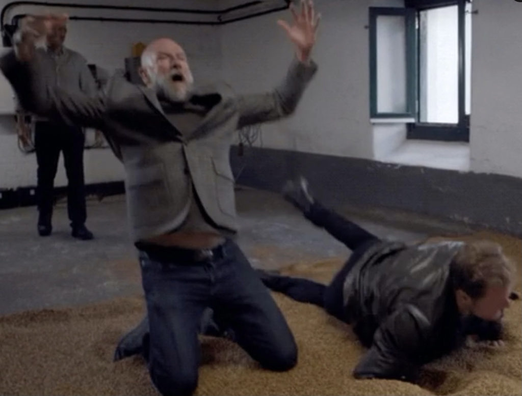
Joy! Joy! Joy! One of their happiest moments involved the delightful daredevils rolling in grain on a malting floor, later to be transformed into whisky! 🥃 Happy dance!!!
But, for me, their most impressive accomplishment was not the history lessons, geography tidbits, driving (sort of) tips, clan warfare, or cultural subtleties. Nope, it was the inventive bits of “slander” furiously lobbed between Sam and Graham. The first one appears on p. 9 of Clanlands:
“I’m not a ginger [Graham:You kind of are] or a virgin, either!”
Alrighty, then! 😲
Having grown up with three brothers, I well understand, that such defamations often represent the highest form of affection expressed between bros. (Sad isn’t it? 😜)
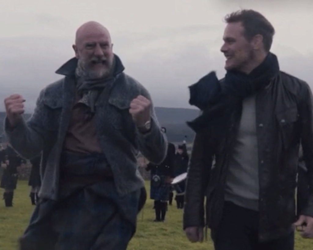
So what did these two jokesters do? Well, they plastered each other with various and sundry Clan-slams: zingers, insults, slanders, one-liners, jokes, barbs, quips, ribs, ripostes, giggles, boffo, waggery, drollery, disparagements, jeers, taunts, pokes, prods, nudges and digs! All were meant to gently malign and disparage but, most of all, to entertain with the gentlest of intents! 💓
Sam and Graham have always sparred with each other, even as their respective characters in the Outlander series!
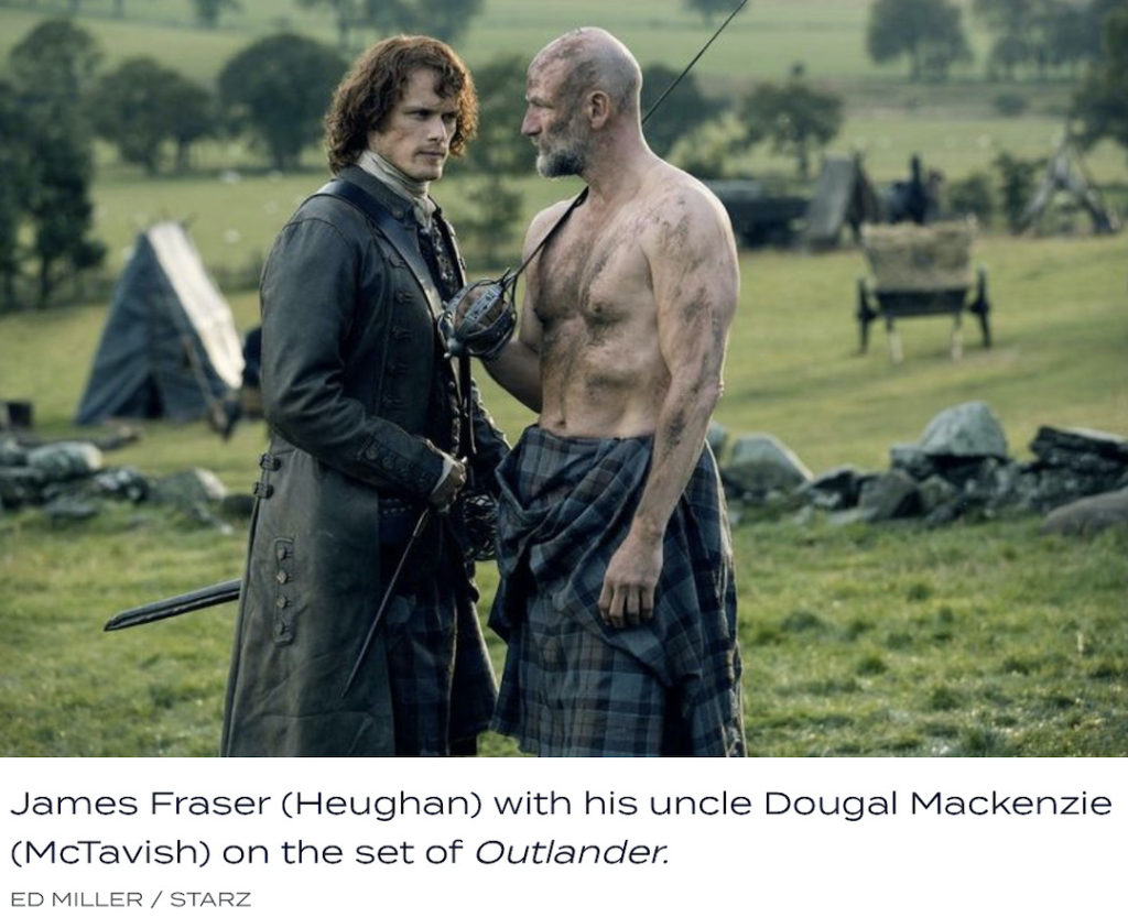
Take this faceoff from Starz, Outlander episode 209, Je Suis Prest. (Psst….Mine is bigger than yours! aka, who’s the man?) 😂
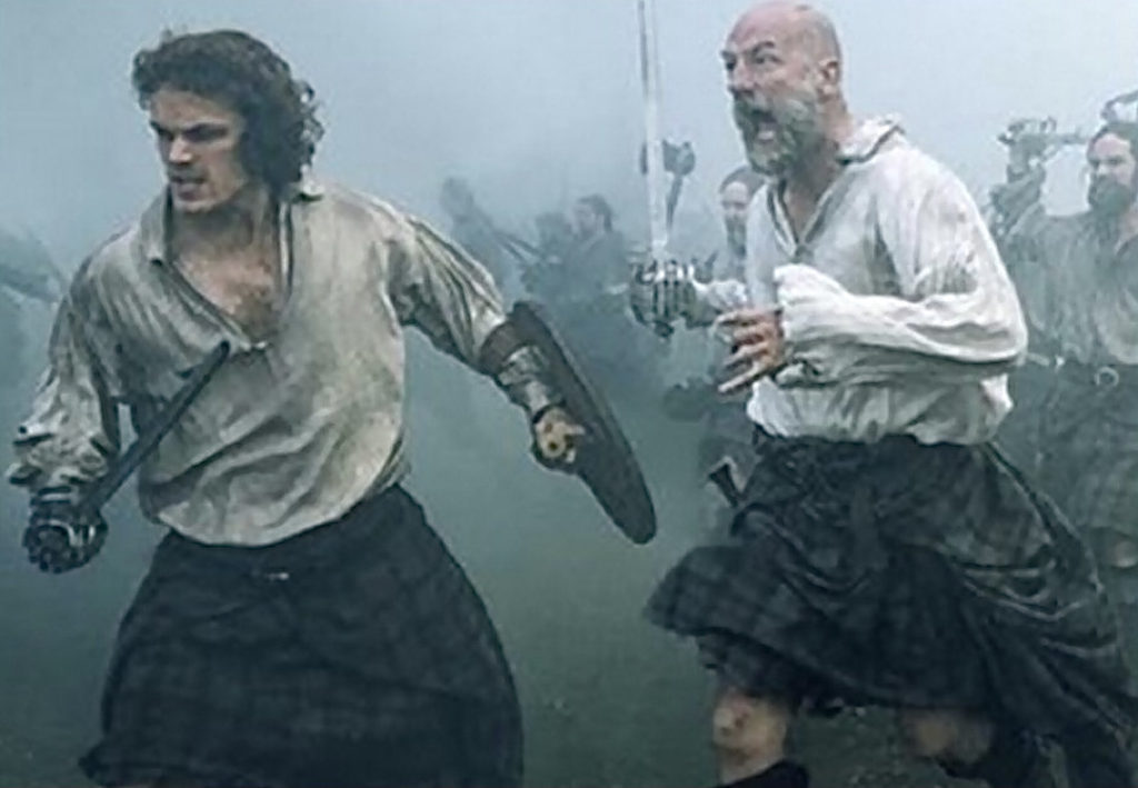
Dougal “pursues” Jamie at Prestonpans (Starz, Outlander episode 210, Prestonpans)! Watch yer back, McDubh! 😉
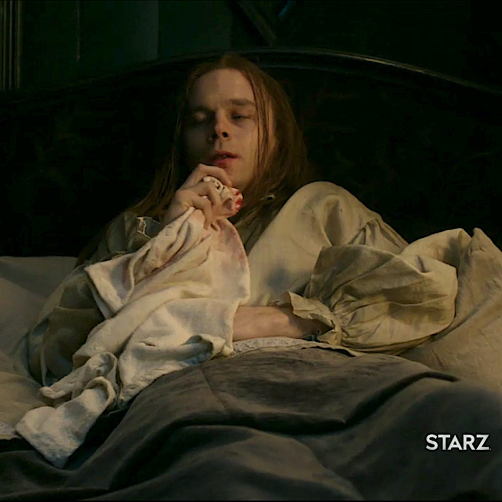
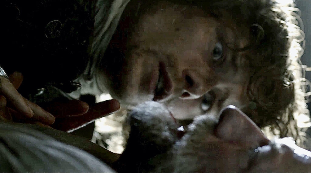
Oops, Jamie finishes off Dougal. My apologies, uncle! 🗡 (Starz, Outlander episode 213, Dragonfly in Amber) 😱
The lingual gymnastics executed by the two stars immediately grabbed my attention prompting me to start a running tab of their salty jibes. Sharing my initial compendium via Twitter, I was gobsmacked by this response from Sam:
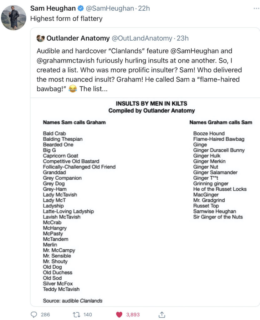
Which was quickly followed by Graham’s reply!
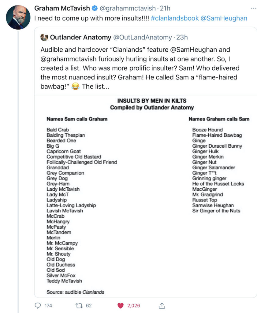
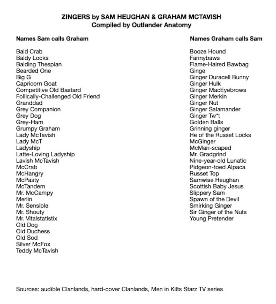
After publishing my first list, I have expanded it, adding a few “insults” missed the first time around. Below, is my latest compilation. Doubtless, I have missed a few so will update if I find more.
Which are your favorites? I am very fond of Mr. Shouty for Graham and Booze Hound for Sam (clothes hound might also work). Other favs are Scottish Baby Jesus (re Sam’s birth place) and Capricorn Goat (Graham’s a January babe).
Now, a quick bit of math reveals that Clan-Man, Graham, is a less prolific insulter than Sam, producing only 75% as many grand-slams as those inflicted by the Hot Scot. This may mean Graham is a nicer guy, or perhaps more gentlemanly not to express every thought that rattles his grey matter, or mayhap he has more important things to do. What do you think?
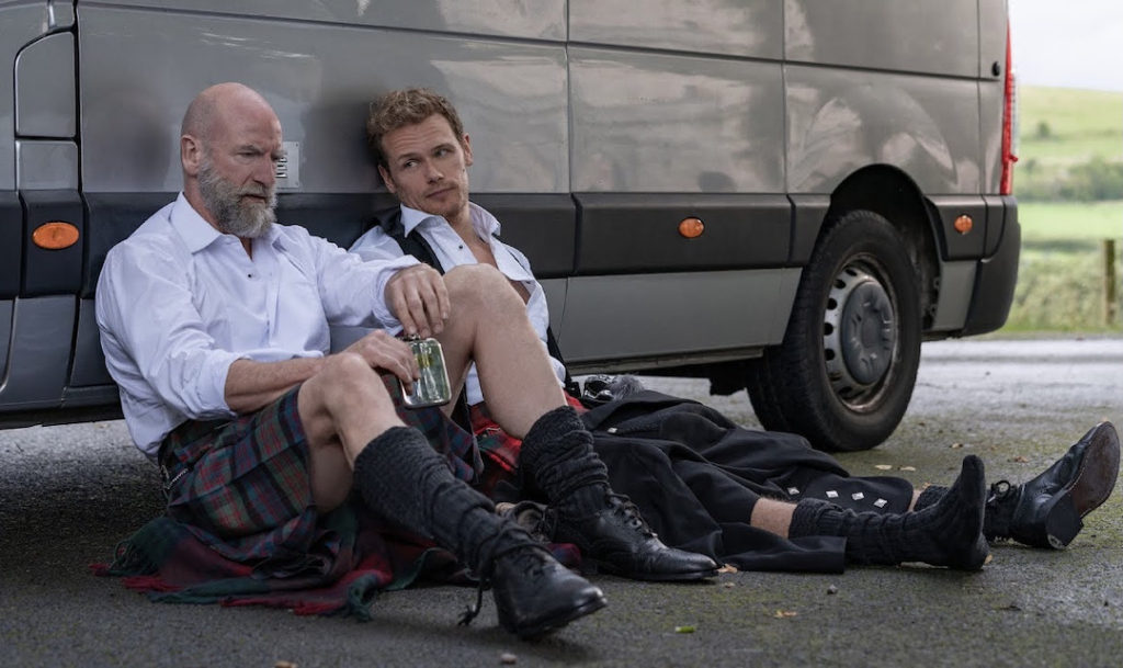
Ahhhh! Finally at journey’s end, Sam and Graham take a well-earned rest, backs resting against the trusty camper van and cheeks on the as-phalt 😜. (Psst… Honestly, they look a wee bit wasted!)
In the end, let us recall, these two blokes harbor great affection for one another. Sam makes this abundantly clear on the final page of Clanlands… His touching tribute to Graham:
Lord, ye gave me a rare friend,
and God! I loved the old sod well.”
Sam Heughan on behalf of Diana Gabaldon,
Dragonfly in Amber
As for me, I remain a die-hard, Clan-Fan!
The deeply grateful,
Outlander Anatomist
Follow me on:
Photo Credits: www.Starz.com; Clanlands; Ed Miller; www.brainstudy.info; www.heraldscotland.com; www.monstersandcritics.com; www.oprahdaily.com, www.outlanderanatomy.com; www.scotsman.com; www.sundaypost.com; www.thescottishsun.co.uk; www.thedipp.com; www.vulture.com; You tube