“If a Tree Falls – The Ear”

Summertime greetings to all Outlander anatomy students! Anatomy Lesson #24 covered the outer ear so today’s Anatomy Lesson #25 is the Ear – Part 2, or to be more exacting the middle and inner ears.

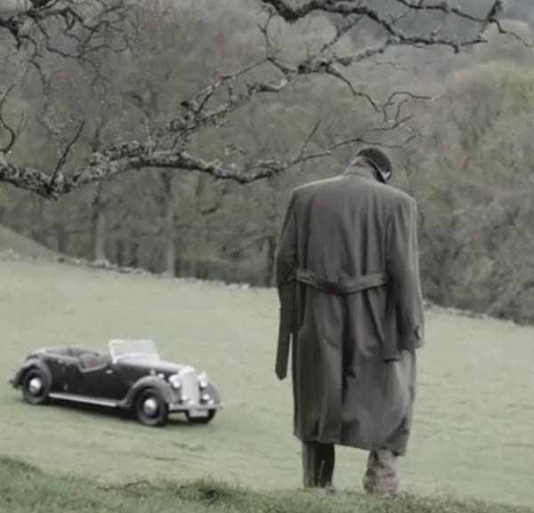
But first, what does a tree have to do with the ear? The title of this lesson derives from a 300 year old philosophical thought: “If a tree falls in a forest and no one is around to hear it, does it make a sound?” An 1884 issue of Scientific American correctly addressed this question. What do you think? Watch for the answer later on!

And to emphasize the importance of the ear in society, do you ken that the English language is replete with many idioms of all things ear? One website lists 120+ idioms including: a tin ear, all ears, music to the ears, up to the ears in, wet behind the ears, bend one’s ears, can’t make a silk purse out of a sow’s ear (who would try?), cute as a bugs ear (didn’t know bugs had ‘em), fall on deaf ears, in one ear and out the other, turn a deaf ear, and blow it out your ear (here’s to you, BJR!).

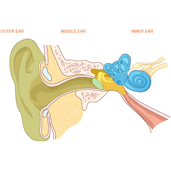
Now, onto the lesson. To review, in anatomy lesson #24 we learned that the human ear is divided into three ears: an outer (Photo A- green), a middle (Photo A- yellow) and an inner ear (Photo A- blue). That lesson dwelt almost exclusively on the outer ear. So, now we move to the middle and inner ears.

Ear-KLS-edited

Photo A

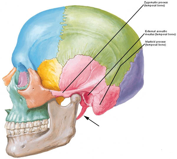
Let’s begin with the middle ears. Each middle ear is housed inside a cavity within one of our two temporal bones. The human skull includes 22 bones two of which are os temporale (Latin meaning temporal bone). The temporal bone is weirdly shaped (Photo B – pink bone). From a side view, it comprises most of the skull around the external acoustic meatus or EAM (Anatomy Lesson #24). The temporal bone also includes the zygomatic arch, part of the cheek bone (Anatomy Lesson # 8) and the gothic-looking styloid process marked by the black arrow (Anatomy Lesson #12).  Let’s add a new part of the temporal bone which is pertinent to today’s lesson, the rounded mastoid process.

Try this: Place your fingers behind the pinna of one ear and move downward until you feel a rounded mound of bone; this is your mastoid process. It typically lies just below the level of the EAM or ear hole as Claire called it (ha ha). Its outer layer is compact bone, but inside it is riddled with air-filled spaces. Well done folks!

Figure0004A-temporal-bone-KLS-edited

Photo B

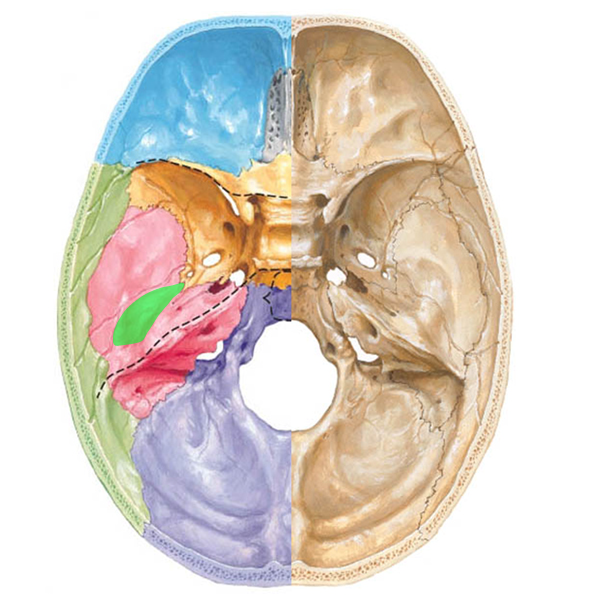
A different image of the skull helps us appreciate the location of the middle ear. With the top of the skull and brain removed, Photo C shows the cranial base. The right side of the image shows the tortuous shape of the bony floor upon which the brain rests. Nerves and blood vessels pass through the many holes in the skull bones. The left side of the image is color coded so once again, the temporal bone is pink. See the bright green area? This is the location of the middle ear – it lies inside the petrous (Latin meaning stone-like) part of the temporal bone, one of the densest bones of the human body.

skull

Photo C

The middle ear is small but it contains a large number of components including tympanic cavity, inner leaflet of tympanic membrane, three bones, the opening of a throat tube, a posterior “attic door,” two wee windows, two tiny muscles, a nerve and a nerve plexus. Wow! That’s quite a list for such a small space! Let’s examine the components.

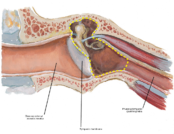
The tympanic cavity is an air-filled chamber (Photo D, yellow dashed line) inside the petrous temporal bone; its shape is so difficult to describe that many anatomists compare it to a small room with four walls, a roof and floor.  So let’s do that: the roof and floor are petrous temporal bone. The outer (lateral) wall is the tympanic membrane. The inner (medial) wall will be discussed shortly. The back wall has an “attic door” leading to the mastoid air cells (see below). The front wall receives the opening of the throat tube or pharyngotympanic or Eustachian tube (photo D) that extends between the back of the throat and the tympanic cavity.

Please understand this: normally, air pressure between the tympanic cavity and the EAM is equal. However, as we climb in altitude, air pressure becomes lower in the EAM than in the tympanic cavity which pushes the tympanic membrane outward causing discomfort, even pain. As we descend in altitude, air pressure is higher in the EAM than in the tympanic cavity which pushes the tympanic membrane inward, again causing pain. Air pressure equalizes on each side of the tympanic membrane when we open the Eustachian tubes: with altitude, air escapes from the tympanic cavity and with descent, air enters the tympanic cavity. Got it? Chewing and swallowing activates a pair of itsy, bitsy, teeny weeny, tensor veli palatini muscles that open the Eustachian tubes to equalize the air pressure. I won’t show an image of these wee muscles because it will clutter the lesson. Just know that they work well unless the Eustachian tubes are congested.

D.Figure0088D-tympanic-cavity-KLS-edite

Photo D

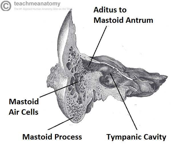
Remember the “attic door” in the back wall of the tympanic cavity? It has a longish Latin name, the aditus ad antrum, but the short of it is that the passageway leads to air cells which riddle the mastoid process (Photo E). These little spaces are thought to reduce the mass of the skull bones and provide physical protection.


Coronal-Section-of-the-Ear-Showing-the-Mastoid-Air-Cells KLS edited

Photo E

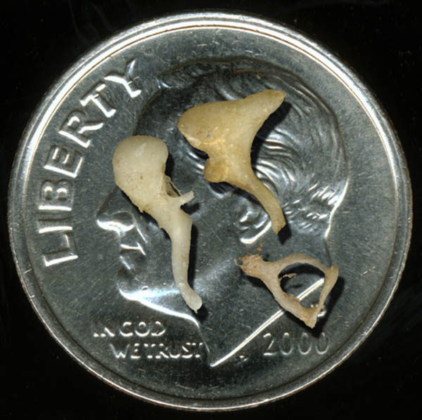
The middle ear bones bring us back to the anatomical rule of three! Three ossicles meaning tiny bones are the smallest bones of the human body. How small are they? Well, all three easily fit on a U.S. dime with plenty of wiggle room (Photo F)! There are three ossicles per middle ear and they are not included in the count of 22 skull bones.

ossicles 3 KLS edited

Photo F

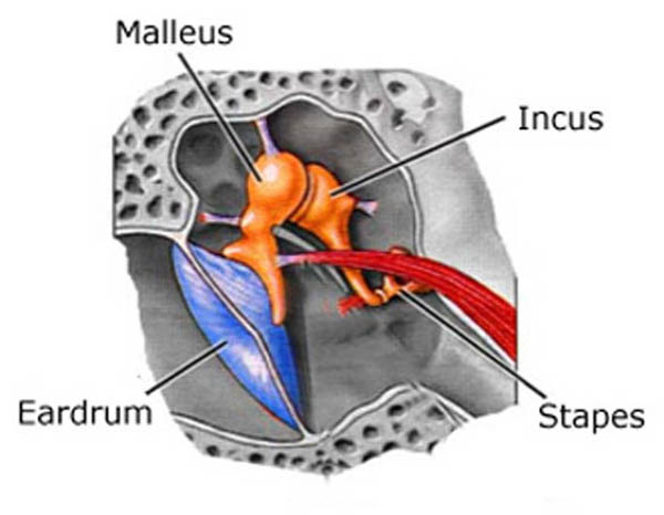
The ossicles form a tortuous bridge spanning the tympanic cavity from outer to inner walls (Photo G). Each ossicle resembles the object for which it is named: the malleus (Latin meaning hammer) has a handle firmly attached to the inner leaflet of the tympanic membrane and a head that articulates (forms a joint with) the incus. The incus (Latin meaning anvil) articulates with the stapes (Latin meaning stirrup) and the stapes inserts into the oval window, an opening in the inner wall of the tympanic cavity. Although tiny, the joints or articulations between the ossicles are moveable.

Please understand this: imagination is sometimes required to relate the Latin names to their corresponding anatomical objects. However, ancient anatomists used objects found in nature to name the body parts. The names such as stapes or malleus seem quaint but I actually prefer them to the current naming trend which often includes meaningless words containing lots of “cs”, “xs” and “zs.”

ossicles KLS edited

Photo G

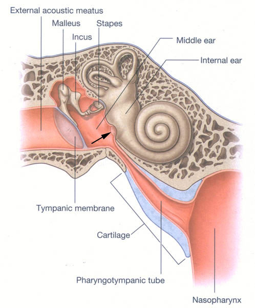
The inner wall of the tympanic membrane bears two holes piercing the temporal bone. One hole, the oval window, is plugged by the stapes (Photo H); the other hole or round window is closed by a membrane (Photo H – black arrow). The inner wall has several other features too, but these are beyond the scope of this lesson

middle-ear-KLS-edited

Photo H

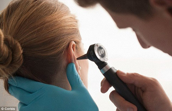
Let’s stop for a short Clinical Correlation: The oral cavity and throat contain oodles of bacteria (Rupert and Angus consider germs in this hilarious video: Angus and Rupert Go Through The Stones). All is well unless they follow the Eustachian tube into the tympanic cavity where they can set up housekeeping to our detriment. Otitis media (Latin meaning inflammation of middle ear) is a rather common condition which typically includes bacterial (or viral) infections of the middle ear. Over 30 million doctor visits per year in the U.S. are due to otitis media. Symptoms include: pain, pulling at the pinna, irritability, sleeplessness, crying, etc. A trip to the doctor and an otoscope exam is in order (Photo I).

otoscope KLS edited

Photo I

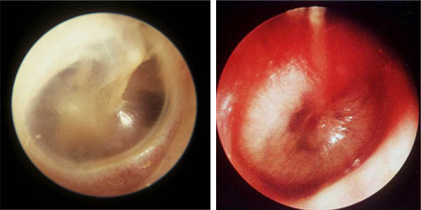
Anatomy Lesson #24, showed the photo of a normal tympanic membrane (Photo J – left). But, with otitis media, the tympanic membrane is red, bulging because there is fluid in the middle ear and it hurts (Photo J – right)! Proper treatment is imperative.

ear-drums

Photo J

Unresolved otitis media can lead to mastoiditis (Latin meaning inflammation of mastoid bone). Now, dinna confuse mastoiditis with mastitis which is inflammation of the breast. Och, we are discussing ears, not mammary glands!

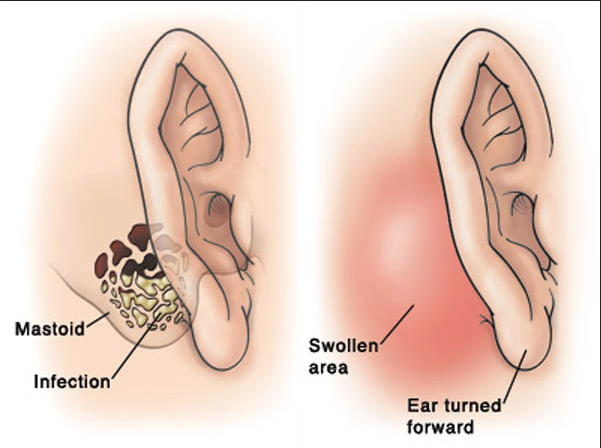
Here’s how mastoiditis works: recall that little attic door in the back wall of the middle ear that leads to the mastoid air cells (photo E)? Well, that door is a perfect conduit for bacteria to make their way from the middle ear into the mastoid air cells causing serious health complications in children and adults. Photo K (left side) shows bacterial invasion of the mastoid air cells and its presentation at the body surface (Photo K – right side). Signs include fever, redness, swelling, and tenderness behind the pinna which is pushed outward and forward.

photo-K

Photo K

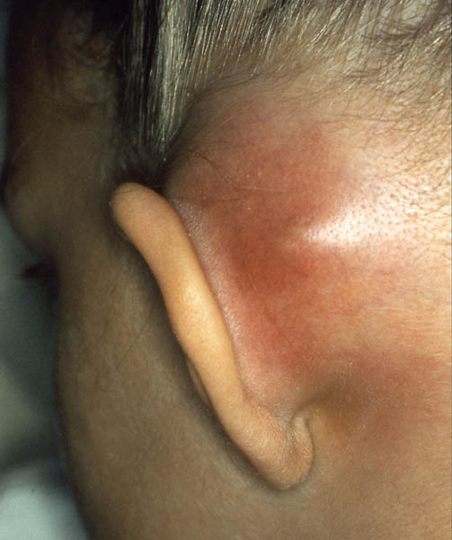
A picture is worth a thousand words, so this photo shows a case of mastoiditis (photo L – left mastoid process). Ouch, that hurts!

Mastoiditis1 KLS edited

Photo L

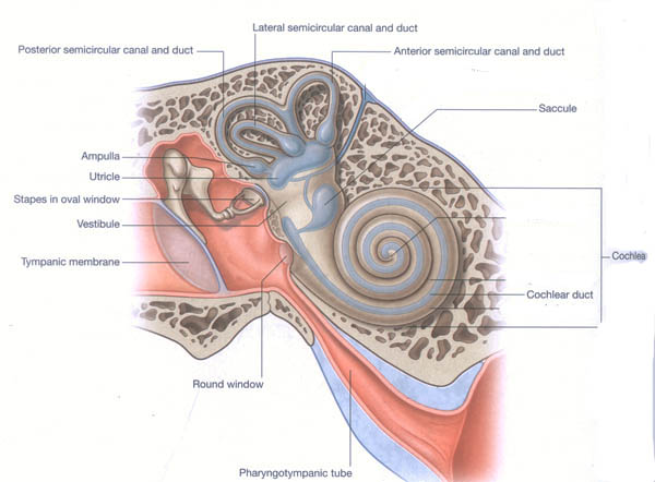
Now back to anatomy. The inner ear is the last and by far the most complex of the three “ears”. At first blush it resembles a mutant squid or snail. It contains both bony and membranous elements. Bony parts include cochlea, vestibule and three (yes, three!)  semicircular canals (Photo M- tan structures) filled with fluid (perilymph). Membranous elements (Photo M – blue structures) are suspended within the bony parts; these are the cochlear duct, utricle, saccule and three semicircular ducts; these are surrounded by perilymph and are filled with endolymph. Only the cochlear duct is involved in hearing, the utricle, saccule and semicircular ducts are necessary for balance and equilibrium.

inner ear KLS edited

Photo M

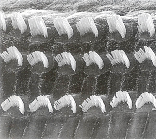
The cochlea and its cochlear duct spiral 2.5 times much like a snail shell. Within each cochlear duct lies the organ of Corti, a strip of 15,000-18,000 specialized “hair” cells arranged in rows like soldiers. “Bristles” project from the surfaces of the “hair” cells although these are unlike the hairs of skin. When examined by scanning electron microscopy (Anatomy Lesson #6), the bristles resemble the pipes of an organ (Photo N – cat hair cells). The bristles are covered by a gelatinous membrane (not shown in Photo N). The organ of Corti is also our organ of hearing.

hair cells of cochlea KLS edited

Photo N

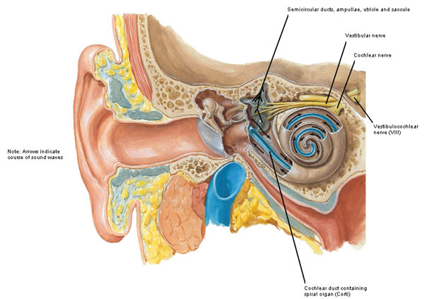
The following is a simplified version of how we hear, as the full vocabulary and structural and functional details would fill yet another anatomy lesson. Sound waves travelling through the air are gathered by the pinna and EAM; they strike the drum-like surface of the tympanic membrane pushing it inward with a thrust equal to the intensity of the sound. Ergo, loud noises push the eardrum inward more than soft sounds. The tympanic membrane vibrations are transferred through malleus, incus and stapes. With each vibration the stapes pushes inward at the oval window creating corresponding shockwaves through perilymph and endolymph of the cochlear duct (Photo O – black arrows). Movements of the fluids rub the bristles of the hair cells against the gelatinous membrane creating an excitation which is transfer to nerve cells forming the cochlear nerve (Photo O). The cochlear nerve joins with the vestibular nerve (see below) to form Cranial Nerve VIII (vestibulocochlear nerve). Impulses of each Cranial Nerve VIII follow auditory pathways into the brain. Hair cells near the base of the cochlea detect high-pitched sounds, such as ringing of a cell phone; those closer to the apex detect lower-pitched sounds, such as barking of a large dog.

Figure0087 inner ear KLS edited

Photo O

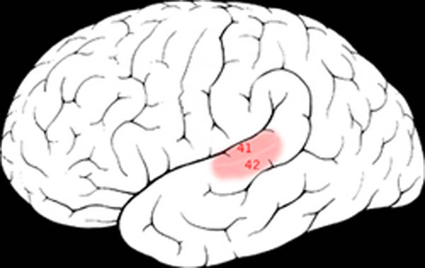
Electrical signals carried by the cochlear nerve make their way to the primary auditory cortex of the brain (Photo P – pink zone) where electrical signals are converted into “sounds” that we learn to recognize and understand.

In summary, outer, middle and inner ears work together to transfer sound waves through air (outer ear), solid (middle ear) and liquid (inner ear) where the good, good, good, good vibrations are converted into electrical signals that make their way to the brain for interpretation of sound. Go Beach boys!

250px-Brodmann_41_42 KLS edited

Photo P

Do you recall this lesson began with “If a tree falls in a forest and no one is around to hear it, does it make a sound?” Take a moment to think of the answer and then read on.

The answer to this philosophical riddle is that if a tree falls in the forest it creates sound waves but a receptor must be present to convert those sound waves into sound. If any creature is present with an organ than can perceive and interpret sound waves, then the falling tree does make a sound otherwise it only makes sound waves. Make sense? Good!

Now, every anatomy lesson must tie into all things Outlander and hearing is no exception. So next is a jolly good quote from Outlander book when Claire tells Jamie she is from a waaay different time zone (Starz episode 111, The Devil’s Mark). Well, Jamie knew there was something unique about this braw and bonny lassie!

“Do you know when I was born?” I asked, looking up. I knew my hair was wild and my eyes staring, and I didn’t care. “On the twentieth of October, in the Year of Our Lord nineteen hundred and eighteen. Do you hear me?” I demanded, for he was blinking at me unmoving, as though paying no attention to a word I said.

ep 111 he hears you 01 KLS edited

“I said nineteen eighteen! Nearly two hundred years from now! Do you hear?” I was shouting now, and he nodded slowly. “I hear,” he said softly.

Hmmm, Jamie is thinking: I ken now why the Sassynach didna take to my leather belt spankin’! It also ‘splains her twitchy-witchy “know how.”

ep 111 do you hear me 02 KLS edited

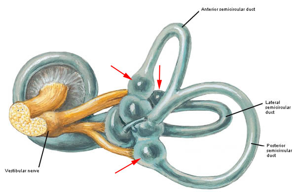
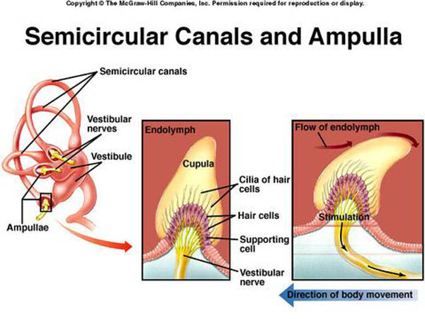
Now we must move on to how the inner ear provides balance and equilibrium. Our ability to balance is independent of outer ear, middle ear and cochlear parts of the inner ear but it is dependent on function of the three semicircular ducts (Photo Q – bone removed). The semicircular ducts contain endolymph and each bears an ampulla, a swelling at one end (Photo Q – red arrows). Each ampulla contains a patch of “hair cells” similar to those of the organ of Corti. Once again, the bristles are covered with a gelatinous membrane.

Anterior and posterior semicircular ducts are oriented vertically at right angles to each other. The lateral semicircular duct is slightly off the horizontal plane. Orientation of the ducts cause each duct to be stimulated by angular rotation of the head in a given plane.


Figure0090C-semicircular-ducts-KLS-edited


Photo Q

Turning your head from left to right sides (as in no) moves endolymph in the lateral semicircular duct. Nodding your head (as in yes) moves endolymph in the anterior semicircular duct. Moving your head to touch one shoulder or as in doing a cartwheel moves endolymph in the posterior semicircular duct. In aviation terms, the semicircular ducts are oriented such that they detect pitch, roll and yaw.

Here is how the semicircular ducts work: As the head moves in angular rotation, endolymph moves in the opposite direction bending the gelatinous membrane (cupula) and exciting the hair cells (Photo R). The hair cells transfer the signal to nerve cells of the vestibular nerve (part of Cranial Nerve VIII). The signal is carried to the brain which interprets it as angular motion of the head. The information can be used to activate various muscles to adjust the head and/or body positions.

ampullae KLS edited

Photo R

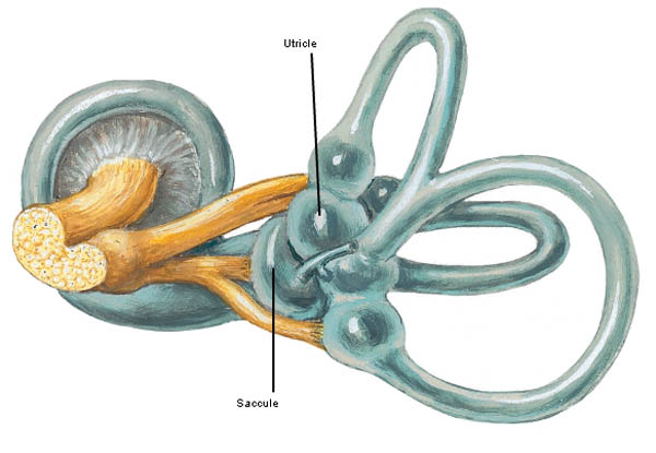
The final anatomical elements of the inner ear are utricle (Latin meaning leather bag) and saccule (Latin meaning money bag –  Dougal is into this one!) located between the semicircular duct and cochlear duct. These elements are also filled with endolymph (Photo S) and are designed to detect changes in linear acceleration or linear deceleration.

Figure0090C utricle and saccule KLS edited

Photo S

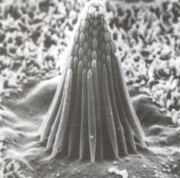
Utricle and saccule each contain a patch of hair cells with surface bristles (Photo T – bullfrog hair cell) covered by a gelatinous membrane (absent in Photo T). The hair cells of the utricle are oriented horizontally and those of the saccule are vertically oriented.

macula stereocilia KLS edited

Photo T

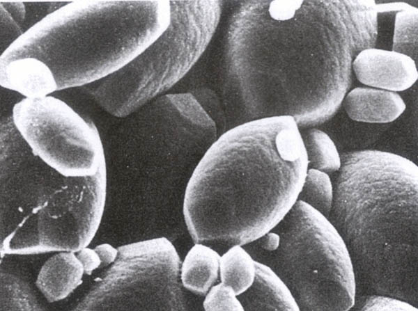
Seated atop the gelatinous membrane are wee otoliths (ear stones or ear rocks) made of calcium carbonate (Photo U); these add weight to the gelatinous membrane thus enhancing our sense of  gravitational pull.

otoliths KLS edited

Photo U

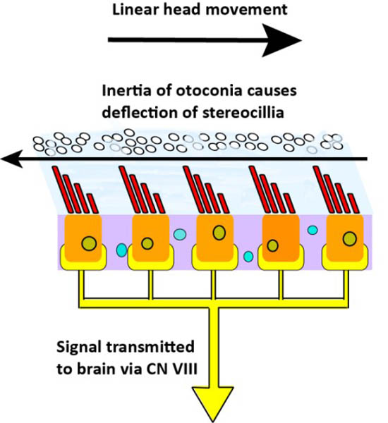
The utricle and saccule work this way: As our head undergoes linear acceleration or linear deceleration (forward, backward, upward, downward), endolymph, the gelatinous membrane and otoconia move in the opposite direction (Photo V). This bends bristles of the hair cells causing them to activate nerve cells of the vestibular nerve of Cranial Nerve VIII. The electrical impulses are carried to the brain. The utricle detects horizontal changes and the saccule detects vertical changes in linear movements of the head. This information arrives at the brain which then determines if and how much the head is tilted and if the body needs to be reoriented in space.

otoconia%20displacement KLS edited


Photo V

This brings us to the end of today’s important lesson but we must tie balance and equilibrium into Outlander. Here are a couple of great quotes from Outlander book. The book quote and the Starz images (Starz, episode 108, Both Sides Now) don’t quite match because the book scenarios weren’t filmed. Nevertheless, it is fun and you will get the idea. The first scene takes place as Frank and Claire descend from Craigh na Dun after watching the Druid’s dance. Frank, the soon-to-be Oxford professor, is so absorbed in thought he doesn’t watch where he plants his feet!

“He dropped into one of his scholarly trances … The trance was broken only when he stumbled unexpectedly over an obstacle near the bottom of the hill. He flung his arms out with a startled cry as his feet went out from under him and he rolled untidily down the last few feet of the path, fetching up in a clump of cow parsley… “Are you all right?” … “I think so.” He passed a hand dazedly over his brow, smoothing back the dark hair. “What did I trip over?” “This.” I held up a sardine tin…”

ep 108 Frank balance KLS edited

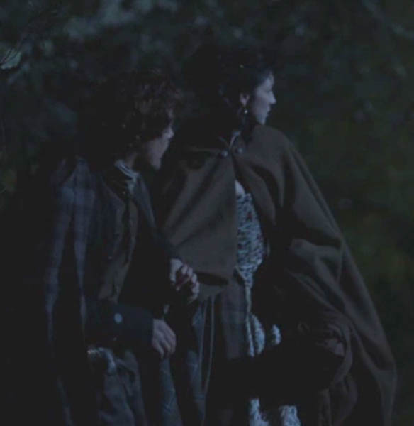
Compare and contrast Frank’s stumble-tumble with Jamie’s sure footedness (Outlander book). In this scene, it is nighttime and Jamie unerringly hauls Claire through the night:

“Jamie kept a tight hold on my arm, hauling me upright when I stumbled over rocks and plants. He himself walked as though the stubbled heath were a paved road in broad daylight. He has cat blood, I reflected sourly, no doubt that was how he managed to sneak up on me in the darkness.”

Yes, Jamie has grace, balance, and equilibrium (Starz episode 104, The Gathering). Nothing will trip-flip this kilt! Hang on tight, Claire! Snort!

ep 104 holding hands KLS edited

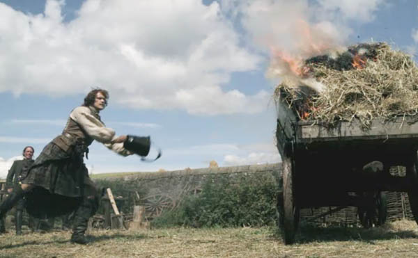
You may have seen this arabesque in the teaser but let’s appreciate it anyway. Jamie throws water on burning hay, a fire set by a Watch weasle! Our highlander fireman even makes tossing water a thing of beauty. Balance and equilibrium on one foot; 17th century ballet!

ep 112 Jamie balance 01 KLS edited

At last, the time has come to report the results of Lesson #24 and the Pinna Poll! Six images of pinnae from the Starz cast (episodes #103, #109, #115, #116) were shown and votes tallied. All the ear flaps are fabulous!

Not sure if you are surprised, but the winner is:…………..drum roll………………..Jamie! Congratulations Big Red! And it was an honest count, too; no stuffing of the ballot box!  Jamie garnered 38% of the votes while the remaining five pinnae were in almost a dead heat with a slight edge by bad boy BJR!

Great comments were shared by many readers including these: exquisite, lovely, charming, manly, rugged, beautiful, elfin-like, fawn-like, nibble-worthy, snuggle-worthy, elegant, awesome, symmetrical, balanced, proportionate and yum! Let’s thank our six wonderful characters and all of you for playing the Pinna Poll!

pinna-poll

That’s it for the ear. Complex and elegant! Oh, and a word to the wise: keep music turned down a notch or two. Over the years, hair cells of the organ of Corti become damaged by excessive noise. For years, the damage has been considered irreversible although a recent study shows some promise using a gene-activating drug regime. Stay tuned.

Closing with this fun poem from Mr. R’s World of Math and Science:

An ear splitting sound!

A crash and a boom!

Ringing so loudly,

It shook the whole room!

But I didn’t hear it,

I couldn’t at all,

My left and right ear,

Had gone to the mall!


My left and right ear,

My organs that hear,

Had gone to go shopping,

And that’s what I fear…

Without my 2 ears,

That spectacular pair,

I can’t hear sound waves,

Move through the air!


The deeply grateful,

Outlander Anatomist

Photo creds: Starz, Basic Histology, Junqueira & Carneiro, 11th ed., Concise Histology, Bloom & Fawcett, 2nd ed., Netter’s Atlas of Human Anatomy, 4th ed., Clinically Oriented Anatomy, 5th ed., www.aviationknowledge.wikidot.com (ampullae), www.clearwaterclinic.com (otoliths), www.fairview.org (mastoiditis), www.kids-ond.com (ossicles), www.mhhe.com (ampula hair cells), www.sciencepoems.net (Mr. R’s ear poem), www.student.com (ossicles with inner ear), www.teachmeanatomy.info (mastoid air cells), www.otopathologynetwork.org (ossicles on dime), www.wallpaper.com (fallen tree), www.wikipedia.org (mastoiditis with subperiosteal abscess)

Anatomy Lesson #22: Jamie’s Hand – Symbol of Sacrifice

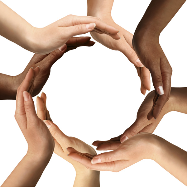
Welcome, anatomy students, to Anatomy Lesson #22: The Hand. Anatomy of the human hand is daunting because it is complex. The hand is a true engineering marvel, containing four of the six classical simple machines as defined by Renaissance scientists: wedges, planes, pulleys, levers. This structural diversity makes the hand arguably the most mobile part of the entire musculoskeletal system (Photo A).

Languages include abundant references to hands so over emphasizing their importance might be a challenge. One English source on hand idioms records almost 300 common references: bird in hand, give me a hand, firm hand, hand-me-down, safe hands, helping hands, tie ones hands (shade #1 of BJR), throwing up one’s hands, wandering hands (shade #2 of BJR), wash ones hands of, lift a hand, lay hands on (shade #3 of BJR) to name but a few.

Brand-Congruence-Hands1 KLS edited

Photo A

In human anatomy, structure and function go “hand-in-hand” (another idiom): if a body part performs a particular function(s) its structure reflects that task and vice versa a body part that has a particular structure will reflect the function(s) it is designed to fulfil. There is no finer example of this axiom than the human hand.
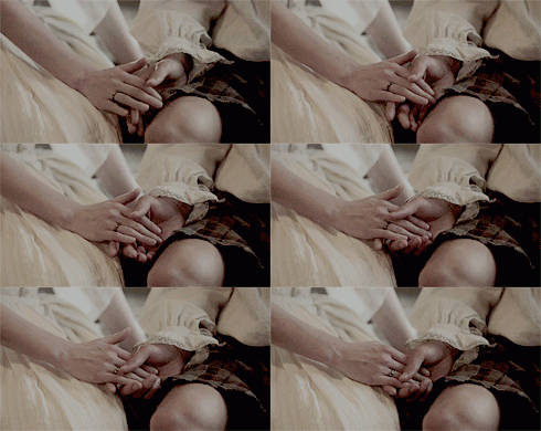
More pertinent to our shared interest, holding hands and touching are important elements of the Outlander books and the Starz series (a.k.a. hand sex). Herself’s own words from Jamie and Claire’s wedding night (Outlander book):

Somewhat tentatively, he reached out and took my hands between his own … I felt a slight shock at the touch … “Aye. More scairt than you, I expect. That’s why I’m holdin’ your hands; to keep my own from shaking.” I didn’t believe this, but squeezed his hands tightly in appreciation. “It’s a good idea. It feels a little easier to talk while we’re touching.”

holding hands

And, from Neil Diamond:

Hands, touchin’ hands

Reachin’ out, touchin’ me, touchin’ you

Sadly, this lesson must include images and analysis of Jamie’s hand after BJR adds his “aesthetic” twist. There will be a WARNING ahead of these images to give you the opportunity to skip them. They appear near the lesson’s end. Look for the warning sign before those images and the kitten when it’s safe to come out. I must also confess, that this chapter in the book and Starz episode 115 Wentworth Prison made it difficult for my usual sass to fly in this lesson. My sass is sad and worried about Jamie and Claire and ready to pull out a can of whoop ass on BJR. Here BJR, this is for YOU!

whoopass

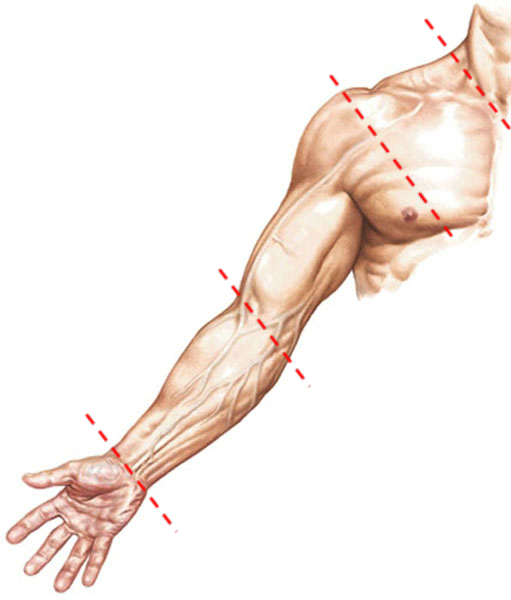
Back to the lesson at hand (haha)…Photo B of the upper limb has appeared in several prior Outlander Anatomy Lessons. But, to reiterate: the shoulder is from base of neck to shoulder joint, the arm is between shoulder and elbow joints, the forearm between elbow and wrist joints and the hand is the terminus of the upper limb. More precisely, the hand lies distal to (away from) the forearm.

Figure0401---upper-limb-KLS-edited

Photo B

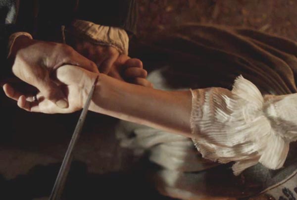
To better understand the hand, we must further consider the forearm. Remember supination? From Starz episode 107 The Wedding, we see a fine example of forearm supination (and elbow joint extension) as Dougal slices Claire’s wrist for her blood vow with Jamie. With the forearm in supination, ulna and radius are parallel, palm faces forward or up, thumb is directed away from body.

ep 107 forearm supination KLS edited

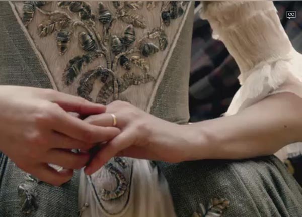
Recall pronation? Next is a great example of forearm pronation (and elbow joint flexion) as Claire removes Frank’s gold wedding band prior to marrying Jamie (Starz episode 107, The Wedding). In forearm pronation, distal radius crosses distal ulna, palm faces downward or back and thumb is directed toward the body.

ep 107 elbow flexion KLS edited

Try this: To review, flex your elbow joint and alternately rotate forearm between supination and pronation. Review positions of radius, ulna and thumb in each of the two movements.

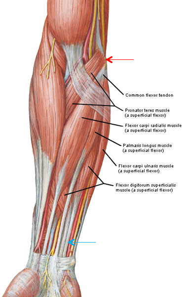
The forearm contains a whopping 20 muscles: eight in a flexor compartment and 12 in an extensor compartment! Flexor muscles lie anterior to ulna and radius and are best demonstrated with a supinated forearm. Photo C shows five muscles of the flexor compartment which arise from the medial epicondyle of humerus (red arrow). I’ll not drown you with the intricate names which reflect function and position. Suffice it to say that although the muscle bellies of the flexors lie in the forearm, they become tendons near the wrist (blue arrow) and then insert into bones of the hand and cause movement.

Figure0429-common-flexor-tendon-muscles-KLS-edited

Photo C

Try this: Examine your wrist with palm up and see or feel various tendons: these are the flexor tendons that arise in the anterior forearm but insert on bones of wrist, thumb and fingers.

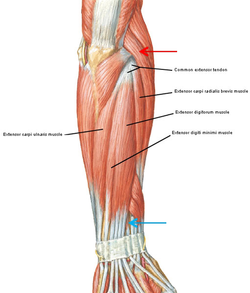
The 12 extensor muscles of the forearm lie posterior to radius and ulna and are best viewed from the back of a supinated forearm. Photo D (forearm viewed from the back) shows four extensor muscles which arise from the lateral epicondyle of humerus (red arrow). Again, naming all 12 extensors along with their actions truly would be tedious. However, the same theme repeats itself: the muscular part of the extensors lie in the forearm, they become tendons near the wrist (blue arrow) and insert into bones of wrist, thumb and fingers and cause movement. 

Figure0427-common-extensor-tendon-muscles-KLS-edited

Photo D

Try this: Examine your hand with palm down and see or feel various extensor tendons on the dorsum of hand. Next, grip your supinated forearm and make a strong fist. Now bend the wrist toward (flexion) and then away (extension) from your torso. Feel muscles of flexor and extensor compartments alternately contract and relax with wrist movement? Just doing their jobs!

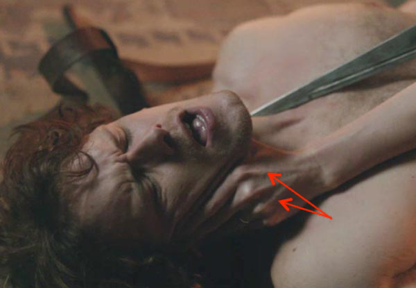
Want to see an excellent example of extensor tendons (Starz episode 109, The Reckoning)? Here Claire threatens a shocked Jamie within an inch of his carotid arteries! The taut tendons (red arrows) on the dorsum of her hand belong to an extensor forearm muscle (extensor digitorum). Mmphm, she doesna even break rhythm. Jamie croaks “You have my word!” No surprise there!  Erm, that’s one impressive looking dirk!
ep-109-extensor-tendons-01-KLS-edited

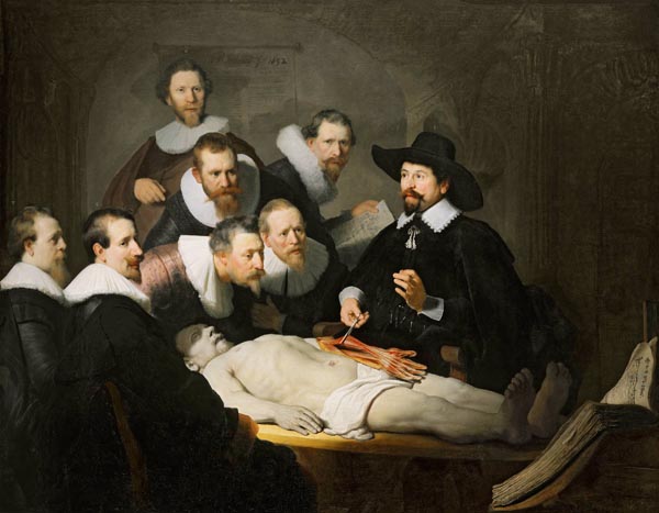
You may recognize Rembrandt’s famous 1632 painting The Anatomy Lesson of Dr. Nicholaes Tulp (Photo E – body likely a condemned criminal). As demonstrator of anatomy (a position I held for many years), Dr. Tulp was demonstrating left forearm and hand muscles to eager viewers. The painting records a real event: in that era, public dissections were not uncommon in Europe. But, did you know this fabulous work of art is anatomically questionable? Indeed, its anatomical accuracy has been debated for decades with advocates as well as detractors. One meticulous study recreated Dr. Tulp’s lesson and concluded that the work was anatomically incorrect, but posited that it was intended to be a symbolic rather than an accurate depiction of the dissection.

What is anatomically incorrect about Rembrandt’s masterpiece? Let’s reason together. The body’s right forearm is pronated but the left forearm is supinated (palm up and thumb directed away from body). Now, which humeral epicondyle gives origin to forearm flexors? If you answered medial epicondyle, congrats! But, the painting shows left forearm flexors arising from the lateral epicondyle and therein lays its major (there are minor ones too) anatomical inaccuracy! As such, the work is a valuable teaching aid for lectures and exams to promote observational skills.

anatomy lesson 01 KLS edited

Photo E

Now for the hand: in science, the term hand or manus (Latin for hand) is used to identify the termini of our upper limbs.

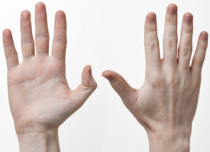
The front of the hand is its palmar surface (Photo F); it is fleshy because it includes intrinsic hand muscles (in a later lesson), forearm flexor tendons, blood vessels, nerves etc. Palmar skin is thick, hairless and lacks sebaceous glands (Anatomy Lesson #5 & Anatomy Lesson #6).  The dermis is thrown into deep ridges which increase friction and help secure grip. Dermal ridges are also the anatomical basis for fingerprints. Compared with other skin, palm skin is lighter in color because its cells have genes inhibiting melanin production and the ability to tan (Anatomy Lesson #6).

Try this: Examine the whorls of one fingertip and realize that dermal ridges create the valleys and elevations of a finger print.

The hand back is its dorsum or dorsal surface (Photo F): this is covered with hair but the skin is thin, soft and pliable and it tans. This surface also has little flesh because it contains no intrinsic hand muscles and contains flat extensor tendons, blood vessels and nerves.

Try this: Alternately supinate and pronate (Anatomy Lesson #20) your forearm to compare and contrast palmar and dorsal skin, the presence or absence of hair and the relative thickness of the skin.

SONY DSC

Photo F

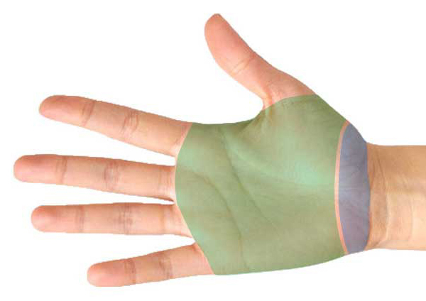
The hand is divided into wrist (blue overlay), mid-hand (green overlay) and five digits (Photo G). Fingers can be folded individually or together over the palm allowing objects to be grasped. With the palm facing up or forward, the thumb or pollex (Latin meaning thumb) points away from the body. Also, our thumbs are opposable meaning they can be brought toward each or all fingers as in a pinch.

Hand-KLS-edited

Photo G

An interesting tidbit about digits: in anatomy, the digits are numbered 1-5 beginning with thumb and ending with little finger (Table 1 -1st column). But, the English word “finger” includes the thumb (Table 1 -2nd column) and matches the anatomical digit number. Now in the U.S., digits are defined as thumb and four fingers (Table 1 -3rd column) meaning the 1st finger is also the 2nd digit! This can be a problem. Why? Let’s say a patient is scheduled for a digital procedure – the correct digit must be designated: do written instructions refer to a finger or a digit?

The good news: nowadays U.S. anatomists and clinicians preferentially use the digit name (Table 1- last column): thumb for 1st digit, index for 2nd, middle for 3rd, ring for 4th and little/small for 5th. This practice helps minimize medical error and most importantly, protects the patient! In this lesson, I use the digit naming system.

ANATOMICAL DIGIT NUMBER ENGLISH FINGER NUMBERING SYSTEM U.S. FINGER NUMBERING SYSTEM DIGIT NAME SYSTEM
1st 1st (thumb) Thumb Thumb
2nd 2nd 1st Index
3rd 3rd 2nd Middle
4th 4th 3rd Ring
5th 5th 4th Little/small

Table 1

Try this: Starting with the thumb, assign numbers to digits and repeat using digit name.

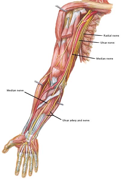
Next, let’s consider nerves of the hand. Three large nerves innervate skin, blood vessels, muscles, ligaments, joints and bone coverings (periosteum) of the hand. Some fibers provide sensory information to the brain while others stimulate muscles to contract and glands to secrete. The three nerves of the hand are: radial, median and ulnar (Photo H).

Figure0456 hand nerves KLS edited

Photo H

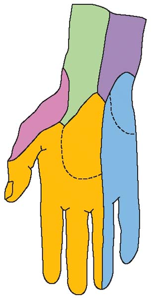
Palmar skin has one of the densest arrays of nerve endings in the human body and is an exquisite source of tactile info to the brain. Here, sensation in the form of pain, temperature, vibratory sense, pressure and touch is mostly provided by median (gold) and ulnar (blue) nerves (Photo I). And, yes, believe it or not sensation to the palmar surface of ring finger is split down the midline between median and ulnar nerves!

Figure0455A hand nerves KLS edited

Photo I

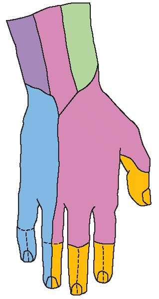
Sensation to the dorsum of the hand is supplied mostly by radial (pink) and ulnar (blue) nerves but the median nerve (gold) supplies some fingertips (Photo J).

Figure0455B hand nerves KLS edited

Photo J

Try this: Place index and middle fingers in the groove behind the medial epicondyle of one humerus (Anatomy Lesson #20) and move the fingers up and down. Feel a faint shocking sensation? Congrats! You found your ulnar nerve! Supinate the forearm and press deeply with the thumb just above the wrist. Feel the discomfort? You just found your median nerve (involved in carpal tunnel syndrome)!

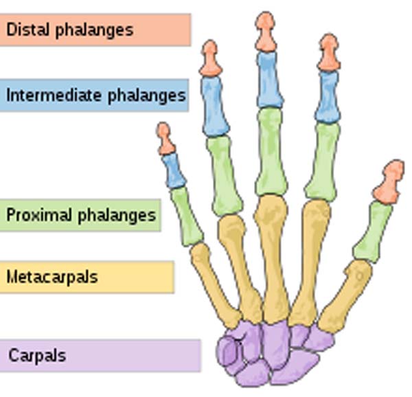
Next, let’s examine the hand skeleton: each hand contains 27 bones. Together both hands contain more than 25% of the 206 bones of the entire human skeleton! This is not only amazing it also accounts for hand movements: more bones = more joints = more movements. Hand bones include (Photo K – palm faces the face): 8 carpal bones (purple) of wrist; 5 metacarpal bones (orange) of mid-hand; 14 phalanges of the digits. Finger phalanges are further named by position: proximal phalanges (green) articulate with metacarpal bones and intermediate phalanges; intermediate phalanges (blue) articulate with proximal and distal phalanges; distal phalanges (peach) articulate with intermediate phalanges. Note that the thumb has only two (proximal and distal) phalanges.

hand bones_KLS edited

Photo K

Try this: Palpate your hand and locate all 5 groups of hand bones.

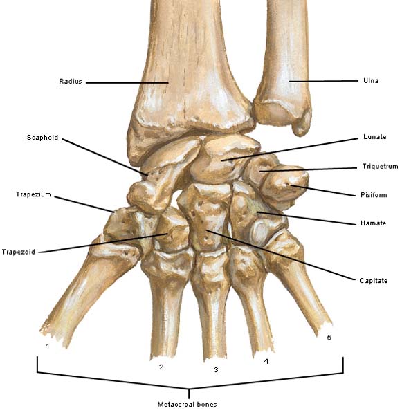
The carpus (Latin for wrist) consists of eight interlocking carpal bones arranged in two rows of four bones each (Photo L – supinated hand points away from body). The main roles of the carpus are to facilitate effective positioning of the hand, increase freedom of movement at the wrist, and form an arch (carpal tunnel) through which passes tendons and a nerve.

Carpal bones have Latin names meaning moon-shaped, boat-shaped, three-cornered, etc. They are named beginning on the thumb (metacarpal #1) side of the near row and moving to the thumb side of the far row.

Figure0435A carpal bones KLS edited

Photo L: Figure0435A carpal bones KLS edited

How does one recall carpal names and positions? This is a fun and popular mnemonic used by anatomists and one designed to keep student attention: Some Lovers Try Positions That They Can’t Handle. This one is suitable for public consumption – some are not: anatomists being naughty!

ROW NUMBER BONE NAME BONE NAME BONE NAME BONE NAME
Proximal Row Some (scaphoid) Lovers (lunate) Try (triquetrum) Positions (pisiform)
Distal Row That (trapezium) They (trapezoid) Can’t (capitate) Handle (hamate)

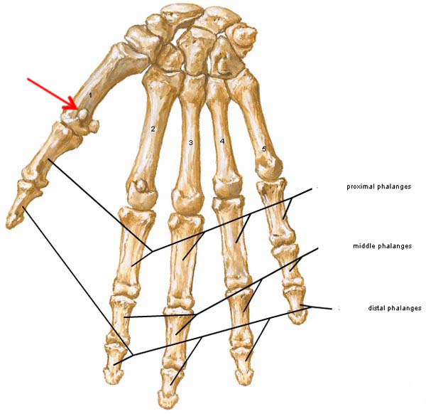
The mid-hand (palm) region contains five metacarpals one for each digit and labelled #1- #5 beginning with the thumb (Photo M).

The digits contain phalanges: proximal and distal for thumb and proximal, middle and distal for each finger (Photo M). Do the math: yep, adds up to 27 bones! The hand also contains a few small sesamoid bones (red arrow) but their numbers are inconsistent and not included in the bone count.

Figure0439A-phalanges-KLS-edited

Photo M

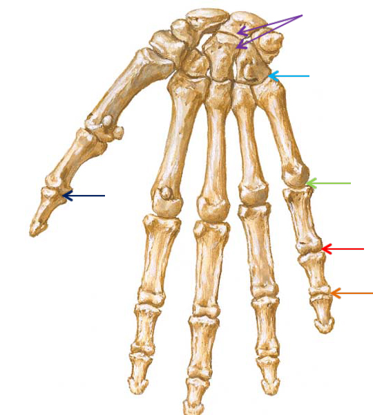
The number of joints in the hand is mind boggling! Here they are (Photo N):

  • Intercarpal (IC) Joints: between and among the carpal bones (purple arrows).
  • Carpometacarpal (CM) Joints: between distal row of carpal bones and metacarpal bones (blue arrow).
  • Metacarpophalangeal joints (MP) Joints: between metacarpals and proximal phalanges (green arrow).
  • Proximal interphalangeal (PIP) joints: between proximal and middle phalanges of fingers (red arrow).
  • Distal interphalangeal (DIP) joints: between middle and distal phalanges of fingers (orange arrow).
  • Interphalangeal (IP) Joint: Between two phalanges of thumb (black arrow).

Figure0439A-joints-KLS-edited

Photo N

Try this: with your own hand, identify as many of these joints as possible.

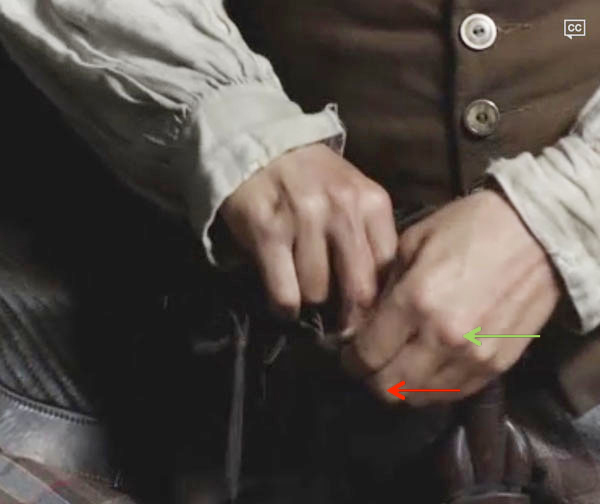
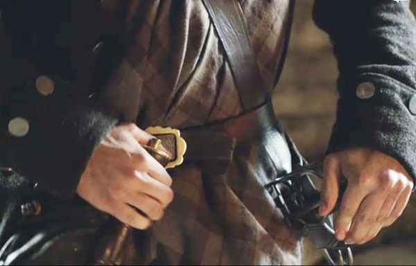
This is a terrific example from Starz episode 107 The Wedding showing some of hand joints as Jamie reaches into his sporran to retrieve his brooch (green arrow = MP joint; red arrow = PIP joint). Murtagh just wants to spit-polish the pin. To be that pin…sigh…

ep-107-finger-flexion-02-KLS-edited

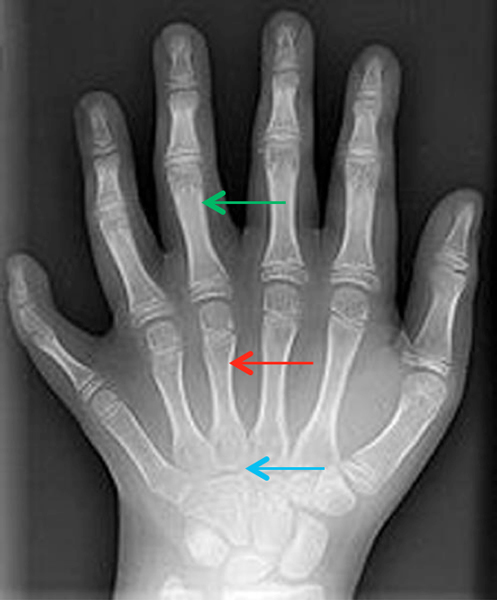
An interesting and not uncommon congenital condition is the presence of supernumerary digits on hands/feet. Known as polydactyly, the additional digit(s) may or may not be functional. Photo O is an x-ray of a child’s hand with an additional normal metacarpal (red arrow) and digit (green arrow). Here, metacarpals crowd at the CM joints (blue arrow) to make room for the extra bone. Supernumerary digits may be surgically treated. The world record holder was born with 34 digits on hands and feet. Our Jamie could use another one of these for sure!

polydactyly-KLS-edited

Photo O

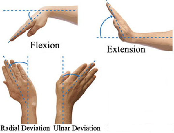
As already noted, the hand is capable of many movements because of the numerous bones and joints. There are 16 classical hand movements although many more combos are possible. Five movements occur at the wrist (Photo P) giving it considerable flexibility: flexion, extension, radial deviation, ulnar deviation and circumduction. Contraction of forearm flexors causes wrist flexion and contraction of forearm extensors causes wrist extension. Interestingly, radial deviation (hand draws toward radius) occurs if radial extensor and radial flexor contract simultaneously. Ulnar deviation (hand draws toward ulna) occurs when ulnar extensor and ulnar flexor contract simultaneously. Circumduction (not shown) occurs as the wrist rolls in a circular motion.

Flexion_Extension_Pronation_Supination KLS edited

Photo P

Try this: Mimic the four wrist movements as shown In Photo P. Try circumduction which is not shown.

How about examples of wrist movements from Outlander episodes? Here are two to keep you focused!

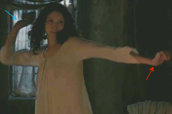
Claire has one major throwing arm! Here she extends her left wrist and elbow joint for balance (red arrow); her right wrist is also extended (blue arrow) but the same elbow joint is flexed as she launches crockery at Jamie! She’ll not let him beat her (Starz episode 109, The Reckoning) without opening up her own can of whoop ass!

ep-109-wrist-extension-KLS-edited

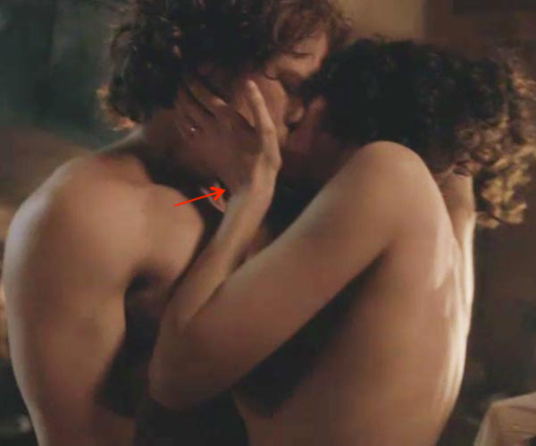
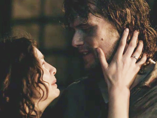
Here in a steamy scene of make-up sex, Claire’s left wrist is in ulnar deviation (hand moves toward ulna – red arrow) as she gently touches the face of her warrior husband (Starz episode 109, The Reckoning). Gah! Scoop me up with a spoon…what a scene!

ep-109-ulnar-deviation-KLS-edited

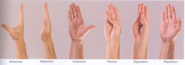
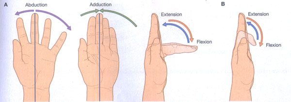
Next, let’s examine movements of the thumb, an extremely versatile digit. Photo Q shows 6 classic thumb movements: abduction (thumb moves 90° from palm and fingers); adduction (thumb returns to resting position); extension (thumb moves away from the fingers but remains in the same plane); flexion (thumb curls in front of the palm); opposition (thumb pinches one or all fingers). In the resting position or reposition, thumb is adjacent to palm and fingers. One last thumb movement is circumduction (not shown) wherein the thumb is rolled in a circular motion.

thumb movements KLS edited

Photo Q

Try This: Move your thumb through all 7 movements as described above.

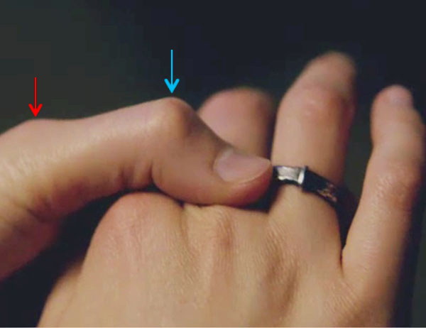
Thumb movements are plentiful in Starz episodes. Here is Claire using her left thumb to steady the beautiful ring placed on her right ring finger by The Big Red One (Starz episode 107, The Wedding). Her thumb IP joint (blue arrow) is flexed; MP joint (red arrow) is mostly extended.

ep-107-IP-joint-KLS-edited

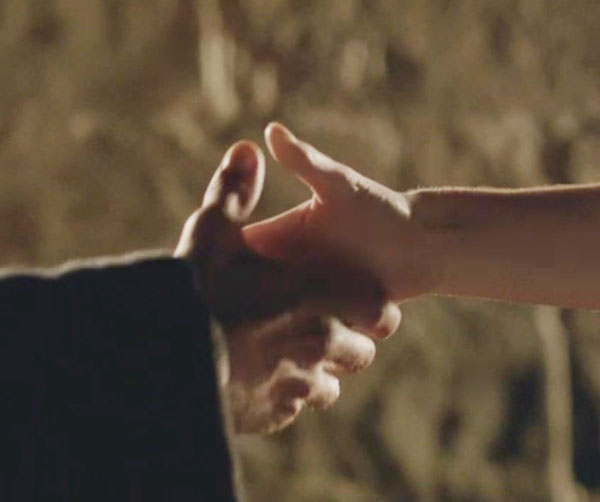
We havena see Dougal for a while so here he executes one of his devious, seal-a-deals with Claire: if she canna free Jamie or he is dead, she will marry him (Starz episode 114, The Search). Uhhh…I dinna think Claire will marry Dougal –no way, no how – but a clever ruse to access his men for a rescue attempt.

ep 114 The Search KLS edited

Finally there are four finger movements: abduction wherein index, ring and little fingers move away from the middle finger (Photo R –purple arrows). By definition the middle finger abducts no matter which direction it sweeps (think windshield wiper). Adduction is returning fingers to the resting position (Photo R –green arrows). Finger abduction and adduction are carried out by intrinsic muscles of the hand which will be cover in a later lesson.

Finger flexion occurs when one or more fingers are curled toward the palm and includes some or all MP, PIP and DIP joints (Photo R –red arrows). Contraction of forearm flexors (and intrinsic hand muscles) produces these movements. Finally, finger extension (Photo R -blue arrows) occurs when one or more fingers are straightened. Contraction of forearm extensors produces these movements.

finger movements KLS edited

Photo R

Try this: Using the fingers, try flexion, extension, abduction and adduction.

Claire is in our sights once again as she circles Jamie (Starz episode 107, The Wedding). Her left fingers are extended and abducted; thumb is extended. Not only is this a great example of hand movement butt, oops, but, it is also…well, we all ken this hand gesture!

ep 107 finger extension & abduction 02 KLS edited

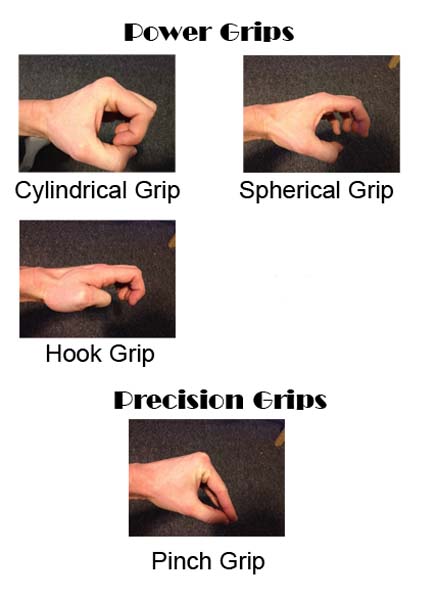
Finally, thumb and fingers provide various power grips the muscular force generated by the hands (Photo S). Grip is important in everyday life and for most athletic ventures.

    • Cylindrical power grip uses thumb abduction and finger flexion to tightly grasp a rounded object.
    • Spherical power grip is similar but thumb abduction and finger flexion is looser as in gripping a larger object.
    • Hook power grip uses extended MP finger joints but flexed DIP and PIP joints (thumb may be flexed or abducted) as in carrying the handle of a bucket.
    • Precision pinch grip is used to grasp small or delicate items.

types of grips KLS edited

Photo S

Jamie exhibits a hook power grip as his right hand clutches his dirk: MP joints extended, PIP and DIP joints flexed. He’s headed for Fort William with an empty gun and his own bare hands to rescue Claire from the redcoats (Starz episode 109, The Reckoning)!

ep 109 grip KLS edited

Next, is a great example of precision pinch grip in which right thumb opposes right index finger as Jamie clutches his doomed petition of complaint for the Duke of Sandringham. Humm…wonder what those bloody MacDonalds are doing here…

ep 110 opposition KLS edited

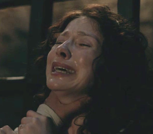
Next is a terrifying example of a cylindrical power grip with BJR’s left thumb abducted and fingers flexed around Claire’s swanlike neck (Anatomy Lesson #12). He is hurting her and her face is sheened with tears and “the sweat of exertion” as narrated by BJR (Starz episode 115, Wentworth Prison).

ep 115 grip Claire KLS edited

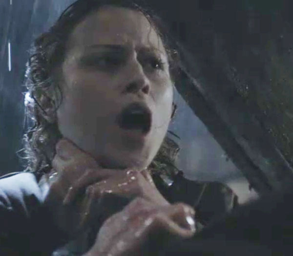
Yet another example of the cylindrical power grip. Look familiar (Starz episode 108, Both Sides Now)? Remind you of anyone you know? Puir not-her-name Sally! She messed with the wrong guy…iffy spherical power-grip genes in that line.

ep 108 grip on Sally KLS edited

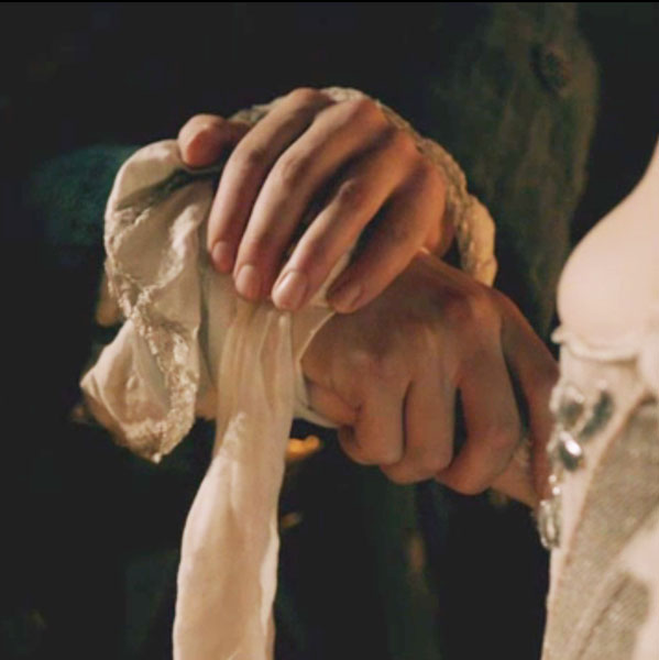
As if we need a reminder, our beloved Jamie’s hands are nimble and elegant (Starz episode 107, The Wedding). In fact, his fingers and hands play their own characters just as Claire’s hair does (Anatomy Lesson #6) throughout the books. This was especially evident during his marriage to Claire. Here his left fingers drape gracefully over his right wrist as he utters his blood vow. His left thumb is slight abducted. His right hand executes a loose spherical grip around Claire’s right wrist and forearm. Stunning imagery!

ep 107 hand holding 02 KLS edited


warning

WARNING: the time has come to examine the damage to Jamie’s hand. After the next few paragraphs, his torture will begin so skip to the kitten if you must.

Fast forward to Starz episode 115, Wentworth Prison, an episode thoughtfully rated “graphic violence”!

About midway through the episode, Jack makes an offer Jamie canna refuse: a swift and honorable death in exchange for free use of his body. But, Jamie does refuse: “I’ll not surrender to you or any man.” The foul fiend vows “I will have your surrender before you leave this world.”

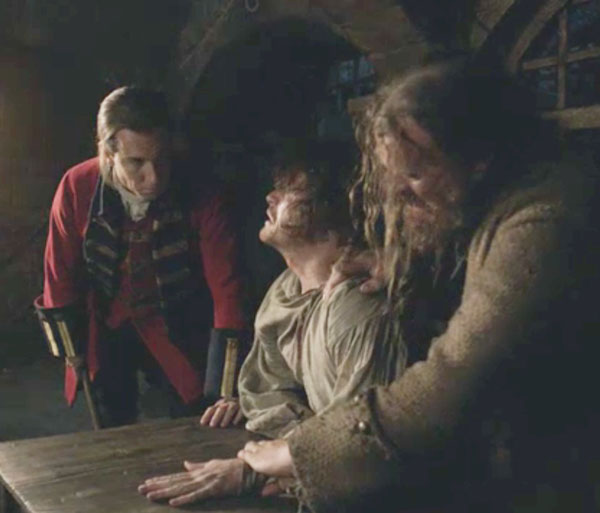
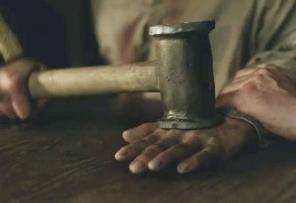
Next, a great scuffle ensues given that Jamie is half starved, chained by an ankle manacle and weaponless against two men, one a minor league giant. Black Jack asks “why do you force me to hurt you?” Oh yeah, Jamie’s forcing your hand! As instructed, genius Marley slams Jamie’s left hand on the wooden table as Jack grasps the mallet (aye, he has it in a spherical power grip).

ep 115 Jamies hand 11 KLS edited

Jack smashes the mallet on Jamie’s left mid-hand followed by about three more strikes. The vulgar villain then launches a volley of totally insane queries: Why do you force me to treat you in such an abominable way? Why do you choose to spend the few hours left to you as a miserable cripple? Why do you force me to hurt you? Och, he’s a delusional devil!

ep 115 Jamies hand 05 KLS edited

Soon, Claire finds Jamie alone (from Outlander book):

“What has he done to you?” I asked … Moving with exquisite care, he used his left hand to lift the object he had been cradling. It was his right hand, almost unrecognizable as a human appendage. Grotesquely swollen, it was now a bloated bag, blotched with red and purple, the fingers dangling at crazy angles. A white shard of bone poked through the torn skin of the middle finger, and a trickle of blood stained the knuckles, puffed into shapeless dimples.”

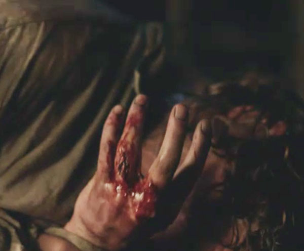
But a minor detail, the left hand is mashed, otherwise the destruction is akin to the Outlander book’s version: swollen fingers, black and blue skin, blood, bone/tendon exposed. A heavy mallet messes with delicate hand anatomy!

ep 115 mangled hand KLS edited

Unfortunately the effing, sadistic piece of shite and his Igor return to find Claire with Jamie. An engrossing exchange ensues between the black bastard and Claire as Marley adds his own singular brand of visual horror. With a flare of desperate strength and courage, Claire shoves Marley and pushes BJR into a wall in a vain attempt to strangle him. Marley ends up dead and Claire ends up hostage.

Next arrives the pivotal moment in this entire episode: Jamie, accepting that he will die on the morrow offers himself in exchange for Claire’s life. “Let her go in safety and you can have me. I won’t struggle. You, you can do as you wish. You have my word!” His body is the only card he has left to play in this gruesome game and he uses it to ransom Claire. Diana explains (Outlander book):

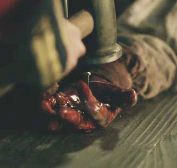
Randall walked slowly…picking up the mallet as he went. He held it up, ironically questioning. “You’ll allow me a brief test of your sincerity?” “Aye.” Jamie’s voice was as steady as his hands, flat and motionless on the table. I tried to speak, to utter some protest, but my throat had dried to a sticky silence. Moving without haste, Randall… positioned the point with care and brought the mallet down, driving the nail through Jamie’s right hand into the table with four solid blows.

In the Starz episode, Jamie’s left palm faces up as Jack impales it to the table. Intentional or not and regardless of your philosophical bent, the shocking image of Jamie’s palm pierced by a nail driven into wood is the ultimate symbol of sacrifice in the Western world. And, a costly sacrifice it is.

ep 115 Jamies hand 02 KLS edited

peeking

peeking

It’s safe to come out now! Had to use two kittens just to make sure…

“I love you, mo nighean donn.”

ep 115 Jamie and Claire kiss 01 KLS edited

What bolsters us, is the knowledge that Claire’s loving kiss will help sustain Jamie trapped in the vortex of Jack’s dark madness.

Again, from Neil:

Warm, touchin’ warm

Reachin’ out, touchin’ me, touchin’ you

ep 115 Claires hand KLS edited

Now, a few final comments for the sake of science: take heart all! in Outlander book Claire observes that Jamie handles a fifteen pound Claymore as though it were a mere flywhisk. Jamie is a warrior, an athlete whose limbs are heavily muscled. Many studies have shown that muscular activity thickens bones; ergo, Jamie’s hand bones will tolerate more abuse than most because he is so fit and fine.

Further, will the nail wound greatly damage Jamie’s hand? Actually, not so much! In the living, nerves, tendons and larger blood vessels are slippery and will likely slide away from the nail path. But, the pain is agonizing: pain receptors of the mangled hand are already on fire from the mallet and the nailing adds fuel to that inferno. But, of the two tools, the mallet is by far the greater destroyer.

Farewell for this lesson. Let us join hands in the hope that Starz episode 116 sees Claire redeem Jamie’s hand and his soul!

The deeply grateful,

Outlander Anatomist

Follow me on:

Photo Credits Starz, Gray’s Atlas of Anatomy for Students, 2005, Netter’s Atlas of Human Anatomy, 4th ed., Clinically Oriented Anatomy, 5th ed., www.breakingmuscle.comwww.britannica.comwww.cs.cmu.edu (types of grips) , www.pinterest.com, www.physioadvisor.com.au, www.pixshark.com, www.researchgate.net, www.slice-works.com, www.the anatomist.wordpress.com (thumb movements), www.ittcs.wordpress.com (arches of hand with grip), www.wikipedia.orgwww.wikiradiography.com, www.webmd.com, www.heroesandheartbreakers.com, www.englishstackexchange.com.

Anatomy Lesson #6: “Claire’s Hair – Jamie’s Mane” or “Jesus H. Roosevelt Christ!”

Hallo again, friends of Outlander Anatomy! Today’s Anatomy Lesson #6:  The Skin – Part 2, Hair, will continue with skin but, today, will focus on hair, hair follicles, arrector pili muscles and sebaceous glands, all of which you learned from Skin Part 1 are made by skin and are therefore appendages of this organ.

Now, before we get on with today’s lesson, I must confess that I did a quiet switcheroo on you in the last anatomy lesson. My first four lessons were confined to that part of human anatomy known as gross anatomy, the field revealed by human dissection.

image

Nay…not that kind of gross, Rupert!  It is termed “gross” not because it is yucky, but because it deals with structures visible to the naked eye. In Anatomy Lesson #5, I switched (without telling) to another field of human anatomy, that of microscopic anatomy.

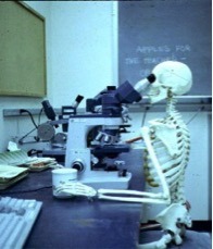
Microscopes are used to magnify structures too wee for us to see with eyes unaided by magnifying lenses. Many of today’s images are drawings made from images observed with a compound microscope such as this one (photo A):

image

photo A

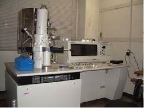
Once again there are 3-D images taken with powerful SEM/scanning electron microscope (Photo B). I have used both types of microscopes many times in teaching and various research projects!

image

photo B

Now, getting in the mood for today’s Anatomy Lesson: Skin 2 – the Hair! As with skin, Herself often writes about hair in the Outlander books, offering her audience a more intimate glimpse into characters and situations through vivid use of this physical trait. So, once again, I begin our lesson with images from the Starz Outlander series and with words from the Outlander books.

Let’s begin with our heroine. Early in Starz episode 1, Sassenach, Claire emerges from the roadster standing in the picturesque village of Inverness.  We can clearly appreciate her dark brown hair – very full and very curly.

image

Later, during a lighting storm, Herself writes

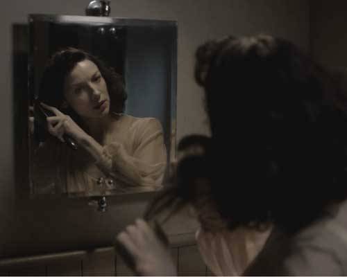
The wind was rising and the very air of the bedroom was prickly with electricity. I drew the brush through my hair, making the curls snap with static and spring into knots and furious tangles!

The humid air makes Claire’s hair wildly curly and disobedient (Starz, episode 101, Sassenach) to which she exclaims: Jesus H. Roosevelt Christ!!!

image

All the while, someone is awatching her futile struggles through the window of her room.

image

Nay, it isn’t a peeping tom, it is a keeking Jamie! Ha!

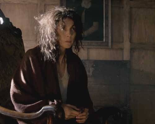
This next image of Claire always makes me laugh! In Starz episode 102, Castle Leoch, Mrs. Fitz unceremoniously rouses Claire from her sleep, seats her in a chair and hands her a cup ‘o brakfast fer her empty belly. Mrs. Fitz then whisks it away afore Claire even finishes! Look at Claire’s hair! It is absolutely fabulous! She certainly looks like the “wee milkweed” Jamie affectionately calls her later in the Outlander book.

“Fretful porpentine, was it?” he asked. He tilted his head, examining me inquisitively. “Mmm,” he said, running a hand over his head to smooth down his own hair. “Fretful, at least. You’re a fuzzy wee thing when ye wake, to be sure.” He rolled over toward me, reaching out a hand. “Come here, my wee milkweed.

image

With these great images to set the mood, it is time for our anatomy lesson on hair and with it a lot of  juicy tidbits to share!

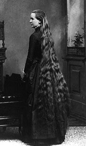
First, the length of body hair varies a lot – from less than 1 mm (.04 in) on the forehead to well over 1 m (3.3 ft) with long scalp hair (Photo C)! But, the wee hairs of the eyelids (not the eyelashes) are so short they barely reach the skin surface! And, you should know that most hair grows very rapidly, about 0.3 mm/day or 1 cm/per month.

image

photo C

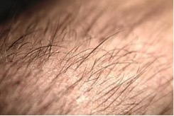
You should also know that hair does not grow straight out of the skin; it emerges at a slant (Photo D).

Try this: Check the angle of growth of your own hair: place your forearm on a flat surface with the palm down. Examine your forearm hairs and see that they are angled toward the little finger side of the forearm. That’s the slant I’m a talking about.

image

photo D

Hair is also denser in some skin areas than in others: the face has about 600 hairs/cm2 (.16 in2) compared to about 60 hairs/cm2 on the rest of the body. 

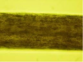
Hair diameter also varies greatly but even the coarsest hair is only about .5 mm (.02 in) in diameter (Photo E). Even so, a scalp hair is strong enough to support the weight of 100 gm (3.5 oz)!

image

photo E

Another interesting tidbit: Human hair grows autonomously; each hair cycles at its own pace through periods of growth and periods of quiescence. If all our hair were on the same cycle, we would molt!

image

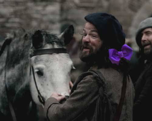
And sometimes our hair does unspeakable things and we just have to pull it outta the way like Angus here who does prefer a wee bit o’ purple ribbon fer his scalp hairs!

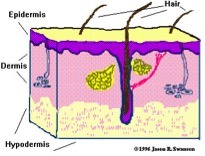
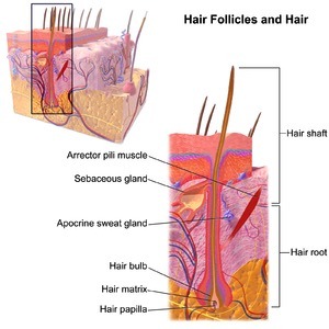
Now back to microscopic anatomy! Using the same image from Skin – Part 1, I’ll be reminding ye that skin is divided into a thin outer epidermis that overlies a thicker dermis. And, although not part of skin the hypodermis lies deeper still. The dermis and hypodermis also anchor structures that we’ll cover in this anatomy lesson: hair, hair follicles, arrector pili muscles, and sebaceous glands (Photo F).

image

photo F

Hairs emerge from hair follicles which are down growths of the epidermis (Photo G). The internal anatomy of each follicle is verra complex so I’m simplifying it: the hair and its follicle are divided into a hair root and a hair shaft. At the root is a bulb where cells divide and push older cells toward the surface to form the hair shaft!

image

photo G

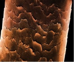
Along the way, hair cells harden and get plastered together so by the time the hair clears the skin surface, the cells are dead, flat and stiff with their free edges pointing toward the hair tip. They also overlap each other like shingles on a roof (Photo H). This is a SEM image of a single hair!

image

Photo H

Ye should also ken that hair follicles are verra sensitive to the influence of hormones! These chemicals produce secondary sex characteristics such as hair distribution. In fact, the distribution of hair between the two sexes play an important role in socio-sexual communications!

In women, estrogens (oestrogens) cause most body hair to develop as short, thin vellus hairs that are anchored in the dermis. Both genders exhibit the coarse terminal hairs of scalp, eyelashes, eyebrows, axilla and pubis that are embedded deep in the hypodermis.

In men, androgens (testosterone being the most important) also convert facial and chest hairs into terminal hairs. Now then, isna this the right place to offer praises to Dougal MacKenzie who won Saturday’s Starz contest with his comely beard? Congrats! It looks mighty fine on ye, man! Tulach Ard!

image

And, no anatomy lecture is ever complete without at least one image of a half-dressed Jamie! So here is his chest hair just in case ye be forgettin’!  No verra damn likely! Gawd!

image

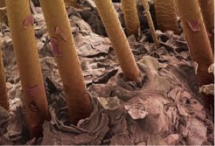
Something else: When viewed by SEM, straight hair has a round shaft as seen in this photo of scalp hair (Photo I –computer generated color); the surrounding dead skin cells look like scatter leaves on a forest floor.

image

Photo I

Murtagh’s scalp offers a perfect example of straight hair – here he is explaining to Claire why Jamie is nowhere to be seen (Starz episode 5, Rent)! Plus, he has mighty fine eyes and braw eyebrows just in case ye been so focused on Jamie that ye havena been noticing!

image

Scalp hair that is curly like Claire’s…

image

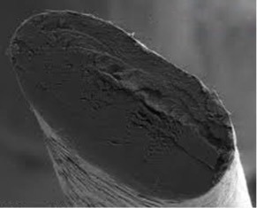
…has a shaft that is flattened in cross-section as shown in this SEM image (Photo J). The flatter the shaft, the curlier the hair!

image

Photo J

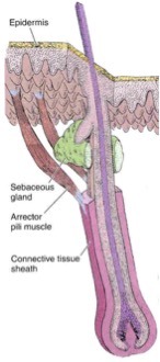
Now, onto a couple of other structures associated with the hair follicle. First, stretched between the follicle and the dermis is a thin band of tissue, the arrector pili muscle. Second, between the hair follicle and the arrector pili muscle lays one or more sebaceous glands with ducts opening into the hair follicle (Photo K). Sebaceous glands produce sebum, a complex mixture of fats, waxes and other materials.

image

photo K

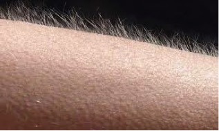
The arrector pili muscles are made of smooth muscle cells that are not under conscious control. They contract in response to cold or the fright, flight, fight reflex! Contractions of this muscle elevate the hair, forming goose bumps or goose flesh and help squeeze sebum from the sebaceous glands into the hair follicle and onto the hair shaft (Photo L).

image

Photo L

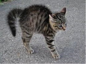
Contraction of the arrector pili muscles in animals traps air between the erect hairs to retain body heat or to help the creature appear more fierce (Photo M)! This adaptation isn’t of much use to us short haired humans but the release of sebum does help lubricate and protect the hair itself.

image

Photo M

Finally, on to hair color! Like the epidermis, hair color requires the presence of melanin; melanocytes in the hair bulb synthesize melanin and package it into granules that move up the hair shaft as it forms. Now, it turns out that there are a couple of different types of melanin!

Like Claire, most hair color is due to the presence of varying amounts of brown or black eumelanin. But, now, ye are in fer a BIG surprise! I bet ye dinna ken this! Flaming red hair in one such as our Great Scott, Jamie, contains a chemically different type of melanin known as pheomelanin and this molecule is red (or red-brown)! Thus, Jamie’s gorgeous mane of red hair is due to the presence of pheomelanin as seen from the back in this image (Starz episode 7, The Wedding)!

image

And just so ye won’t ferget it, here is Jamie’s hair from the front! We can literally see the words Herself wrote in Outlander about his hair:

…a mass of auburn, copper, cinnamon and gold all gleaming together in the morning sun…

image

And one last point fer yer eddycation: Check out both upper corners of Jamie’s forehead. See how the hair line is squared off? This is known as the temporal notch; it is a secondary sexual characteristic in men brought about by the influence of testosterone. Women typically have an oval hairline in the corresponding areas of the forehead!

And now, folks, our journey through the skin and its appendages has come to an end! I do hope you have enjoyed learning about the skin ye are in and that of the Outlander cast while we are at it! At some point in the future, I will post Skin 3 – The Breast.

In the meantime, I’ll be leaving ye with these lovely words from Herself in the Outlander book and an image from Starz episode 7 (The Wedding):

You’ve the loveliest hair,” said Jamie, watching me.  ….”But it’s so .…curly,” I said, blushing a little….“Aye, of course.” ….He sat up and tugged gently on one curl, stretching it down so that, uncurled, it reached nearly to my breast…

And:

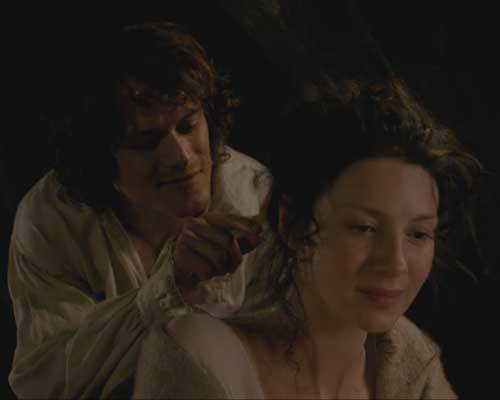
 “Mo duinne?”…“It means ’my brown one.’ ”He raised a lock of hair to his lips and smiled, with a look in his eyes that started all the drops of my own blood chasing each other through my veins. Rather a dull color, brown, I’ve always thought,”….”No, I’d not say that, Sassenach. Not dull at all.”  He lifted the mass of my hair with both hands and fanned it out. “It’s like the water in a bern, where it ruffles over the stones. Dark in the wavy spots, with bits of silver (auburn on Starz) on the surface where the sun catches it.”

image

Gah, this man has a way with words! Does he ever say anything wrong?  Just look at the look on Claire’s face! She’s both enchanting and enchanted!

Psst…next time, I will be writing about someone’s thighs and knees (guess whose?)! Stay tuned!

The deeply grateful,

Outlander Anatomist

Follow me on:

Photo credit: Starz, Cat photo from goosecam Edmonton Journal, Goosebumps from genius.com, Basic Histology by Junqueira and Carneiro, 11th ed., University of Leeds, Rochester education Foundation, Wikipedia, WebMD, Loyola University Dermatology website, Histology Guide, University of Leeds, Wikimedia.org. CSIR – Council for Scientific and Industrial Research, South Africa. OA archival photos, Aersol Research – Washington University St. Louis